|
Papillomatosis Of Breasts
Papillomatosis of the breast (PB) is a rare, benign, epitheliosis-like lesion, i.e. an overgrowth of the cells lining the ducts of glands that resembles a papilla (i.e. small rounded protuberance) or nipple-like nodule/tumor. PB tumors develop in the apocrine glands of the breast. PB is also termed juvenile papillomatosis because of its frequent occurrence in younger women (including, in uncommon cases, children and adolescent females) and Swiss cheese disease because of its microscopic appearance. Rarely, PB has also been diagnosed in very young, adolescent, and adult males. A PB tumor is typically an asymptomatic lesion that is detected on examination as a palpable but otherwise symptomless breast mass or in some cases by routine breast cancer screening methods in individuals unaware of the mass's presence. Although PB tumors are themselves benign, a significant percentage of individuals with these tumors concurrently have or will develop certain types of breast carcinomas and ... [...More Info...] [...Related Items...] OR: [Wikipedia] [Google] [Baidu] |
Breast Surgery
Breast surgery is a form of surgery performed on the breast. Types Types include: *Breast reduction surgery *Augmentation mammoplasty *Mastectomy *Lumpectomy *Breast-conserving surgery, a less radical cancer surgery than mastectomy *Mastopexy, or breast lift surgery * Surgery for breast abscess, including incision and drainage as well as excision of lactiferous ducts * Surgical breast biopsy * Microdochectomy (removal of a lactiferous duct) Complications After surgical intervention to the breast, complications may arise related to wound healing. As in other types of surgery, hematoma (post-operative bleeding), seroma (fluid accumulation), or incision-site breakdown (wound infection) may occur. Breast hematoma due to an operation will normally resolve with time but should be followed up with more detailed evaluation if it does not. Breast abscess can occur as post-surgical complication, for example after cancer treatment or reduction mammaplasty.Noel Weidner, Chapter ''Infectio ... [...More Info...] [...Related Items...] OR: [Wikipedia] [Google] [Baidu] |
Proteus Syndrome
Proteus syndrome is a rare disorder with a genetic background that can cause tissue overgrowth involving all three embryonic lineages. Patients with Proteus syndrome tend to have an increased risk of embryonic tumor development.Freedberg, et al. (2003). ''Fitzpatrick's Dermatology in General Medicine''. (6th ed.). McGraw-Hill. . The clinical and radiographic symptoms of Proteus syndrome are highly variable, as are its orthopedic manifestations. Only a few more than 200 cases have been confirmed worldwide, with estimates that about 120 people are currently alive with the condition.Woman's 11-stone legs may be lost at As attenuated forms of the disease may exist, there could be many people with ... [...More Info...] [...Related Items...] OR: [Wikipedia] [Google] [Baidu] |
HER2/neu
Receptor tyrosine-protein kinase erbB-2 is a protein that in humans is encoded by the ''ERBB2'' gene. ERBB is abbreviated from erythroblastic oncogene B, a gene originally isolated from the avian genome. The human protein is also frequently referred to as ''HER2'' (human epidermal growth factor receptor 2) or CD340 (cluster of differentiation 340). HER2 is a member of the human epidermal growth factor receptor (HER/EGFR/ERBB) family. But contrary to other member of the ERBB family, HER2 does not directly bind ligand. HER2 activation results from heterodimerization with another ERBB member or by homodimerization when HER2 concentration are high, for instance in cancer. Amplification or over-expression of this oncogene has been shown to play an important role in the development and progression of certain aggressive types of breast cancer. In recent years the protein has become an important biomarker and target of therapy for approximately 30% of breast cancer patients. Name ''H ... [...More Info...] [...Related Items...] OR: [Wikipedia] [Google] [Baidu] |
Epidermal Growth Factor Receptor
The epidermal growth factor receptor (EGFR; ErbB-1; HER1 in humans) is a transmembrane protein that is a receptor for members of the epidermal growth factor family (EGF family) of extracellular protein ligands. The epidermal growth factor receptor is a member of the ErbB family of receptors, a subfamily of four closely related receptor tyrosine kinases: EGFR (ErbB-1), HER2/neu (ErbB-2), Her 3 (ErbB-3) and Her 4 (ErbB-4). In many cancer types, mutations affecting EGFR expression or activity could result in cancer. Epidermal growth factor and its receptor was discovered by Stanley Cohen of Vanderbilt University. Cohen shared the 1986 Nobel Prize in Medicine with Rita Levi-Montalcini for their discovery of growth factors. Deficient signaling of the EGFR and other receptor tyrosine kinases in humans is associated with diseases such as Alzheimer's, while over-expression is associated with the development of a wide variety of tumors. Interruption of EGFR signalling, either by ... [...More Info...] [...Related Items...] OR: [Wikipedia] [Google] [Baidu] |
Progesterone Receptor
The progesterone receptor (PR), also known as NR3C3 or nuclear receptor subfamily 3, group C, member 3, is a protein found inside cells. It is activated by the steroid hormone progesterone. In humans, PR is encoded by a single ''PGR'' gene residing on chromosome 11q22, it has two isoforms, PR-A and PR-B, that differ in their molecular weight. The PR-B is the positive regulator of the effects of progesterone, while PR-A serve to antagonize the effects of PR-B. Mechanism Progesterone is necessary to induce the progesterone receptors. When no binding hormone is present the carboxyl terminal inhibits transcription. Binding to a hormone induces a structural change that removes the inhibitory action. Progesterone antagonists prevent the structural reconfiguration. After progesterone binds to the receptor, restructuring with dimerization follows and the complex enters the nucleus and binds to DNA. There transcription takes place, resulting in formation of messenger RNA that is tra ... [...More Info...] [...Related Items...] OR: [Wikipedia] [Google] [Baidu] |
Estrogen Receptor
Estrogen receptors (ERs) are a group of proteins found inside cells. They are receptors that are activated by the hormone estrogen ( 17β-estradiol). Two classes of ER exist: nuclear estrogen receptors (ERα and ERβ), which are members of the nuclear receptor family of intracellular receptors, and membrane estrogen receptors (mERs) (GPER (GPR30), ER-X, and Gq-mER), which are mostly G protein-coupled receptors. This article refers to the former (ER). Once activated by estrogen, the ER is able to translocate into the nucleus and bind to DNA to regulate the activity of different genes (i.e. it is a DNA-binding transcription factor). However, it also has additional functions independent of DNA binding. As hormone receptors for sex steroids (steroid hormone receptors), ERs, androgen receptors (ARs), and progesterone receptors (PRs) are important in sexual maturation and gestation. Proteomics There are two different forms of the estrogen receptor, usually referred to as α a ... [...More Info...] [...Related Items...] OR: [Wikipedia] [Google] [Baidu] |
Immunohistochemistry
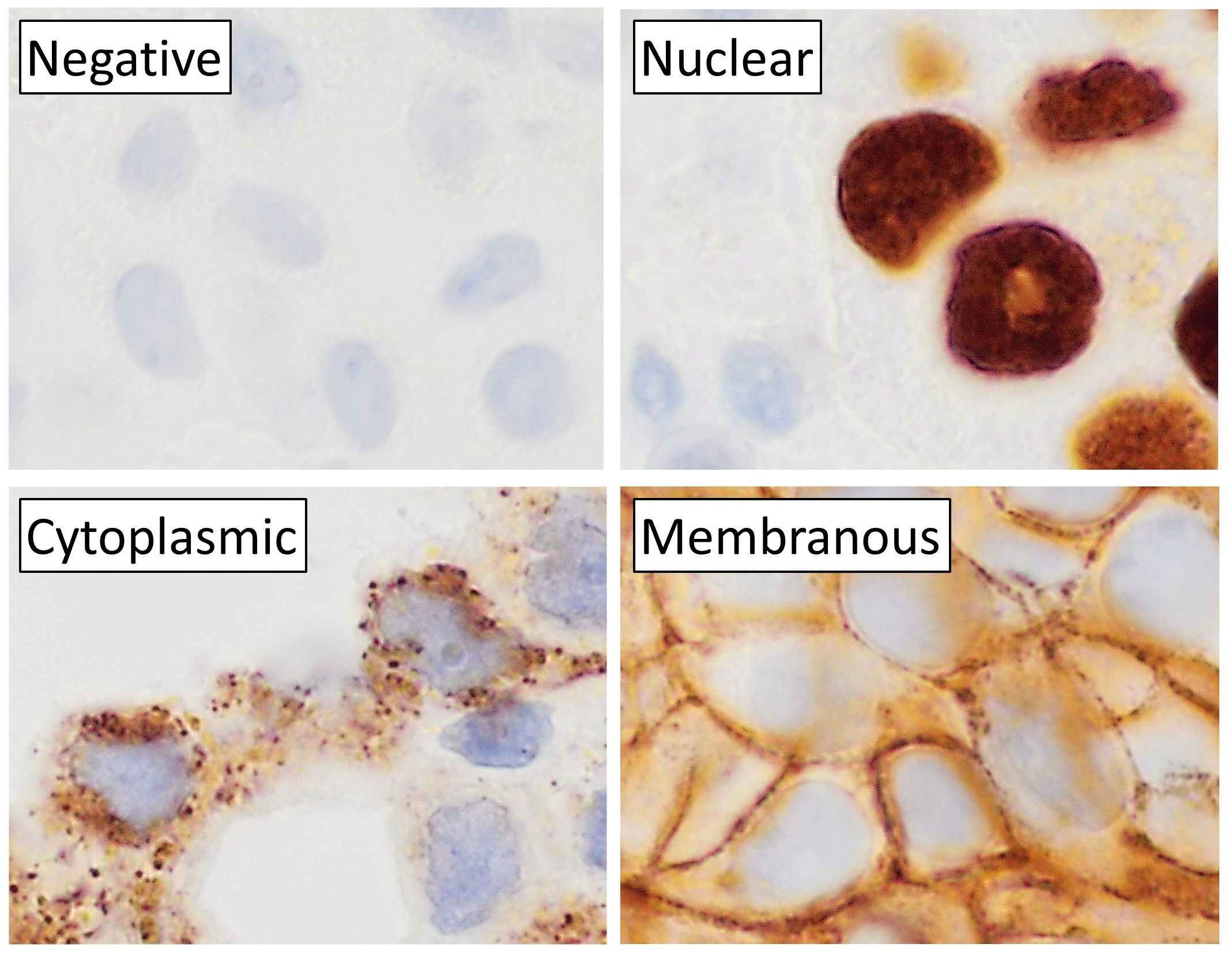
Immunohistochemistry (IHC) is the most common application of immunostaining. It involves the process of selectively identifying antigens (proteins) in cells of a tissue section by exploiting the principle of antibodies binding specifically to antigens in biological tissues. IHC takes its name from the roots "immuno", in reference to antibodies used in the procedure, and "histo", meaning tissue (compare to immunocytochemistry). Albert Coons conceptualized and first implemented the procedure in 1941. Visualising an antibody-antigen interaction can be accomplished in a number of ways, mainly either of the following: * ''Chromogenic immunohistochemistry'' (CIH), wherein an antibody is conjugated to an enzyme, such as peroxidase (the combination being termed immunoperoxidase), that can catalyse a colour-producing reaction. * '' Immunofluorescence'', where the antibody is tagged to a fluorophore, such as fluorescein or rhodamine. Immunohistochemical staining is widely used in the dia ... [...More Info...] [...Related Items...] OR: [Wikipedia] [Google] [Baidu] |
Necrosis
Necrosis () is a form of cell injury which results in the premature death of cells in living tissue by autolysis. Necrosis is caused by factors external to the cell or tissue, such as infection, or trauma which result in the unregulated digestion of cell components. In contrast, apoptosis is a naturally occurring programmed and targeted cause of cellular death. While apoptosis often provides beneficial effects to the organism, necrosis is almost always detrimental and can be fatal. Cellular death due to necrosis does not follow the apoptotic signal transduction pathway, but rather various receptors are activated and result in the loss of cell membrane integrity and an uncontrolled release of products of cell death into the extracellular space. This initiates in the surrounding tissue an inflammatory response, which attracts leukocytes and nearby phagocytes which eliminate the dead cells by phagocytosis. However, microbial damaging substances released by leukocytes would crea ... [...More Info...] [...Related Items...] OR: [Wikipedia] [Google] [Baidu] |
Microcalcification
Microcalcifications are tiny deposits of calcium salts that are too small to be felt but can be detected by imaging. They can be scattered throughout the mammary gland, or occur in clusters. Microcalcifications can be an early sign of breast cancer. Based on morphology, it is possible to classify by radiography how likely microcalcifications are to indicate cancer. Microcalcifications are made up of calcium oxalate and calcium phosphate The term calcium phosphate refers to a family of materials and minerals containing calcium ions (Ca2+) together with inorganic phosphate anions. Some so-called calcium phosphates contain oxide and hydroxide as well. Calcium phosphates are whi .... The mechanism of their formation is not known. Microcalcification was first described in 1913 by surgeon Albert Salomon. References Medical signs {{Med-sign-stub ... [...More Info...] [...Related Items...] OR: [Wikipedia] [Google] [Baidu] |
Mucus
Mucus ( ) is a slippery aqueous secretion produced by, and covering, mucous membranes. It is typically produced from cells found in mucous glands, although it may also originate from mixed glands, which contain both serous and mucous cells. It is a viscous colloid containing inorganic salts, antimicrobial enzymes (such as lysozymes), immunoglobulins (especially IgA), and glycoproteins such as lactoferrin and mucins, which are produced by goblet cells in the mucous membranes and submucosal glands. Mucus serves to protect epithelial cells in the linings of the respiratory, digestive, and urogenital systems, and structures in the visual and auditory systems from pathogenic fungi, bacteria and viruses. Most of the mucus in the body is produced in the gastrointestinal tract. Amphibians, fish, snails, slugs, and some other invertebrates also produce external mucus from their epidermis as protection against pathogens, and to help in movement and is also produced in fish to line the ... [...More Info...] [...Related Items...] OR: [Wikipedia] [Google] [Baidu] |
Cyst
A cyst is a closed sac, having a distinct envelope and cell division, division compared with the nearby Biological tissue, tissue. Hence, it is a cluster of Cell (biology), cells that have grouped together to form a sac (like the manner in which water molecules group together to form a bubble); however, the distinguishing aspect of a cyst is that the cells forming the "shell" of such a sac are distinctly abnormal (in both appearance and behaviour) when compared with all surrounding cells for that given location. A cyst may contain air, fluids, or semi-solid material. A collection of pus is called an abscess, not a cyst. Once formed, a cyst may resolve on its own. When a cyst fails to resolve, it may need to be removed surgically, but that would depend upon its type and location. Cancer-related cysts are formed as a defense mechanism for the body following the development of mutations that lead to an uncontrolled cellular division. Once that mutation has occurred, the affected cell ... [...More Info...] [...Related Items...] OR: [Wikipedia] [Google] [Baidu] |
Histiocytes
A histiocyte is a vertebrate cell that is part of the mononuclear phagocyte system (also known as the reticuloendothelial system or lymphoreticular system). The mononuclear phagocytic system is part of the organism's immune system. The histiocyte is a tissue macrophage or a dendritic cell (histio, diminutive of histo, meaning ''tissue'', and cyte, meaning ''cell''). Part of their job is to clear out neutrophils once they've reached the end of their lifespan. Development Histiocytes are derived from the bone marrow by multiplication from a stem cell. The derived cells migrate from the bone marrow to the blood as monocytes. They circulate through the body and enter various organs, where they undergo differentiation into histiocytes, which are part of the mononuclear phagocytic system (MPS). However, the term ''histiocyte'' has been used for multiple purposes in the past, and some cells called "histocytes" do not appear to derive from monocytic-macrophage lines. The term Histioc ... [...More Info...] [...Related Items...] OR: [Wikipedia] [Google] [Baidu] |


