|
Papillary Urothelial Lesion
Papilla (Latin, 'nipple') or papillae may refer to: In animals * Papilla (fish anatomy), in the mouth of fish * Basilar papilla, a sensory organ of lizards, amphibians and fish * Dental papilla, in a developing tooth * Dermal papillae, part of the skin * Major duodenal papilla, in the duodenum * Minor duodenal papilla, in the duodenum * Genital papilla, a feature of the external genitalia of some animals * Interdental papilla, part of the gums * Lacrimal papilla, on the bottom eyelid * Lingual papillae, small structures on the upper surface of the tongue * Renal papilla, part of the kidney In plants and fungi * Papilla (mycology), a nipple-shaped protrusion in the center of the cap * Stigmatic papilla, part of the stigma (botany) See also * * * Blister, a small pocket of body fluid within the upper layers of the skin * Papillary muscle, a muscle in the heart * Papilloma, a benign epithelial tumor * Papule A papule is a small, well-defined bump in the skin. It may have a ro ... [...More Info...] [...Related Items...] OR: [Wikipedia] [Google] [Baidu] |
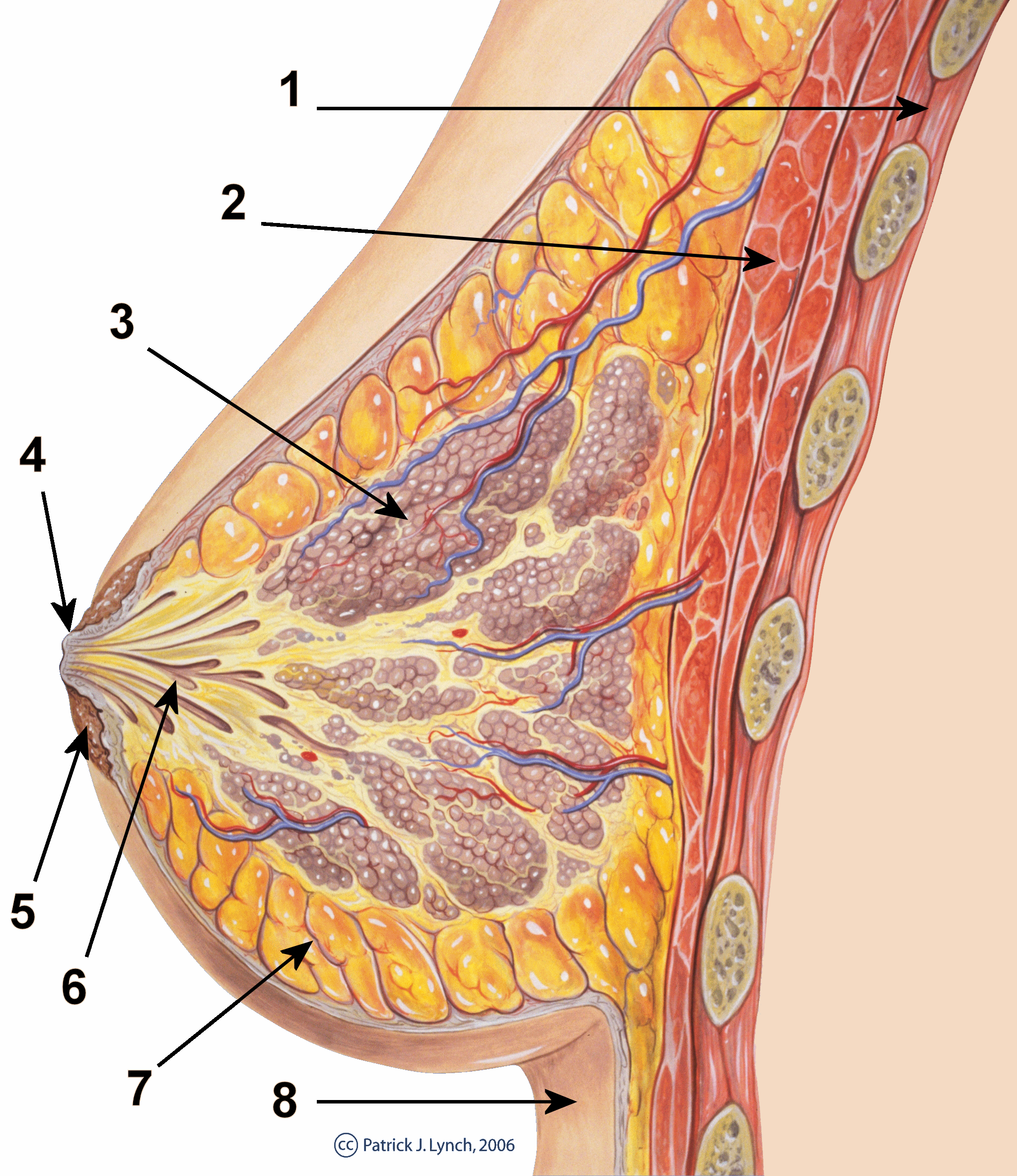
Nipple
The nipple is a raised region of tissue on the surface of the breast from which, in females, milk leaves the breast through the lactiferous ducts to feed an infant. The milk can flow through the nipple passively or it can be ejected by smooth muscle contractions that occur along with the ductal system. The nipple is surrounded by the areola, which is often a darker colour than the surrounding skin. A nipple is often called a teat when referring to non-humans. Nipple or teat can also be used to describe the flexible mouthpiece of a baby bottle. In humans, the nipples of both males and females can be stimulated as part of sexual arousal. In many cultures, human female nipples are sexualized, or "regarded as sex objects and evaluated in terms of their physical characteristics and sexiness." Anatomy In mammals, a nipple (also called mammary papilla or teat) is a small projection of skin containing the outlets for 15–20 lactiferous ducts arranged cylindrically around the tip. Ma ... [...More Info...] [...Related Items...] OR: [Wikipedia] [Google] [Baidu] |
Lacrimal Papilla
The lacrimal papilla is the small rise in the bottom (inferior) and top (superior) eyelid just before it ends at the corner of the eye closest to the nose. At the medial edge of it is the lacrimal punctum, a small hole that lets tears drain into the inside of the nose through the lacrimal canaliculi. In medical terms, the lacrimal papilla is a small conical elevation on the margin of each eyelid at the basal angles of the lacrimal lake. Its apex is pierced by a small orifice, the lacrimal punctum The lacrimal punctum (plural ''puncta'') or lacrimal point, is a minute opening on the summits of the lacrimal papillae, seen on the margins of the eyelids at the lateral extremity of the lacrimal lake. There are two lacrimal puncta in the medial ..., the commencement of the lacrimal canaliculi. It is otherwise known commonly as simply the 'tear duct'. See also * Papilla (other) References External links Description at uams.edu Human eye anatomy {{eye-stub ... [...More Info...] [...Related Items...] OR: [Wikipedia] [Google] [Baidu] |
Papillary Muscle
The papillary muscles are muscles located in the ventricles of the heart. They attach to the cusps of the atrioventricular valves (also known as the mitral and tricuspid valves) via the chordae tendineae and contract to prevent inversion or prolapse of these valves on systole (or ventricular contraction). The papillary muscles constitute about 10% of the total heart mass. Structure There are five total papillary muscles in the heart; three in the right ventricle and two in the left. The anterior, posterior, and septal papillary muscles of the right ventricle each attach via chordae tendineae to the tricuspid valve. The anterolateral and posteromedial papillary muscles of the left ventricle attach via chordae tendineae to the mitral valve.Netter's Atlas of Human Anatomy, plates 216B and 217A Blood supply The mitral valve papillary muscles in the left ventricle are called the anterolateral and posteromedial muscles. * Anterolateral muscle blood supply: left anterior descending a ... [...More Info...] [...Related Items...] OR: [Wikipedia] [Google] [Baidu] |
Blister
A blister is a small pocket of body fluid (lymph, serum, plasma, blood, or pus) within the upper layers of the skin, usually caused by forceful rubbing (friction), burning, freezing, chemical exposure or infection. Most blisters are filled with a clear fluid, either serum or plasma. However, blisters can be filled with blood (known as " blood blisters") or with pus (for instance, if they become infected). The word "blister" entered English in the 14th century. It came from the Middle Dutch and was a modification of the Old French , which meant a leprous nodule—a rise in the skin due to leprosy. In dermatology today, the words ''vesicle'' and ''bulla'' refer to blisters of smaller or greater size, respectively. To heal properly, a blister should not be popped unless medically necessary. If popped, the bacteria can spread. The excess skin should not be removed because the skin underneath needs the top layer to heal properly. Causes A blister may form when the skin has ... [...More Info...] [...Related Items...] OR: [Wikipedia] [Google] [Baidu] |
Stigma (botany)
The stigma () is the receptive tip of a carpel, or of several fused carpels, in the gynoecium of a flower. Description The stigma, together with the style and ovary (typically called the stigma-style-ovary system) comprises the pistil, which is part of the gynoecium or female reproductive organ of a plant. The stigma itself forms the distal portion of the style, or stylodia, and is composed of , the cells of which are receptive to pollen. These may be restricted to the apex of the style or, especially in wind pollinated species, cover a wide surface. The stigma receives pollen and it is on the stigma that the pollen grain germinates. Often sticky, the stigma is adapted in various ways to catch and trap pollen with various hairs, flaps, or sculpturings. The pollen may be captured from the air (wind-borne pollen, anemophily), from visiting insects or other animals ( biotic pollination), or in rare cases from surrounding water (hydrophily). Stigma can vary from long and sle ... [...More Info...] [...Related Items...] OR: [Wikipedia] [Google] [Baidu] |
Papilla (mycology)
'' Cantharellula umbonata'' has an umbo. The cap of '' Psilocybe makarorae'' is acutely papillate.">papillate.html" ;"title="Psilocybe makarorae'' is acutely papillate">Psilocybe makarorae'' is acutely papillate. An umbo is a raised area in the center of a mushroom cap. pileus (mycology), Caps that possess this feature are called ''umbonate''. Umbos that are sharply pointed are called ''acute'', while those that are more rounded are ''broadly umbonate''. If the umbo is elongated, it is ''cuspidate'', and if the umbo is sharply delineated but not elongated (somewhat resembling the shape of a human areola The human areola (''areola mammae'', or ) is the pigmented area on the breast around the nipple. Areola, more generally, is a small circular area on the body with a different histology from the surrounding tissue, or other small circular ar ...), it is called '' mammilate'' or ''papillate''. References {{reflist Fungal morphology and anatomy Mycology ... [...More Info...] [...Related Items...] OR: [Wikipedia] [Google] [Baidu] |
Renal Papilla
The renal medulla is the innermost part of the kidney. The renal medulla is split up into a number of sections, known as the renal pyramids. Blood enters into the kidney via the renal artery, which then splits up to form the segmental arteries which then branch to form interlobar arteries. The interlobar arteries each in turn branch into arcuate arteries, which in turn branch to form interlobular arteries, and these finally reach the glomeruli. At the glomerulus the blood reaches a highly disfavourable pressure gradient and a large exchange surface area, which forces the serum portion of the blood out of the vessel and into the renal tubules. Flow continues through the renal tubules, including the proximal tubule, the Loop of Henle, through the distal tubule and finally leaves the kidney by means of the collecting duct, leading to the renal pelvis, the dilated portion of the ureter. The renal medulla (Latin: ''medulla renis'' 'marrow of the kidney') contains the structures of th ... [...More Info...] [...Related Items...] OR: [Wikipedia] [Google] [Baidu] |
Lingual Papillae
Lingual papillae (singular papilla) are small structures on the upper surface of the tongue that give it its characteristic rough texture. The four types of papillae on the human tongue have different structures and are accordingly classified as circumvallate (or vallate), fungiform, filiform, and foliate. All except the filiform papillae are associated with taste buds. Structure In living subjects, lingual papillae are more readily seen when the tongue is dry. There are four types of papillae present on the tongue: Filiform papillae Filiform papillae are the most numerous of the lingual papillae. They are fine, small, cone-shaped papillae covering most of the dorsum of the tongue. They are responsible for giving the tongue its texture and are responsible for the sensation of touch. Unlike the other kinds of papillae, filiform papillae do not contain taste buds. They cover most of the front two-thirds of the tongue's surface. They appear as very small, conical or cylindrical s ... [...More Info...] [...Related Items...] OR: [Wikipedia] [Google] [Baidu] |
Interdental Papilla
The interdental papilla, also known as the interdental gingiva, is the part of the gums (gingiva) that exists coronal to the free gingival margin on the buccal and lingual surfaces of the teeth A tooth ( : teeth) is a hard, calcified structure found in the jaws (or mouths) of many vertebrates and used to break down food. Some animals, particularly carnivores and omnivores, also use teeth to help with capturing or wounding prey, tear .... The interdental papillae fill in the area between the teeth apical to their contact areas to prevent food impaction; they assume a conical shape for the anterior teeth and a blunted shape buccolingually for the posterior teeth. A missing papilla is often visible as a small triangular gap between adjacent teeth. The relationship of interdental bone to the interproximal contact point between adjacent teeth is a determining factor in whether the interdental papilla will be present. If greater than 8mm exist between the interdental bone and t ... [...More Info...] [...Related Items...] OR: [Wikipedia] [Google] [Baidu] |
Papilla (fish Anatomy)
The papilla, in certain kinds of fish, particularly rays, sharks, and catfish, are small lumps of dermal tissue found in the mouth, where they are "distributed uniformly on the tongue, palate, and pharynx".B. G. Kapoor, H. E. Evans, E. A. Pevzner "The gustatory system in fish" in ''Advances in Marine Biology, Volume 13'' (1976), F. S. Russell, Maurice Yonge (eds). They "project slightly above the surrounding multi-layered epithelium", and the taste buds of the fish are "situated along the crest or at the apex of the papillae". Unlike humans, fish have little or nothing in the way of a tongue The tongue is a muscular organ (anatomy), organ in the mouth of a typical tetrapod. It manipulates food for mastication and swallowing as part of the digestive system, digestive process, and is the primary organ of taste. The tongue's upper surfa ..., and those that have such an organ do not use it for tasting, but merely for cushioning the mouth and manipulating things within it. The papill ... [...More Info...] [...Related Items...] OR: [Wikipedia] [Google] [Baidu] |
Genital Papilla
The genital papilla is an anatomical feature of the external genitalia of some animals. In mammals In mammals, the genital papilla is a part of female external genitalia not present in humans, which appears as a small, fleshy flab of tissue. The papilla covers the opening of the vagina In mammals, the vagina is the elastic, muscular part of the female genital tract. In humans, it extends from the vestibule to the cervix. The outer vaginal opening is normally partly covered by a thin layer of mucosal tissue called the hymen ....Laboratory Manual for General Biology 5th Edition In fish In fish, the genital papilla is a small, fleshy tube behind the anus present in some fishes, from which the sperm or eggs are released; the sex of a fish often can be determined by the shape of its papilla. References Mammal anatomy Mammal reproductive system Fish anatomy {{mammal-stub ... [...More Info...] [...Related Items...] OR: [Wikipedia] [Google] [Baidu] |
Minor Duodenal Papilla
The minor duodenal papilla is the opening of the accessory pancreatic duct into the descending second section of the duodenum. Structure The minor duodenal papilla is contained within the second part of the duodenum. It is situated 2 cm proximal to the major duodenal papilla, and thus 5–8 cm from the opening of the pylorus. The gastroduodenal artery lies posterior. Variation The minor duodenal papilla may or may not contain a functioning sphincter and patent duct. When present, the sphincter is known as the ''sphincter of Helly'', and the duct as the ''accessory pancreatic duct of Santorini''. In 10% of people, the minor duodenal papilla is the prime duct for drainage of the pancreas, although in others it may not be present at all. Pain from the region will be referred to the epigastric region of the abdomen due to its associated dermatomes. Function The duct is an embryological remnant, however in a small majority of people drains the pancreas. Development The m ... [...More Info...] [...Related Items...] OR: [Wikipedia] [Google] [Baidu] |


