|
Ovarian Germ Cell Tumors
Ovarian germ cell tumors (OGCTs) are heterogeneous tumors that are derived from the primitive germ cells of the embryonic gonad, which accounts for about 2.6% of all ovarian malignancies. There are four main types of OGCTs, namely dysgerminomas, yolk sac tumor, teratoma, and choriocarcinoma. Dygerminomas are Malignant germ cell tumor of ovary and particularly prominent in patients diagnosed with gonadal dysgenesis. OGCTs are relatively difficult to detect and diagnose at an early stage because of the nonspecific histological characteristics. Common symptoms of OGCT are bloating, abdominal distention, ascites, and dyspareunia. OGCT is caused mainly due to the formation of malignant cancer cells in the primordial germ cells of the ovary. The exact pathogenesis of OGCTs is still unknown however, various genetic mutations and environmental factors have been identified. OGCTs are commonly found during pregnancy when an adnexal mass is found during a pelvic examination, ultrasound scans ... [...More Info...] [...Related Items...] OR: [Wikipedia] [Google] [Baidu] |
Gonad
A gonad, sex gland, or reproductive gland is a mixed gland that produces the gametes and sex hormones of an organism. Female reproductive cells are egg cells, and male reproductive cells are sperm. The male gonad, the testicle, produces sperm in the form of spermatozoa. The female gonad, the ovary, produces egg cells. Both of these gametes are haploid cells. Some hermaphroditic animals have a type of gonad called an ovotestis. Evolution It is hard to find a common origin for gonads, but gonads most likely evolved independently several times. Regulation The gonads are controlled by luteinizing hormone and follicle-stimulating hormone, produced and secreted by gonadotropes or gonadotrophins in the anterior pituitary gland. This secretion is regulated by gonadotropin-releasing hormone produced in the hypothalamus. Development Gonads start developing as a common primordium (an organ in the earliest stage of development), in the form of genital ridges, which are only l ... [...More Info...] [...Related Items...] OR: [Wikipedia] [Google] [Baidu] |
Lymphocytic Infiltration
Cutaneous lymphoid hyperplasia refers to a groups of benign cutaneous disorders characterized by collections of lymphocytes, macrophages, and dendritic cells in the skin. Conditions included in this groups are: * Cutaneous lymphoid hyperplasia with nodular pattern, a condition of the skin characterized by a solitary or localized cluster of asymptomatic erythematous to violaceous papules or nodules * Cutaneous lymphoid hyperplasia with bandlike and perivascular patterns, a condition of the skin characterized by skin lesions that clinically resemble mycosis fungoides Jessner lymphocytic infiltrate Jessner lymphocytic infiltrate of the skin is a cutaneous condition characterized by a persistent papular and plaque-like skin eruption which can occur on the neck, face and back and may re-occur. This is an uncommon skin disease and is a benign collection of lymph cells. Its cause is not known and can be hereditary. It is named for Max Jessner. It is thought to be equivalent to lupus eryt ... [...More Info...] [...Related Items...] OR: [Wikipedia] [Google] [Baidu] |
Neoplasm
A neoplasm () is a type of abnormal and excessive growth of tissue. The process that occurs to form or produce a neoplasm is called neoplasia. The growth of a neoplasm is uncoordinated with that of the normal surrounding tissue, and persists in growing abnormally, even if the original trigger is removed. This abnormal growth usually forms a mass, when it may be called a tumor. ICD-10 classifies neoplasms into four main groups: benign neoplasms, in situ neoplasms, malignant neoplasms, and neoplasms of uncertain or unknown behavior. Malignant neoplasms are also simply known as cancers and are the focus of oncology. Prior to the abnormal growth of tissue, as neoplasia, cells often undergo an abnormal pattern of growth, such as metaplasia or dysplasia. However, metaplasia or dysplasia does not always progress to neoplasia and can occur in other conditions as well. The word is from Ancient Greek 'new' and 'formation, creation'. Types A neoplasm can be benign, potentially ma ... [...More Info...] [...Related Items...] OR: [Wikipedia] [Google] [Baidu] |
Necrosis
Necrosis () is a form of cell injury which results in the premature death of cells in living tissue by autolysis. Necrosis is caused by factors external to the cell or tissue, such as infection, or trauma which result in the unregulated digestion of cell components. In contrast, apoptosis is a naturally occurring programmed and targeted cause of cellular death. While apoptosis often provides beneficial effects to the organism, necrosis is almost always detrimental and can be fatal. Cellular death due to necrosis does not follow the apoptotic signal transduction pathway, but rather various receptors are activated and result in the loss of cell membrane integrity and an uncontrolled release of products of cell death into the extracellular space. This initiates in the surrounding tissue an inflammatory response, which attracts leukocytes and nearby phagocytes which eliminate the dead cells by phagocytosis. However, microbial damaging substances released by leukocytes would crea ... [...More Info...] [...Related Items...] OR: [Wikipedia] [Google] [Baidu] |
Hemorrhage
Bleeding, hemorrhage, haemorrhage or blood loss, is blood escaping from the circulatory system from damaged blood vessels. Bleeding can occur internally, or externally either through a natural opening such as the mouth, nose, ear, urethra, vagina or anus, or through a puncture in the skin. Hypovolemia is a massive decrease in blood volume, and death by excessive loss of blood is referred to as exsanguination. Typically, a healthy person can endure a loss of 10–15% of the total blood volume without serious medical difficulties (by comparison, blood donation typically takes 8–10% of the donor's blood volume). The stopping or controlling of bleeding is called hemostasis and is an important part of both first aid and surgery. Types * Upper head ** Intracranial hemorrhage – bleeding in the skull. ** Cerebral hemorrhage – a type of intracranial hemorrhage, bleeding within the brain tissue itself. ** Intracerebral hemorrhage – bleeding in the brain caused by the ruptu ... [...More Info...] [...Related Items...] OR: [Wikipedia] [Google] [Baidu] |
Cyst
A cyst is a closed sac, having a distinct envelope and cell division, division compared with the nearby Biological tissue, tissue. Hence, it is a cluster of Cell (biology), cells that have grouped together to form a sac (like the manner in which water molecules group together to form a bubble); however, the distinguishing aspect of a cyst is that the cells forming the "shell" of such a sac are distinctly abnormal (in both appearance and behaviour) when compared with all surrounding cells for that given location. A cyst may contain air, fluids, or semi-solid material. A collection of pus is called an abscess, not a cyst. Once formed, a cyst may resolve on its own. When a cyst fails to resolve, it may need to be removed surgically, but that would depend upon its type and location. Cancer-related cysts are formed as a defense mechanism for the body following the development of mutations that lead to an uncontrolled cellular division. Once that mutation has occurred, the affected cell ... [...More Info...] [...Related Items...] OR: [Wikipedia] [Google] [Baidu] |
Oophorectomy
Oophorectomy (; from Greek , , 'egg-bearing' and , , 'a cutting out of'), historically also called ''ovariotomy'' is the surgical removal of an ovary or ovaries. The surgery is also called ovariectomy, but this term is mostly used in reference to animals, e.g. the surgical removal of ovaries from laboratory animals. Removal of the ovaries of females is the biological equivalent of castration of males; the term ''castration'' is only occasionally used in the medical literature to refer to oophorectomy of women. In veterinary medicine, the removal of ovaries and uterus is called ovariohysterectomy (spaying) and is a form of sterilization. The first reported successful human oophorectomy was carried out by (Sir) Sydney Jones at Sydney Infirmary, Australia, in 1870. Partial oophorectomy or ovariotomy is a term sometimes used to describe a variety of surgeries such as ovarian cyst removal, or resection of parts of the ovaries. This kind of surgery is fertility-preserving, although ... [...More Info...] [...Related Items...] OR: [Wikipedia] [Google] [Baidu] |
Endodermal Sinus Tumor
Endodermal sinus tumor (EST) is a member of the germ cell tumor group of cancers. It is the most common testicular tumor in children under three, and is also known as infantile embryonal carcinoma. This age group has a very good prognosis. In contrast to the pure form typical of infants, adult endodermal sinus tumors are often found in combination with other kinds of germ cell tumor, particularly teratoma and embryonal carcinoma. While pure teratoma is usually benign, endodermal sinus tumor is malignant. Cause Causes for this cancer are poorly understood. Diagnosis The histology of EST is variable, but usually includes malignant endodermal cells. These cells secrete alpha-fetoprotein (AFP), which can be detected in tumor tissue, serum, cerebrospinal fluid, urine and, in the rare case of fetal EST, in amniotic fluid. When there is incongruence between biopsy and AFP test results for EST, the result indicating presence of EST dictates treatment. This is because EST often occurs as s ... [...More Info...] [...Related Items...] OR: [Wikipedia] [Google] [Baidu] |
Yolk Sac Tumor
Endodermal sinus tumor (EST) is a member of the germ cell tumor group of cancers. It is the most common testicular tumor in children under three, and is also known as infantile embryonal carcinoma. This age group has a very good prognosis. In contrast to the pure form typical of infants, adult endodermal sinus tumors are often found in combination with other kinds of germ cell tumor, particularly teratoma and embryonal carcinoma. While pure teratoma is usually benign, endodermal sinus tumor is malignant. Cause Causes for this cancer are poorly understood. Diagnosis The histology of EST is variable, but usually includes malignant endodermal cells. These cells secrete alpha-fetoprotein (AFP), which can be detected in tumor tissue, serum, cerebrospinal fluid, urine and, in the rare case of fetal EST, in amniotic fluid. When there is incongruence between biopsy and AFP test results for EST, the result indicating presence of EST dictates treatment. This is because EST often occurs as s ... [...More Info...] [...Related Items...] OR: [Wikipedia] [Google] [Baidu] |
Yolk Sac Tumour -- Intermed Mag
Among animals which produce eggs, the yolk (; also known as the vitellus) is the nutrient-bearing portion of the egg whose primary function is to supply food for the development of the embryo. Some types of egg contain no yolk, for example because they are laid in situations where the food supply is sufficient (such as in the body of the host of a parasitoid) or because the embryo develops in the parent's body, which supplies the food, usually through a placenta. Reproductive systems in which the mother's body supplies the embryo directly are said to be matrotrophic; those in which the embryo is supplied by yolk are said to be lecithotrophic. In many species, such as all birds, and most reptiles and insects, the yolk takes the form of a special storage organ constructed in the reproductive tract of the mother. In many other animals, especially very small species such as some fish and invertebrates, the yolk material is not in a special organ, but inside the egg cell. As sto ... [...More Info...] [...Related Items...] OR: [Wikipedia] [Google] [Baidu] |
Neuroepithelium
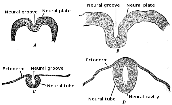
Neuroepithelial cells, or neuroectodermal cells, form the wall of the closed neural tube in early embryonic development. The neuroepithelial cells span the thickness of the tube's wall, connecting with the pial surface and with the ventricular or lumenal surface. They are joined at the lumen of the tube by junctional complexes, where they form a pseudostratified layer of epithelium called neuroepithelium. Neuroepithelial cells are the stem cells of the central nervous system, known as neural stem cells, and generate the intermediate progenitor cells known as radial glial cells, that differentiate into neurons and glia in the process of neurogenesis. Embryonic neural development Brain development During the third week of embryonic growth the brain begins to develop in the early fetus in a process called morphogenesis. Neuroepithelial cells of the ectoderm begin multiplying rapidly and fold in forming the neural plate, which invaginates during the fourth week of embryonic growth ... [...More Info...] [...Related Items...] OR: [Wikipedia] [Google] [Baidu] |
Hemorrhagic
Bleeding, hemorrhage, haemorrhage or blood loss, is blood escaping from the circulatory system from damaged blood vessels. Bleeding can occur internally, or externally either through a natural opening such as the mouth, nose, ear, urethra, vagina or anus, or through a puncture in the skin. Hypovolemia is a massive decrease in blood volume, and death by excessive loss of blood is referred to as exsanguination. Typically, a healthy person can endure a loss of 10–15% of the total blood volume without serious medical difficulties (by comparison, blood donation typically takes 8–10% of the donor's blood volume). The stopping or controlling of bleeding is called hemostasis and is an important part of both first aid and surgery. Types * Upper head ** Intracranial hemorrhage – bleeding in the skull. ** Cerebral hemorrhage – a type of intracranial hemorrhage, bleeding within the brain tissue itself. ** Intracerebral hemorrhage – bleeding in the brain caused by the rupture of a ... [...More Info...] [...Related Items...] OR: [Wikipedia] [Google] [Baidu] |


