|
Ovarian Vein
The ovarian vein, the female gonadal vein, carries deoxygenated blood from its corresponding ovary to inferior vena cava or one of its tributaries. It is the female equivalent of the testicular vein, and is the venous counterpart of the ovarian artery. It can be found in the suspensory ligament of the ovary. Structure It is a paired vein, each one supplying an ovary. * The right ovarian vein travels through the suspensatory ligament of the ovary and generally joins the inferior vena cava. * The left ovarian vein, unlike the right, often joins the left renal vein instead of the inferior vena cava.Lampmann LE, Smeets AJ, Lohle PN. Uterine fibroids: targeted embolization, an update on technique. Abdom Imaging. 2003 Oct 31; . Pathology Thrombosis of ovarian vein is associated with postpartum endometritis, pelvic inflammatory disease, diverticulitis, appendicitis Appendicitis is inflammation of the appendix. Symptoms commonly include right lower abdominal pain, nausea, v ... [...More Info...] [...Related Items...] OR: [Wikipedia] [Google] [Baidu] |
Sheep
Sheep or domestic sheep (''Ovis aries'') are domesticated, ruminant mammals typically kept as livestock. Although the term ''sheep'' can apply to other species in the genus '' Ovis'', in everyday usage it almost always refers to domesticated sheep. Like all ruminants, sheep are members of the order Artiodactyla, the even-toed ungulates. Numbering a little over one billion, domestic sheep are also the most numerous species of sheep. An adult female is referred to as a ''ewe'' (), an intact male as a ''ram'', occasionally a ''tup'', a castrated male as a ''wether'', and a young sheep as a ''lamb''. Sheep are most likely descended from the wild mouflon of Europe and Asia, with Iran being a geographic envelope of the domestication center. One of the earliest animals to be domesticated for agricultural purposes, sheep are raised for fleeces, meat (lamb, hogget or mutton) and milk. A sheep's wool is the most widely used animal fiber, and is usually harvested by shearing. In Comm ... [...More Info...] [...Related Items...] OR: [Wikipedia] [Google] [Baidu] |
Appendicitis
Appendicitis is inflammation of the appendix. Symptoms commonly include right lower abdominal pain, nausea, vomiting, and decreased appetite. However, approximately 40% of people do not have these typical symptoms. Severe complications of a ruptured appendix include widespread, painful inflammation of the inner lining of the abdominal wall and sepsis. Appendicitis is caused by a blockage of the hollow portion of the appendix. This is most commonly due to a calcified "stone" made of feces. Inflamed lymphoid tissue from a viral infection, parasites, gallstone, or tumors may also cause the blockage. This blockage leads to increased pressures in the appendix, decreased blood flow to the tissues of the appendix, and bacterial growth inside the appendix causing inflammation. The combination of inflammation, reduced blood flow to the appendix and distention of the appendix causes tissue injury and tissue death. If this process is left untreated, the appendix may burst, releasing b ... [...More Info...] [...Related Items...] OR: [Wikipedia] [Google] [Baidu] |
Diverticulitis
Diverticulitis, specifically colonic diverticulitis, is a gastrointestinal disease characterized by inflammation of abnormal pouches— diverticula—which can develop in the wall of the large intestine. Symptoms typically include lower abdominal pain of sudden onset, but the onset may also occur over a few days. There may also be nausea; and diarrhea or constipation. Fever or blood in the stool suggests a complication. Repeated attacks may occur. The causes of diverticulitis are unclear. Risk factors may include obesity, lack of exercise, smoking, a family history of the disease, and use of nonsteroidal anti-inflammatory drugs (NSAIDs). The role of a low fiber diet as a risk factor is unclear. Having pouches in the large intestine that are not inflamed is known as diverticulosis. Inflammation occurs in between 10% and 25% at some point in time, and is due to a bacterial infection. Diagnosis is typically by CT scan, though blood tests, colonoscopy, or a lower gastrointes ... [...More Info...] [...Related Items...] OR: [Wikipedia] [Google] [Baidu] |
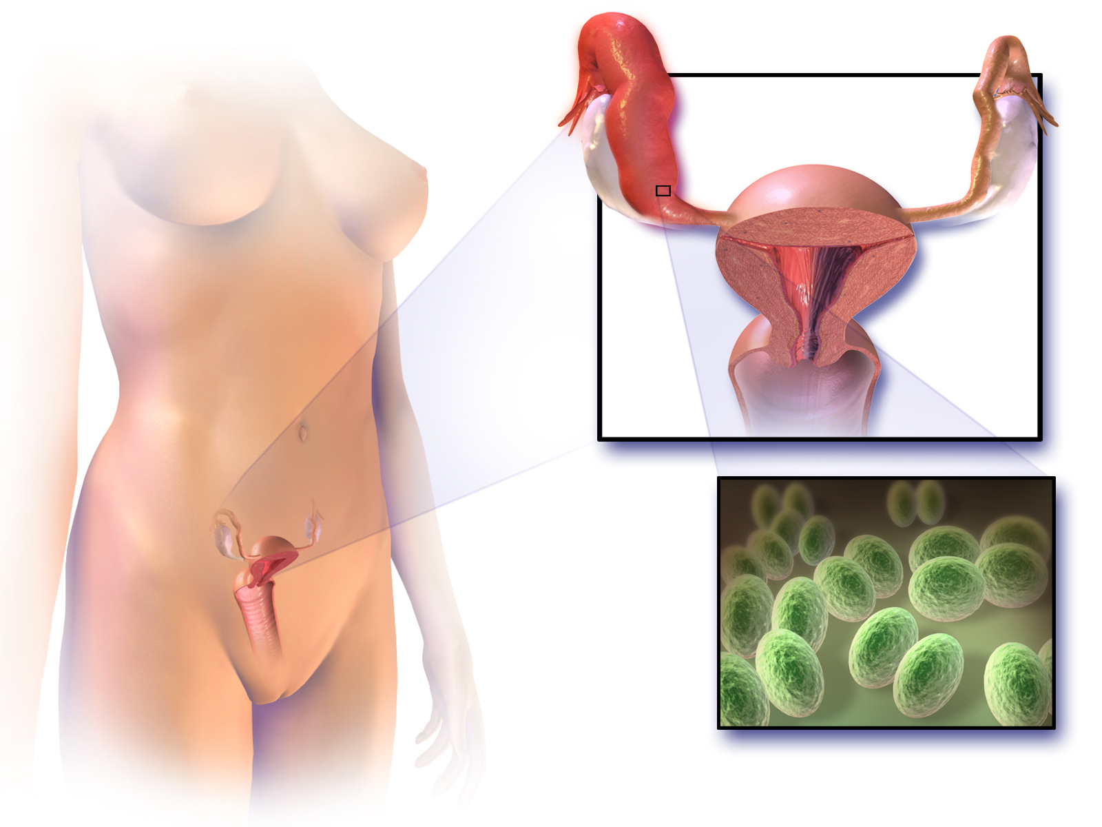
Pelvic Inflammatory Disease
Pelvic inflammatory disease, also known as pelvic inflammatory disorder (PID), is an infection of the upper part of the female reproductive system, namely the uterus, fallopian tubes, and ovaries, and inside of the pelvis. Often, there may be no symptoms. Signs and symptoms, when present, may include lower abdominal pain, vaginal discharge, fever, burning with urination, pain with sex, bleeding after sex, or irregular menstruation. Untreated PID can result in long-term complications including infertility, ectopic pregnancy, chronic pelvic pain, and cancer. The disease is caused by bacteria that spread from the vagina and cervix. Infections by '' Neisseria gonorrhoeae'' or '' Chlamydia trachomatis'' are present in 75 to 90 percent of cases. Often, multiple different bacteria are involved. Without treatment, about 10 percent of those with a chlamydial infection and 40 percent of those with a gonorrhea infection will develop PID. Risk factors are generally similar to thos ... [...More Info...] [...Related Items...] OR: [Wikipedia] [Google] [Baidu] |
Endometritis
Endometritis is inflammation of the inner lining of the uterus ( endometrium). Symptoms may include fever, lower abdominal pain, and abnormal vaginal bleeding or discharge. It is the most common cause of infection after childbirth. It is also part of spectrum of diseases that make up pelvic inflammatory disease. Endometritis is divided into acute and chronic forms. The acute form is usually from an infection that passes through the cervix as a result of an abortion, during menstruation, following childbirth, or as a result of douching or placement of an IUD. Risk factors for endometritis following delivery include Caesarean section and prolonged rupture of membranes. Chronic endometritis is more common after menopause. The diagnosis may be confirmed by endometrial biopsy. Ultrasound may be useful to verify that there is no retained tissue within the uterus. Treatment is usually with antibiotics. Recommendations for treatment of endometritis following delivery includes ... [...More Info...] [...Related Items...] OR: [Wikipedia] [Google] [Baidu] |
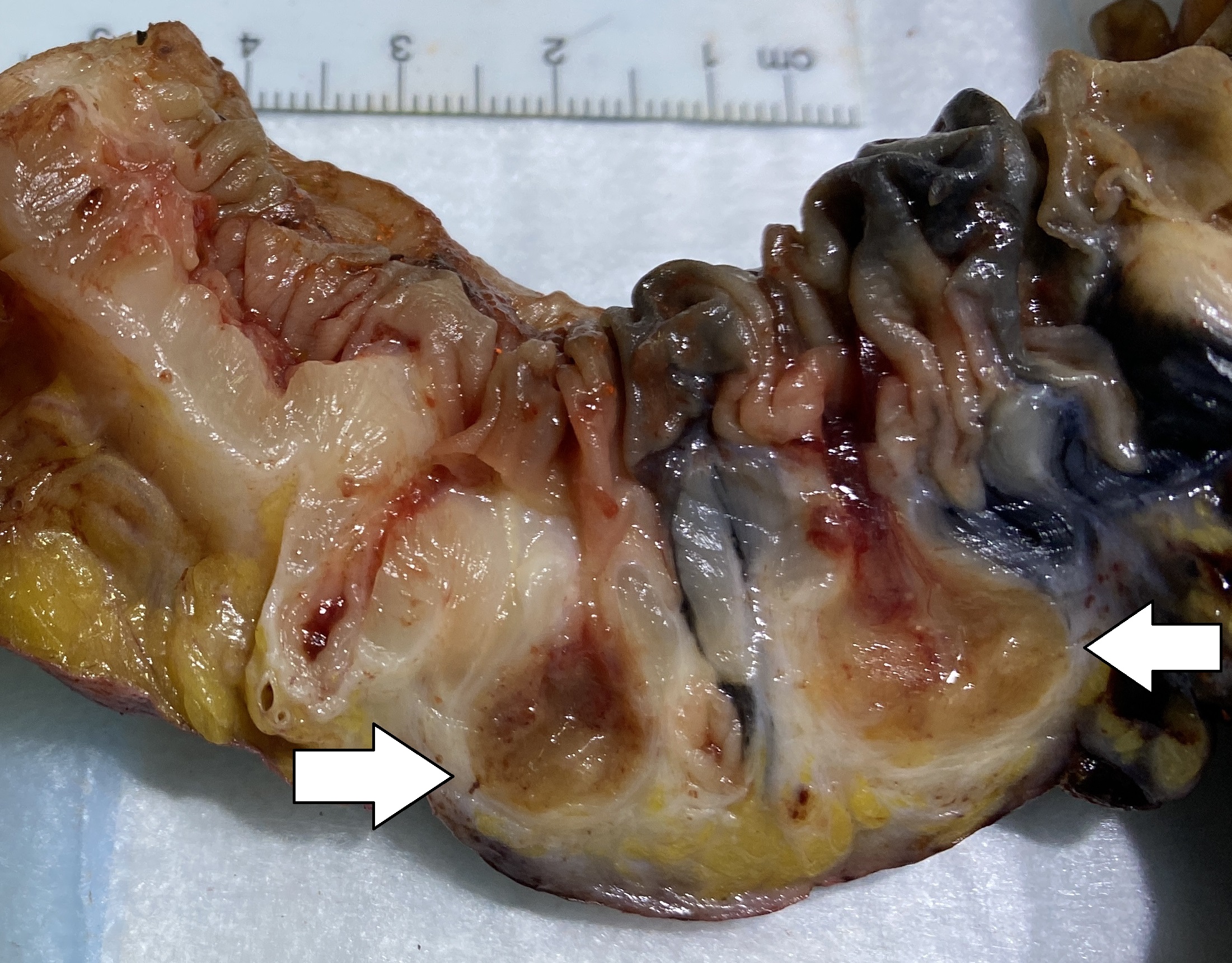
Ovarian Vein Thrombosis
The ovary is an organ in the female reproductive system that produces an ovum. When released, this travels down the fallopian tube into the uterus, where it may become fertilized by a sperm. There is an ovary () found on each side of the body. The ovaries also secrete hormones that play a role in the menstrual cycle and fertility. The ovary progresses through many stages beginning in the prenatal period through menopause. It is also an endocrine gland because of the various hormones that it secretes. Structure The ovaries are considered the female gonads. Each ovary is whitish in color and located alongside the lateral wall of the uterus in a region called the ovarian fossa. The ovarian fossa is the region that is bounded by the external iliac artery and in front of the ureter and the internal iliac artery. This area is about 4 cm x 3 cm x 2 cm in size.Daftary, Shirish; Chakravarti, Sudip (2011). Manual of Obstetrics, 3rd Edition. Elsevier. pp. 1-16. . The ov ... [...More Info...] [...Related Items...] OR: [Wikipedia] [Google] [Baidu] |
Thrombosis
Thrombosis (from Ancient Greek "clotting") is the formation of a blood clot inside a blood vessel, obstructing the flow of blood through the circulatory system. When a blood vessel (a vein or an artery) is injured, the body uses platelets (thrombocytes) and fibrin to form a blood clot to prevent blood loss. Even when a blood vessel is not injured, blood clots may form in the body under certain conditions. A clot, or a piece of the clot, that breaks free and begins to travel around the body is known as an embolus. Thrombosis may occur in veins ( venous thrombosis) or in arteries ( arterial thrombosis). Venous thrombosis (sometimes called DVT, deep vein thrombosis) leads to a blood clot in the affected part of the body, while arterial thrombosis (and, rarely, severe venous thrombosis) affects the blood supply and leads to damage of the tissue supplied by that artery ( ischemia and necrosis). A piece of either an arterial or a venous thrombus can break off as an embolus, whi ... [...More Info...] [...Related Items...] OR: [Wikipedia] [Google] [Baidu] |
Testicular Vein
The testicular vein (or spermatic vein), the male gonadal vein, carries deoxygenated blood from its corresponding testis to the inferior vena cava or one of its tributaries. It is the male equivalent of the ovarian vein, and is the venous counterpart of the testicular artery. Structure It is a paired vein, with one supplying each testis: * the right testicular vein generally joins the inferior vena cava; * the left testicular vein, unlike the right one, joins the left renal vein instead of the inferior vena cava. The veins emerge from the back of the testis, and receive tributaries from the epididymis. They unite and form a convoluted plexus, called the pampiniform plexus, which constitutes the greater mass of the spermatic cord; the vessels composing this plexus are very numerous, and ascend along the cord, in front of the ductus deferens. Below the subcutaneous inguinal ring, they unite to form three or four veins, which pass along the inguinal canal, and, entering the abdome ... [...More Info...] [...Related Items...] OR: [Wikipedia] [Google] [Baidu] |
Ovary
The ovary is an organ in the female reproductive system that produces an ovum. When released, this travels down the fallopian tube into the uterus, where it may become fertilized by a sperm. There is an ovary () found on each side of the body. The ovaries also secrete hormones that play a role in the menstrual cycle and fertility. The ovary progresses through many stages beginning in the prenatal period through menopause. It is also an endocrine gland because of the various hormones that it secretes. Structure The ovaries are considered the female gonads. Each ovary is whitish in color and located alongside the lateral wall of the uterus in a region called the ovarian fossa. The ovarian fossa is the region that is bounded by the external iliac artery and in front of the ureter and the internal iliac artery. This area is about 4 cm x 3 cm x 2 cm in size.Daftary, Shirish; Chakravarti, Sudip (2011). Manual of Obstetrics, 3rd Edition. Elsevier. pp. 1-16. . The ... [...More Info...] [...Related Items...] OR: [Wikipedia] [Google] [Baidu] |
Gonadal Vein
In medicine, gonadal vein refers to the blood vessel that carries blood away from the gonad (testis, ovary) toward the heart. These are different arteries in women (ovarian vein) and men ( testicular vein), but share the same embryological origin. The termination of the two gonadal veins in an individual is usually asymmetrical, with the left one draining into the left renal vein, and the right one draining into the inferior vena cava. Anatomy Fate The left gonadal vein usually empties into (inferior aspect of) the ipsilateral renal vein proximally to where the renal vein crossing over the aorta. The right gonadal vein typically empties directly into the (right anterolateral aspect of) inferior vena cava, joining it at an acute angle, some 2 cm inferior to the ipsilateral renal vein The renal veins are large-calibre veins that drain blood filtered by the kidneys into the inferior vena cava. There is one renal vein draining each kidney. Because the inferior vena cava ... [...More Info...] [...Related Items...] OR: [Wikipedia] [Google] [Baidu] |
Ovarian Artery
The ovarian artery is an artery that supplies oxygenated blood to the ovary in females. It arises from the abdominal aorta below the renal artery. It can be found within the suspensory ligament of the ovary, anterior to the ovarian vein and ureter. Structure The ovarian arteries are paired structures that arise from the abdominal aorta, usually at the level of L2. After emerging from the aorta, the artery travels within the suspensory ligament of the ovary and enters the mesovarium. The ovarian arteries are the corresponding arteries in the female to the testicular artery in the male. They are shorter than the testicular arteries, as the testicular arteries courses through the abdominal wall to the external scrotum. The origin and course of the first part of each artery are the same as those of the testicular artery, but on arriving at the upper opening of the lesser pelvis the ovarian artery passes inward, between the two layers of the ovariopelvic ligament and of the br ... [...More Info...] [...Related Items...] OR: [Wikipedia] [Google] [Baidu] |


