|
Osborn Wave
A J wave — also known as Osborn wave, camel-hump sign, late delta wave, hathook junction, hypothermic wave, K wave, H wave or current of injury — is an abnormal electrocardiogram finding. J waves are positive deflections occurring at the junction between the QRS complex and the ST segment, where the S point, also known as the J point, has a myocardial infarction-like elevation. __TOC__ Causes They are usually observed in people suffering from hypothermia with a temperature of less than 32 °C (90 °F), though they may also occur in people with very high blood levels of calcium (hypercalcemia), brain injury, vasospastic angina, acute pericarditis, or ventricular fibrillation and could also be a normal variant. History The prominent J deflection attributed to hypothermia was first reported in 1938 by Tomaszewski. These waves were then definitively described in 1953 by John J. Osborn (1917–2014) and were named in his honor. Over time, the wave has increasingly been ... [...More Info...] [...Related Items...] OR: [Wikipedia] [Google] [Baidu] |
QRS Complex
The QRS complex is the combination of three of the graphical deflections seen on a typical electrocardiogram (ECG or EKG). It is usually the central and most visually obvious part of the tracing. It corresponds to the depolarization of the right and left ventricles of the heart and contraction of the large ventricular muscles. In adults, the QRS complex normally lasts ; in children it may be shorter. The Q, R, and S waves occur in rapid succession, do not all appear in all leads, and reflect a single event and thus are usually considered together. A Q wave is any downward deflection immediately following the P wave. An R wave follows as an upward deflection, and the S wave is any downward deflection after the R wave. The T wave follows the S wave, and in some cases, an additional U wave follows the T wave. To measure the QRS interval start at the end of the PR interval (or beginning of the Q wave) to the end of the S wave. Normally this interval is 0.08 to 0.10 seconds. When ... [...More Info...] [...Related Items...] OR: [Wikipedia] [Google] [Baidu] |
ST Segment
In electrocardiography, the ST segment connects the QRS complex and the T wave and has a duration of 0.005 to 0.150 sec (5 to 150 ms). It starts at the J point (junction between the QRS complex and ST segment) and ends at the beginning of the T wave. However, since it is usually difficult to determine exactly where the ST segment ends and the T wave begins, the relationship between the ST segment and T wave should be examined together. The typical ST segment duration is usually around 0.08 sec (80 ms). It should be essentially level with the PR and TP segments. The ST segment represents the isoelectric period when the ventricles are in between depolarization and repolarization. Interpretation * The normal ST segment has a slight upward concavity. * Flat, downsloping, or depressed ST segments may indicate coronary ischemia. * ST elevation may indicate transmural myocardial infarction. An elevation of >1mm and longer than 80 milliseconds following the J-point. This measure has a ... [...More Info...] [...Related Items...] OR: [Wikipedia] [Google] [Baidu] |
Myocardial Infarction
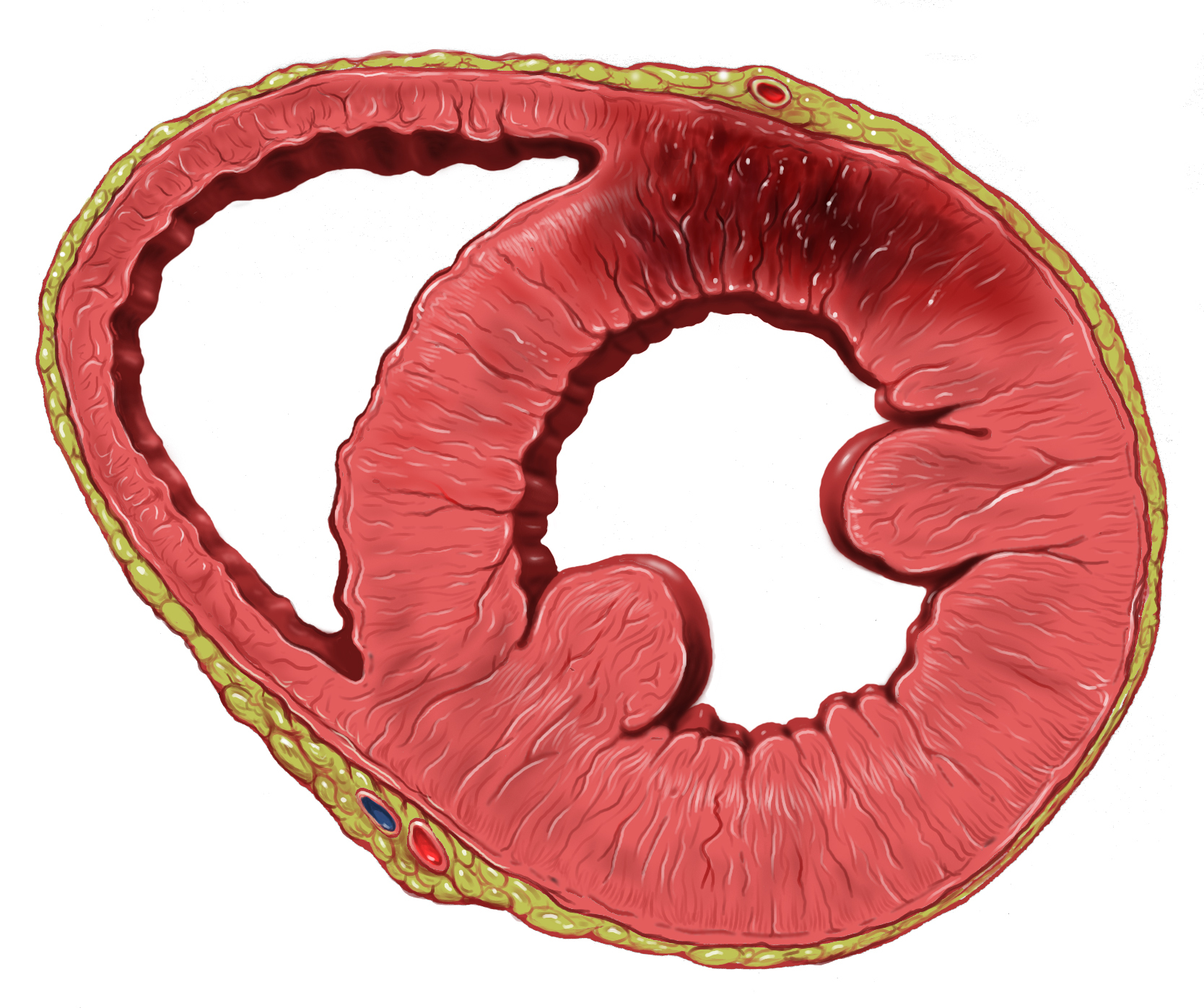
A myocardial infarction (MI), commonly known as a heart attack, occurs when blood flow decreases or stops to the coronary artery of the heart, causing damage to the heart muscle. The most common symptom is chest pain or discomfort which may travel into the shoulder, arm, back, neck or jaw. Often it occurs in the center or left side of the chest and lasts for more than a few minutes. The discomfort may occasionally feel like heartburn. Other symptoms may include shortness of breath, nausea, feeling faint, a cold sweat or feeling tired. About 30% of people have atypical symptoms. Women more often present without chest pain and instead have neck pain, arm pain or feel tired. Among those over 75 years old, about 5% have had an MI with little or no history of symptoms. An MI may cause heart failure, an irregular heartbeat, cardiogenic shock or cardiac arrest. Most MIs occur due to coronary artery disease. Risk factors include high blood pressure, smoking, diabetes, ... [...More Info...] [...Related Items...] OR: [Wikipedia] [Google] [Baidu] |
Hypothermia
Hypothermia is defined as a body core temperature below in humans. Symptoms depend on the temperature. In mild hypothermia, there is shivering and mental confusion. In moderate hypothermia, shivering stops and confusion increases. In severe hypothermia, there may be hallucinations and paradoxical undressing, in which a person removes their clothing, as well as an increased risk of the heart stopping. Hypothermia has two main types of causes. It classically occurs from exposure to cold weather and cold water immersion. It may also occur from any condition that decreases heat production or increases heat loss. Commonly, this includes alcohol intoxication but may also include low blood sugar, anorexia and advanced age. Body temperature is usually maintained near a constant level of through thermoregulation. Efforts to increase body temperature involve shivering, increased voluntary activity, and putting on warmer clothing. Hypothermia may be diagnosed based on either a person ... [...More Info...] [...Related Items...] OR: [Wikipedia] [Google] [Baidu] |
Calcium
Calcium is a chemical element with the symbol Ca and atomic number 20. As an alkaline earth metal, calcium is a reactive metal that forms a dark oxide-nitride layer when exposed to air. Its physical and chemical properties are most similar to its heavier homologues strontium and barium. It is the fifth most abundant element in Earth's crust, and the third most abundant metal, after iron and aluminium. The most common calcium compound on Earth is calcium carbonate, found in limestone and the fossilised remnants of early sea life; gypsum, anhydrite, fluorite, and apatite are also sources of calcium. The name derives from Latin ''calx'' "lime", which was obtained from heating limestone. Some calcium compounds were known to the ancients, though their chemistry was unknown until the seventeenth century. Pure calcium was isolated in 1808 via electrolysis of its oxide by Humphry Davy, who named the element. Calcium compounds are widely used in many industries: in foods and pharma ... [...More Info...] [...Related Items...] OR: [Wikipedia] [Google] [Baidu] |
Hypercalcemia
Hypercalcemia, also spelled hypercalcaemia, is a high calcium (Ca2+) level in the blood serum. The normal range is 2.1–2.6 mmol/L (8.8–10.7 mg/dL, 4.3–5.2 mEq/L), with levels greater than 2.6 mmol/L defined as hypercalcemia. Those with a mild increase that has developed slowly typically have no symptoms. In those with greater levels or rapid onset, symptoms may include abdominal pain, bone pain, confusion, depression, weakness, kidney stones or an abnormal heart rhythm including cardiac arrest. Most outpatient cases are due to primary hyperparathyroidism and inpatient cases due to cancer. Other causes of hypercalcemia include sarcoidosis, tuberculosis, Paget disease, multiple endocrine neoplasia (MEN), vitamin D toxicity, familial hypocalciuric hypercalcaemia and certain medications such as lithium and hydrochlorothiazide. Diagnosis should generally include either a corrected calcium or ionized calcium level and be confirmed after a week. Specific chang ... [...More Info...] [...Related Items...] OR: [Wikipedia] [Google] [Baidu] |
Brain
A brain is an organ that serves as the center of the nervous system in all vertebrate and most invertebrate animals. It is located in the head, usually close to the sensory organs for senses such as vision. It is the most complex organ in a vertebrate's body. In a human, the cerebral cortex contains approximately 14–16 billion neurons, and the estimated number of neurons in the cerebellum is 55–70 billion. Each neuron is connected by synapses to several thousand other neurons. These neurons typically communicate with one another by means of long fibers called axons, which carry trains of signal pulses called action potentials to distant parts of the brain or body targeting specific recipient cells. Physiologically, brains exert centralized control over a body's other organs. They act on the rest of the body both by generating patterns of muscle activity and by driving the secretion of chemicals called hormones. This centralized control allows rapid and coordinated respon ... [...More Info...] [...Related Items...] OR: [Wikipedia] [Google] [Baidu] |
Vasospasm
Vasospasm refers to a condition in which an arterial spasm leads to vasoconstriction. This can lead to tissue ischemia and tissue death (necrosis). Cerebral vasospasm may arise in the context of subarachnoid hemorrhage. Symptomatic vasospasm or delayed cerebral ischemia is a major contributor to post-operative stroke and death especially after aneurysmal subarachnoid hemorrhage. Vasospasm typically appears 4 to 10 days after subarachnoid hemorrhage. Along with physical resistance, vasospasm is a main cause of ischemia. Like physical resistance, vasospasms can occur due to atherosclerosis. Vasospasm is the major cause of Prinzmetal's angina. Pathophysiology Normally endothelial cells release prostacyclin and nitric oxide (NO) which induce relaxation of the smooth muscle cells, and reduce aggregation of platelets. Aggregating platelets stimulate ADP to act on endothelial cells and help them induce relaxation of the smooth muscle cells. However, aggregating platelets also stimulat ... [...More Info...] [...Related Items...] OR: [Wikipedia] [Google] [Baidu] |
Angina Pectoris
Angina, also known as angina pectoris, is chest pain or pressure, usually caused by insufficient blood flow to the heart muscle (myocardium). It is most commonly a symptom of coronary artery disease. Angina is typically the result of obstruction or spasm of the arteries that supply blood to the heart muscle. The main mechanism of coronary artery obstruction is atherosclerosis as part of coronary artery disease. Other causes of angina include abnormal heart rhythms, heart failure and, less commonly, anemia. The term derives from the Latin ''angere'' ("to strangle") and ''pectus'' ("chest"), and can therefore be translated as "a strangling feeling in the chest". There is a weak relationship between severity of angina and degree of oxygen deprivation in the heart muscle, however, the severity of angina does not always match the degree of oxygen deprivation to the heart or the risk of a myocardial infarction (heart attack). Some people may experience severe pain even though the ... [...More Info...] [...Related Items...] OR: [Wikipedia] [Google] [Baidu] |


