|
Obturator Fascia
The obturator fascia, or fascia of the internal obturator muscle, covers the pelvic surface of that muscle and is attached around the margin of its origin. Above, it is loosely connected to the back part of the arcuate line, and here it is continuous with the iliac fascia. In front of this, as it follows the line of origin of the internal obturator, it gradually separates from the iliac fascia and the continuity between the two is retained only through the periosteum. It arches beneath the obturator vessels and nerve, completing the obturator canal, and at the front of the pelvis is attached to the back of the superior ramus of the pubis. Below, the obturator fascia is attached to the falciform process of the sacrotuberous ligament and to the pubic arch, where it becomes continuous with the superior fascia of the urogenital diaphragm. Behind, it is prolonged into the gluteal region. The internal pudendal vessels and pudendal nerve cross the pelvic surface of the inter ... [...More Info...] [...Related Items...] OR: [Wikipedia] [Google] [Baidu] |
Internal Obturator Muscle
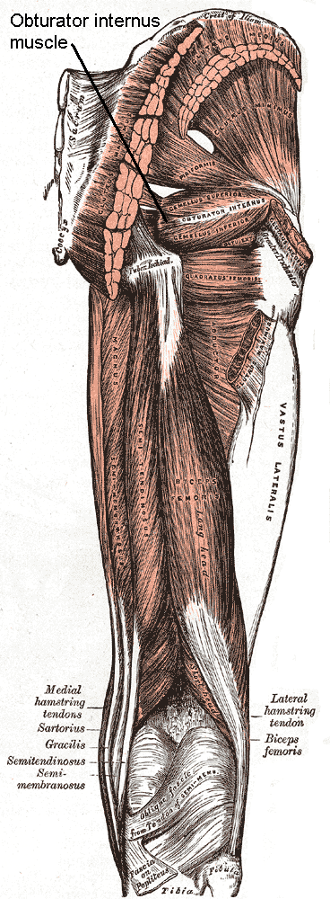
The internal obturator muscle or obturator internus muscle originates on the medial surface of the obturator membrane, the ischium near the membrane, and the rim of the pubis. It exits the pelvic cavity through the lesser sciatic foramen. The internal obturator is situated partly within the lesser pelvis, and partly at the back of the hip-joint. It functions to help laterally rotate femur with hip extension and abduct femur with hip flexion, as well as to steady the femoral head in the acetabulum. Structure Origin The internal obturator muscle arises from the inner surface of the antero-lateral wall of the pelvis. It surrounds the obturator foramen. It is attached to the inferior pubic ramus and ischium, and at the side to the inner surface of the hip bone below and behind the pelvic brim. It reaches from the upper part of the greater sciatic foramen above and behind to the obturator foramen below and in front. It also arises from the pelvic surface of the obturator mem ... [...More Info...] [...Related Items...] OR: [Wikipedia] [Google] [Baidu] |
Pubic Arch
The pubic arch, also referred to as the ''ischiopubic arch'', is part of the pelvis. It is formed by the convergence of the inferior rami of the ischium and pubis on either side, below the pubic symphysis. The angle at which they converge is known as the subpubic angle. Function The pubic arch is one of three notches (the one in front) that separate the eminences of the lower circumference of the true pelvis. Clinical significance Subpubic angle The subpubic angle (or pubic angle) is the angle in the human body as the apex of the pubic arch, formed by the convergence of the inferior rami of the ischium and pubis on either side. The subpubic angle is important in forensic anthropology, in determining the sex of someone from skeletal remains. A subpubic angle of 50-82 degrees indicates a male; an angle of 90 degrees indicates a female.Anthony J. Bertino. Forensic Science - Fundamentals and Investigations. South-Western Cengage Learning, 2000. . Page 368 Other sources operate ... [...More Info...] [...Related Items...] OR: [Wikipedia] [Google] [Baidu] |
Levator Ani
The levator ani is a broad, thin muscle group, situated on either side of the pelvis. It is formed from three muscle components: the pubococcygeus, the iliococcygeus, and the puborectalis. It is attached to the inner surface of each side of the lesser pelvis, and these unite to form the greater part of the pelvic floor. The coccygeus muscle completes the pelvic floor, which is also called the ''pelvic diaphragm''. It supports the viscera in the pelvic cavity, and surrounds the various structures that pass through it. The levator ani is the main pelvic floor muscle and painfully contracts during vaginismus. It also contracts rhythmically during orgasm. Structure The levator ani is made up of 3 parts: * Iliococcygeus muscle * Pubococcygeus muscle * Puborectalis muscle The iliococcygeus arises from the inner side of the ischium (the lower and back part of the hip bone) and from the posterior part of the tendinous arch of the obturator fascia, and is attached to the coccyx ... [...More Info...] [...Related Items...] OR: [Wikipedia] [Google] [Baidu] |
Iliococcygeus
The levator ani is a broad, thin muscle group, situated on either side of the pelvis. It is formed from three muscle components: the pubococcygeus, the iliococcygeus, and the puborectalis. It is attached to the inner surface of each side of the lesser pelvis, and these unite to form the greater part of the pelvic floor. The coccygeus muscle completes the pelvic floor, which is also called the ''pelvic diaphragm''. It supports the viscera in the pelvic cavity, and surrounds the various structures that pass through it. The levator ani is the main pelvic floor muscle and painfully contracts during vaginismus. It also contracts rhythmically during orgasm. Structure The levator ani is made up of 3 parts: * Iliococcygeus muscle * Pubococcygeus muscle * Puborectalis muscle The iliococcygeus arises from the inner side of the ischium (the lower and back part of the hip bone) and from the posterior part of the tendinous arch of the obturator fascia, and is attached to the coccyx an ... [...More Info...] [...Related Items...] OR: [Wikipedia] [Google] [Baidu] |
Alcock’s Canal
The pudendal canal (also called Alcock's canal) is an anatomical structure in the pelvis through which the internal pudendal artery, internal pudendal veins, and the pudendal nerve pass. Structure The pudendal canal is formed by the fascia of the Internal obturator muscle, obturator internus muscle, or obturator fascia. It encloses the following: * Internal pudendal artery. * Internal pudendal veins. * Pudendal nerve. These vessels and nerve cross the pelvic surface of the obturator internus. Clinical significance Pudendal nerve entrapment can occur when the pudendal nerve is compressed while it passes through the pudendal canal. History The pudendal canal is also known as Alcock's canal, named after Benjamin Alcock. Additional images Image:Gray542.png , The superficial branches of the internal pudendal artery. (Canal not labeled, but pudendal nerve and internal pudendal artery labeled at bottom right.) See also * Femoral canal * Inguinal canal References External ... [...More Info...] [...Related Items...] OR: [Wikipedia] [Google] [Baidu] |
Pudendal Nerve
The pudendal nerve is the main nerve of the perineum. It carries sensation from the external genitalia of both sexes and the skin around the anus and perineum, as well as the motor supply to various pelvic muscles, including the male or female external urethral sphincter and the external anal sphincter. If damaged, most commonly by childbirth, lesions may cause sensory loss or fecal incontinence. The nerve may be temporarily blocked as part of an anaesthetic procedure. The pudendal canal that carries the pudendal nerve is also known by the eponymous term "Alcock's canal", after Benjamin Alcock, an Irish anatomist who documented the canal in 1836. Structure The pudendal nerve is paired, meaning there are two nerves, one on the left and one on the right side of the body. Each is formed as three roots immediately converge above the upper border of the sacrotuberous ligament and the coccygeus muscle. The three roots become two cords when the middle and lower root join to ... [...More Info...] [...Related Items...] OR: [Wikipedia] [Google] [Baidu] |
Gluteal
The gluteal muscles, often called glutes are a group of three muscles which make up the gluteal region commonly known as the buttocks: the gluteus maximus, gluteus medius and gluteus minimus. The three muscles originate from the ilium and sacrum and insert on the femur. The functions of the muscles include extension, abduction, external rotation, and internal rotation of the hip joint. Structure The gluteus maximus is the largest and most superficial of the three gluteal muscles. It makes up a large part of the shape and appearance of the hips. It is a narrow and thick fleshy mass of a quadrilateral shape, and forms the prominence of the buttocks. The gluteus medius is a broad, thick, radiating muscle, situated on the outer surface of the pelvis. It lies profound to the gluteus maximus and its posterior third is covered by the gluteus maximus, its anterior two-thirds by the gluteal aponeurosis, which separates it from the superficial fascia and skin. The gluteus minimus is the ... [...More Info...] [...Related Items...] OR: [Wikipedia] [Google] [Baidu] |
Urogenital Diaphragm
Older texts have asserted the existence of a urogenital diaphragm, also called the triangular ligament, which was described as a layer of the pelvis that separates the deep perineal sac from the upper pelvis, lying between the inferior fascia of the urogenital diaphragm (perineal membrane) and superior fascia of the urogenital diaphragm. While this term is used to refer to a layer of the pelvis that separates the deep perineal sac from the upper pelvis, such a discrete border of the sac probably does not exist. While it has no official entry in Terminologia Anatomica, the term is still used occasionally to describe the muscular components of the deep perineal pouch. The urethra and the vagina, though part of the pouch, are usually said to be passing through the urogenital diaphragm, rather than part of the diaphragm itself. Some researchers still assert that such a diaphragm exists, and the term is still used in the literature. The urethral diaphragm is an anatomic landmark ... [...More Info...] [...Related Items...] OR: [Wikipedia] [Google] [Baidu] |
Sacrotuberous Ligament
The sacrotuberous ligament (great or posterior sacrosciatic ligament) is situated at the lower and back part of the pelvis. It is flat, and triangular in form; narrower in the middle than at the ends. Structure It runs from the sacrum (the lower transverse sacral tubercles, the inferior margins sacrum and the upper coccyx) to the tuberosity of the ischium. It is a remnant of part of Biceps femoris muscle. The sacrotuberous ligament is attached by its broad base to the posterior superior iliac spine, the posterior sacroiliac ligaments (with which it is partly blended), to the lower transverse sacral tubercles and the lateral margins of the lower sacrum and upper coccyx. Its oblique fibres descend laterally, converging to form a thick, narrow band that widens again below and is attached to the medial margin of the ischial tuberosity. It then spreads along the ischial ramus as the falciform process, whose concave edge blends with the fascial sheath of the internal pudendal vessels ... [...More Info...] [...Related Items...] OR: [Wikipedia] [Google] [Baidu] |
Arcuate Line (ilium)
The arcuate line of the ilium is a smooth rounded border on the internal surface of the ilium. It is immediately inferior to the iliac fossa and Iliacus muscle. It forms part of the border of the pelvic inlet. In combination with the pectineal line, it comprises the iliopectineal line. The arcuate line marks the border between the body (''corpus'') and the wing (''ala'') of the ilium, and, running inferior, anterior, and medial from the auricular surface to the area corresponding to the acetabulum, it also indicates where weight is transferred from the sacroiliac joint to the hip joint In vertebrate anatomy, hip (or "coxa"Latin ''coxa'' was used by Celsus in the sense "hip", but by Pliny the Elder in the sense "hip bone" (Diab, p 77) in medical terminology) refers to either an anatomical region or a joint. The hip region is .... Additional images Arcuate line of ilium 02 animation.gif, Position of arcuate line of ilium. Shown in red. Arcuate line of ilium 03 anima ... [...More Info...] [...Related Items...] OR: [Wikipedia] [Google] [Baidu] |
Falciform Process
The ''falx'' was a weapon with a curved blade that was sharp on the inside edge used by the Thracians and Dacians. The name was later applied to a siege hook used by the Romans. Etymology ''Falx'' is a Latin word originally meaning 'sickle' but was later used to mean any of a number of tools that had a curved blade that was sharp on the inside edge like a sickle. ''Falx'' was thus also used to mean the weapon of the Thracians and Dacians, and the Roman siege hook. Dacian ''falx'' In Latin texts, the weapon was described as an ' (whence ''falcata'') by Ovid in ''Metamorphose'' and as a ' by Juvenal in ''Satiriae''. The Dacian ''falx'' came in two sizes: one-handed and two-handed. The shorter variant was called ''sica'' (sickle) in the Dacian language (Valerius Maximus, III, 2.12) with a blade length that varied but was usually around long with a handle 1/3 longer than the blade. The two-handed ''falx'' was a polearm. It consisted of a long wooden shaft with a long curved ... [...More Info...] [...Related Items...] OR: [Wikipedia] [Google] [Baidu] |
Pubis (bone)
In vertebrates, the pubic region ( la, pubis) is the most forward-facing (ventral and anterior) of the three main regions making up the coxal bone. The left and right pubic regions are each made up of three sections, a superior ramus, inferior ramus, and a body. Structure The pubic region is made up of a ''body'', ''superior ramus'', and ''inferior ramus'' (). The left and right coxal bones join at the pubic symphysis. It is covered by a layer of fat, which is covered by the mons pubis. The pubis is the lower limit of the suprapubic region. In the female, the pubic region is anterior to the urethral sponge. Body The body forms the wide, strong, middle and flat part of the pubic region. The bodies of the left and right pubic regions join at the pubic symphysis. The rough upper edge is the pubic crest, ending laterally in the pubic tubercle. This tubercle, found roughly 3 cm from the pubic symphysis, is a distinctive feature on the lower part of the abdominal wall; important ... [...More Info...] [...Related Items...] OR: [Wikipedia] [Google] [Baidu] |


