|
Owl's Eye Appearance
Owl's eye appearance, also known as owl's eye sign, is a term used to describe a pattern that is found in the study of histology, radiology, and pathology cases. This pattern resembles the shape of a real owl's eye. The pattern is used for the analysis of symptoms on patients within the medical field. They may refer to: *Cells with perinuclear vacuolization around centrally located pyknotic nuclei, such as typically seen in flat warts. Last Update: August 11, 2020. *Owl's eye appearance of inclusion bodies, which is highly specific for cytomegalovirus infection. *Owl's eye appearance of entire nucleus – a finding in Reed–Sternberg cells in individuals with Hodgkin's lymphoma. *Owl's eye appearance of the Lentiform nucleus of the basal ganglia on head CT scan images in individuals with cerebral hypoxia. __TOC__ Pathological cases The owl's eye appearance has a relationship with Reed–Sternberg cells, in regards to cytomegalovirus infection. Owl's eye appearance was used ... [...More Info...] [...Related Items...] OR: [Wikipedia] [Google] [Baidu] |
Orange Eye (6959548877)
''Buddleja davidii'' (spelling variant ''Buddleia davidii''), also called summer lilac, butterfly-bush, or orange eye, is a species of flowering plant in the family (biology), family Scrophulariaceae, native plant, native to Sichuan and Hubei provinces in central China, and also Japan. It is widely used as an ornamental plant, and many named varieties are in cultivation. The genus was named ''Buddleja'' after Reverend Adam Buddle, an English botanist. The species name ''davidii'' honors the French missionary and explorer in China, Father Armand David, who was the first European to report the shrub. It was found near Ichang by Dr Augustine Henry about 1887 and sent to St Petersburg. Another botanist-missionary in China, Jean-André Soulié, sent seed to the French nursery Vilmorin, and ''B. davidii'' entered commerce in the 1890s. ''B. davidii'' was accorded the Royal Horticultural Society, RHS Award of Merit (AM) in 1898, and the Award of Garden Merit (AGM) in 1941.Hillier & Sons ... [...More Info...] [...Related Items...] OR: [Wikipedia] [Google] [Baidu] |
Cerebral Hypoxia
Cerebral hypoxia is a form of hypoxia (reduced supply of oxygen), specifically involving the brain; when the brain is completely deprived of oxygen, it is called ''cerebral anoxia''. There are four categories of cerebral hypoxia; they are, in order of increasing severity: diffuse cerebral hypoxia (DCH), focal cerebral ischemia, cerebral infarction, and global cerebral ischemia. Prolonged hypoxia induces neuronal cell death via apoptosis, resulting in a hypoxic brain injury. Cases of total oxygen deprivation are termed "anoxia", which can be hypoxic in origin (reduced oxygen availability) or ischemic in origin (oxygen deprivation due to a disruption in blood flow). Brain injury as a result of oxygen deprivation either due to hypoxic or anoxic mechanisms are generally termed hypoxic/anoxic injuries (HAI). Hypoxic ischemic encephalopathy (HIE) is a condition that occurs when the entire brain is deprived of an adequate oxygen supply, but the deprivation is not total. While HIE is as ... [...More Info...] [...Related Items...] OR: [Wikipedia] [Google] [Baidu] |
Inflammation
Inflammation (from la, wikt:en:inflammatio#Latin, inflammatio) is part of the complex biological response of body tissues to harmful stimuli, such as pathogens, damaged cells, or Irritation, irritants, and is a protective response involving immune cells, blood vessels, and molecular mediators. The function of inflammation is to eliminate the initial cause of cell injury, clear out necrotic cells and tissues damaged from the original insult and the inflammatory process, and initiate tissue repair. The five cardinal signs are heat, pain, redness, swelling, and Functio laesa, loss of function (Latin ''calor'', ''dolor'', ''rubor'', ''tumor'', and ''functio laesa''). Inflammation is a generic response, and therefore it is considered as a mechanism of innate immune system, innate immunity, as compared to adaptive immune system, adaptive immunity, which is specific for each pathogen. Too little inflammation could lead to progressive tissue destruction by the harmful stimulus (e.g. b ... [...More Info...] [...Related Items...] OR: [Wikipedia] [Google] [Baidu] |
Organs
In biology, an organ is a collection of tissues joined in a structural unit to serve a common function. In the hierarchy of life, an organ lies between tissue and an organ system. Tissues are formed from same type cells to act together in a function. Tissues of different types combine to form an organ which has a specific function. The intestinal wall for example is formed by epithelial tissue and smooth muscle tissue. Two or more organs working together in the execution of a specific body function form an organ system, also called a biological system or body system. An organ's tissues can be broadly categorized as parenchyma, the functional tissue, and stroma, the structural tissue with supportive, connective, or ancillary functions. For example, the gland's tissue that makes the hormones is the parenchyma, whereas the stroma includes the nerves that innervate the parenchyma, the blood vessels that oxygenate and nourish it and carry away its metabolic wastes, and the connecti ... [...More Info...] [...Related Items...] OR: [Wikipedia] [Google] [Baidu] |
Tissue (biology)
In biology, tissue is a biological organizational level between cells and a complete organ. A tissue is an ensemble of similar cells and their extracellular matrix from the same origin that together carry out a specific function. Organs are then formed by the functional grouping together of multiple tissues. The English word "tissue" derives from the French word "tissu", the past participle of the verb tisser, "to weave". The study of tissues is known as histology or, in connection with disease, as histopathology. Xavier Bichat is considered as the "Father of Histology". Plant histology is studied in both plant anatomy and physiology. The classical tools for studying tissues are the paraffin block in which tissue is embedded and then sectioned, the histological stain, and the optical microscope. Developments in electron microscopy, immunofluorescence, and the use of frozen tissue-sections have enhanced the detail that can be observed in tissues. With these tools, the c ... [...More Info...] [...Related Items...] OR: [Wikipedia] [Google] [Baidu] |
Enterovirus
''Enterovirus'' is a genus of positive-sense single-stranded RNA virus An RNA virus is a virusother than a retrovirusthat has ribonucleic acid (RNA) as its genetic material. The nucleic acid is usually single-stranded RNA ( ssRNA) but it may be double-stranded (dsRNA). Notable human diseases caused by RNA viruses ...es associated with several human and mammalian diseases. Enteroviruses are named by their transmission-route through the intestine ('enteric' meaning intestinal). Serologic studies have distinguished 71 human enterovirus serotypes on the basis of antibody neutralization tests. Additional antigenic variants have been defined within several of the serotypes on the basis of reduced or nonreciprocal cross-neutralization between variant strains. On the basis of their pathogenesis in humans and animals, the enteroviruses were originally classified into four groups, polioviruses, Coxsackie A viruses (CA), Coxsackie B viruses (CB), and echoviruses, but it was quickly rea ... [...More Info...] [...Related Items...] OR: [Wikipedia] [Google] [Baidu] |
Acute Flaccid Paralysis
Flaccid paralysis is a neurological condition characterized by weakness or paralysis and reduced muscle tone without other obvious cause (e.g., trauma). This abnormal condition may be caused by disease or by trauma affecting the nerves associated with the involved muscles. For example, if the somatic nerves to a skeletal muscle are severed, then the muscle will exhibit flaccid paralysis. When muscles enter this state, they become limp and cannot contract. This condition can become fatal if it affects the respiratory muscles, posing the threat of suffocation. It also occurs in spinal shock stage in complete transection of spinal cord occurred in injuries like gunshots injuries.Saladin, Kenneth S. Anatomy & Physiology: The Unity of Form and Function. McGraw-Hill. 6th Edition. 2012. Causes Polio and other viruses The term ''acute flaccid paralysis'' (AFP) is often used to describe an instance with a sudden onset, as might be found with polio. AFP is the most common sign of acu ... [...More Info...] [...Related Items...] OR: [Wikipedia] [Google] [Baidu] |
Retina
The retina (from la, rete "net") is the innermost, light-sensitive layer of tissue of the eye of most vertebrates and some molluscs. The optics of the eye create a focused two-dimensional image of the visual world on the retina, which then processes that image within the retina and sends nerve impulses along the optic nerve to the visual cortex to create visual perception. The retina serves a function which is in many ways analogous to that of the film or image sensor in a camera. The neural retina consists of several layers of neurons interconnected by synapses and is supported by an outer layer of pigmented epithelial cells. The primary light-sensing cells in the retina are the photoreceptor cells, which are of two types: rods and cones. Rods function mainly in dim light and provide monochromatic vision. Cones function in well-lit conditions and are responsible for the perception of colour through the use of a range of opsins, as well as high-acuity vision used for task ... [...More Info...] [...Related Items...] OR: [Wikipedia] [Google] [Baidu] |
White Blood Cells
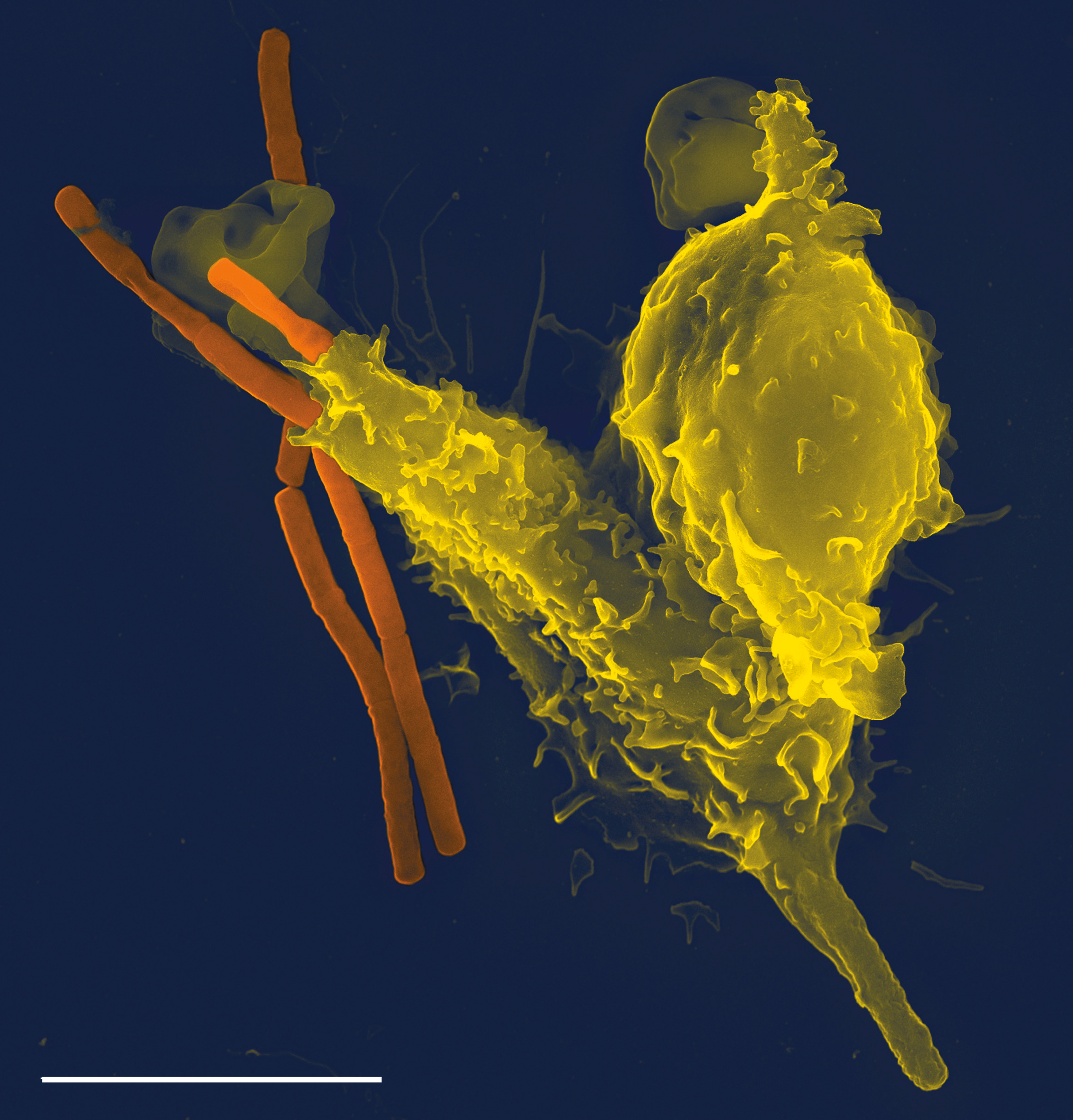
White blood cells, also called leukocytes or leucocytes, are the cells of the immune system that are involved in protecting the body against both infectious disease and foreign invaders. All white blood cells are produced and derived from multipotent cells in the bone marrow known as hematopoietic stem cells. Leukocytes are found throughout the body, including the blood and lymphatic system. All white blood cells have nuclei, which distinguishes them from the other blood cells, the anucleated red blood cells (RBCs) and platelets. The different white blood cells are usually classified by cell lineage (myeloid cells or lymphoid cells). White blood cells are part of the body's immune system. They help the body fight infection and other diseases. Types of white blood cells are granulocytes (neutrophils, eosinophils, and basophils), and agranulocytes (monocytes, and lymphocytes (T cells and B cells)). Myeloid cells (myelocytes) include neutrophils, eosinophils, mast cells, bas ... [...More Info...] [...Related Items...] OR: [Wikipedia] [Google] [Baidu] |
Immune System
The immune system is a network of biological processes that protects an organism from diseases. It detects and responds to a wide variety of pathogens, from viruses to parasitic worms, as well as cancer cells and objects such as wood splinters, distinguishing them from the organism's own healthy tissue. Many species have two major subsystems of the immune system. The innate immune system provides a preconfigured response to broad groups of situations and stimuli. The adaptive immune system provides a tailored response to each stimulus by learning to recognize molecules it has previously encountered. Both use molecules and cells to perform their functions. Nearly all organisms have some kind of immune system. Bacteria have a rudimentary immune system in the form of enzymes that protect against virus infections. Other basic immune mechanisms evolved in ancient plants and animals and remain in their modern descendants. These mechanisms include phagocytosis, antimicrobial pe ... [...More Info...] [...Related Items...] OR: [Wikipedia] [Google] [Baidu] |
Histopathological
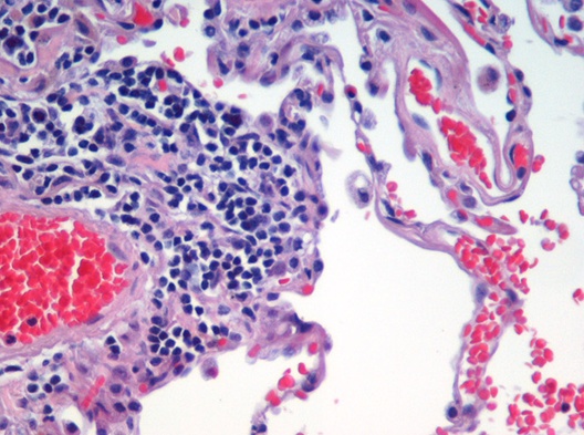
Histopathology (compound of three Greek words: ''histos'' "tissue", πάθος ''pathos'' "suffering", and -λογία ''-logia'' "study of") refers to the microscopic examination of tissue in order to study the manifestations of disease. Specifically, in clinical medicine, histopathology refers to the examination of a biopsy or surgical specimen by a pathologist, after the specimen has been processed and histological sections have been placed onto glass slides. In contrast, cytopathology examines free cells or tissue micro-fragments (as "cell blocks"). Collection of tissues Histopathological examination of tissues starts with surgery, biopsy, or autopsy. The tissue is removed from the body or plant, and then, often following expert dissection in the fresh state, placed in a fixative which stabilizes the tissues to prevent decay. The most common fixative is 10% neutral buffered formalin (corresponding to 3.7% w/v formaldehyde in neutral buffered water, such as phosphate buff ... [...More Info...] [...Related Items...] OR: [Wikipedia] [Google] [Baidu] |
Immunohistochemistry
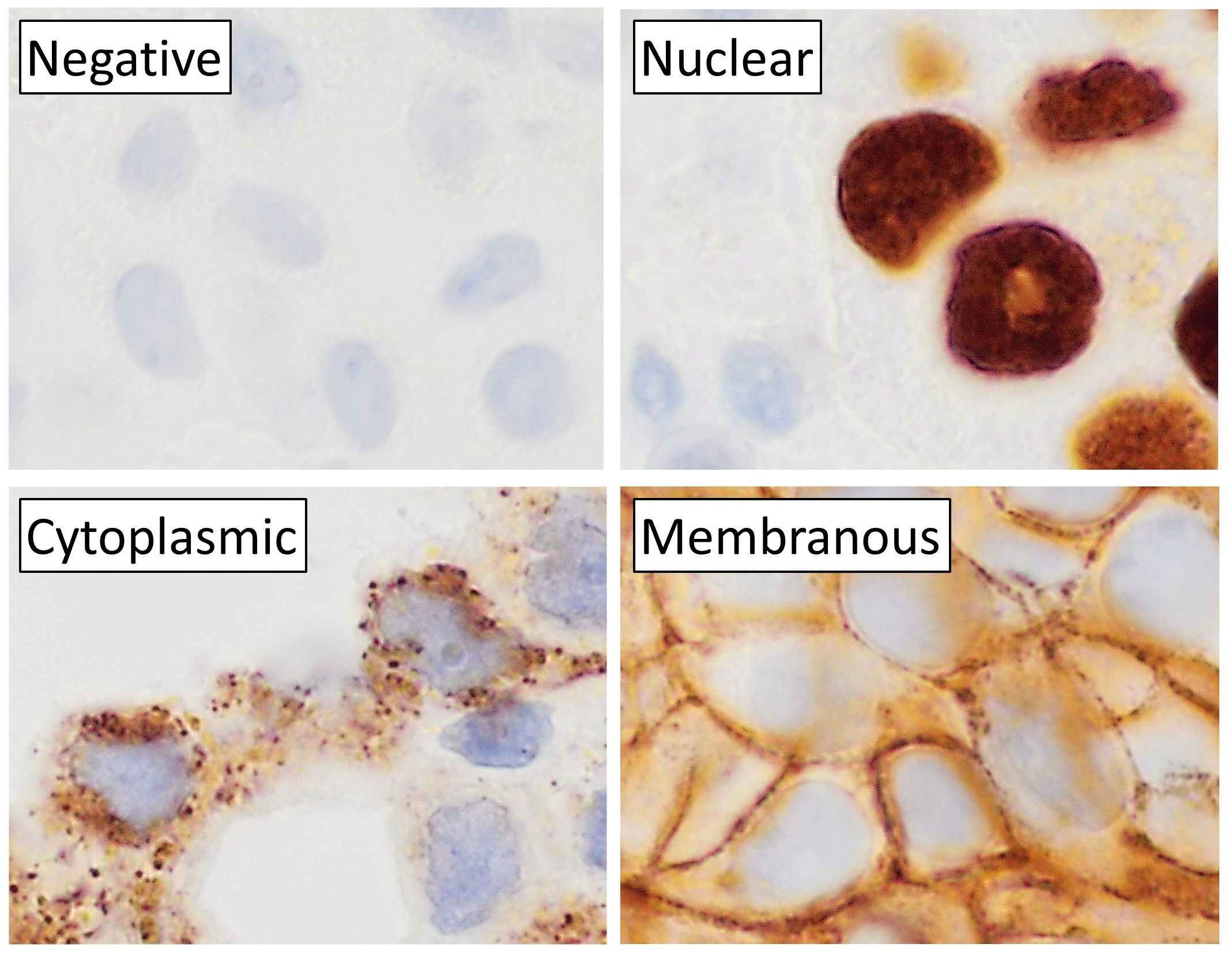
Immunohistochemistry (IHC) is the most common application of immunostaining. It involves the process of selectively identifying antigens (proteins) in cells of a tissue section by exploiting the principle of antibodies binding specifically to antigens in biological tissues. IHC takes its name from the roots "immuno", in reference to antibodies used in the procedure, and "histo", meaning tissue (compare to immunocytochemistry). Albert Coons conceptualized and first implemented the procedure in 1941. Visualising an antibody-antigen interaction can be accomplished in a number of ways, mainly either of the following: * ''Chromogenic immunohistochemistry'' (CIH), wherein an antibody is conjugated to an enzyme, such as peroxidase (the combination being termed immunoperoxidase), that can catalyse a colour-producing reaction. * '' Immunofluorescence'', where the antibody is tagged to a fluorophore, such as fluorescein or rhodamine. Immunohistochemical staining is widely used in the dia ... [...More Info...] [...Related Items...] OR: [Wikipedia] [Google] [Baidu] |
.jpg)


