|
Oral Pathology
Oral and maxillofacial pathology refers to the diseases of the mouth ("oral cavity" or "stoma"), jaws ("maxillae" or "gnath") and related structures such as salivary glands, temporomandibular joints, facial muscles and perioral skin (the skin around the mouth). The mouth is an important organ with many different functions. It is also prone to a variety of medical and dental disorders. The specialty oral and maxillofacial pathology is concerned with diagnosis and study of the causes and effects of diseases affecting the oral and maxillofacial region. It is sometimes considered to be a specialty of dentistry and pathology. Sometimes the term head and neck pathology is used instead, which may indicate that the pathologist deals with otorhinolaryngologic disorders (i.e. ear, nose and throat) in addition to maxillofacial disorders. In this role there is some overlap between the expertise of head and neck pathologists and that of endocrine pathologists. Diagnosis The key to any dia ... [...More Info...] [...Related Items...] OR: [Wikipedia] [Google] [Baidu] |
Dentistry
Dentistry, also known as dental medicine and oral medicine, is the branch of medicine focused on the teeth, gums, and mouth. It consists of the study, diagnosis, prevention, management, and treatment of diseases, disorders, and conditions of the mouth, most commonly focused on dentition (the development and arrangement of teeth) as well as the oral mucosa. Dentistry may also encompass other aspects of the craniofacial complex including the temporomandibular joint. The practitioner is called a dentist. The history of dentistry is almost as ancient as the history of humanity and civilization with the earliest evidence dating from 7000 BC to 5500 BC. Dentistry is thought to have been the first specialization in medicine which have gone on to develop its own accredited degree with its own specializations. Dentistry is often also understood to subsume the now largely defunct medical specialty of stomatology (the study of the mouth and its disorders and diseases) for which reas ... [...More Info...] [...Related Items...] OR: [Wikipedia] [Google] [Baidu] |
Microscopy
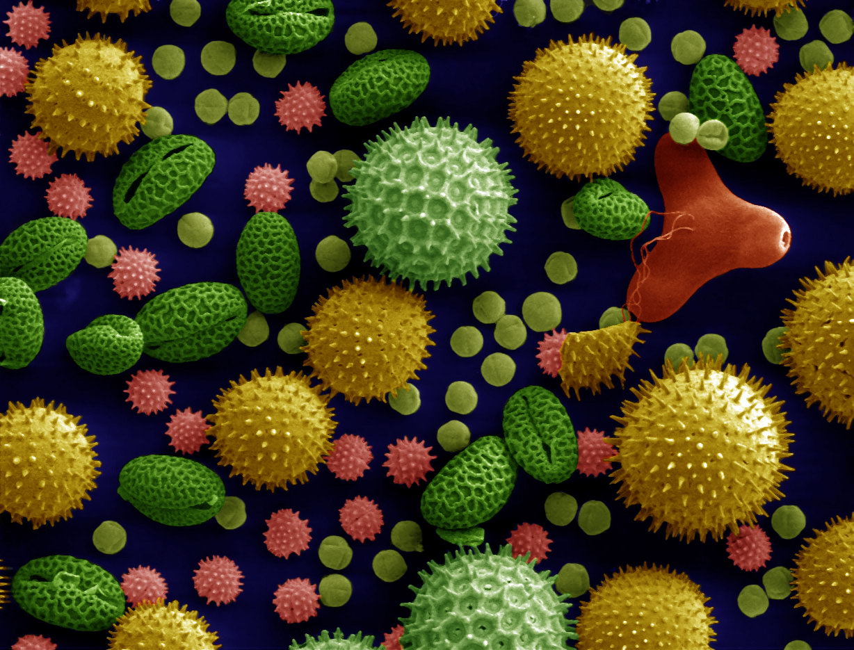
Microscopy is the technical field of using microscopes to view objects and areas of objects that cannot be seen with the naked eye (objects that are not within the resolution range of the normal eye). There are three well-known branches of microscopy: optical, electron, and scanning probe microscopy, along with the emerging field of X-ray microscopy. Optical microscopy and electron microscopy involve the diffraction, reflection, or refraction of electromagnetic radiation/electron beams interacting with the specimen, and the collection of the scattered radiation or another signal in order to create an image. This process may be carried out by wide-field irradiation of the sample (for example standard light microscopy and transmission electron microscopy) or by scanning a fine beam over the sample (for example confocal laser scanning microscopy and scanning electron microscopy). Scanning probe microscopy involves the interaction of a scanning probe with the surface of the o ... [...More Info...] [...Related Items...] OR: [Wikipedia] [Google] [Baidu] |
Dental Tartar
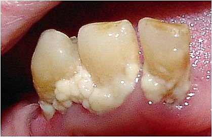
In dentistry, calculus or tartar is a form of hardened dental plaque. It is caused by precipitation of minerals from saliva and gingival crevicular fluid (GCF) in plaque on the teeth. This process of precipitation kills the bacterial cells within dental plaque, but the rough and hardened surface that is formed provides an ideal surface for further plaque formation. This leads to calculus buildup, which compromises the health of the gingiva (gums). Calculus can form both along the gumline, where it is referred to as supragingival ("above the gum"), and within the narrow sulcus that exists between the teeth and the gingiva, where it is referred to as subgingival ("below the gum"). Calculus formation is associated with a number of clinical manifestations, including bad breath, receding gums and chronically inflamed gingiva. Brushing and flossing can remove plaque from which calculus forms; however, once formed, calculus is too hard (firmly attached) to be removed with a toothbr ... [...More Info...] [...Related Items...] OR: [Wikipedia] [Google] [Baidu] |
Dental Plaque
Dental plaque is a biofilm of microorganisms (mostly bacteria, but also fungi) that grows on surfaces within the mouth. It is a sticky colorless deposit at first, but when it forms tartar, it is often brown or pale yellow. It is commonly found between the teeth, on the front of teeth, behind teeth, on chewing surfaces, along the gumline (supragingival), or below the gumline cervical margins (subgingival). Dental plaque is also known as microbial plaque, oral biofilm, dental biofilm, dental plaque biofilm or bacterial plaque biofilm. Bacterial plaque is one of the major causes for dental decay and gum disease. Progression and build-up of dental plaque can give rise to tooth decay – the localised destruction of the tissues of the tooth by acid produced from the bacterial degradation of fermentable sugar – and periodontal problems such as gingivitis and periodontitis; hence it is important to disrupt the mass of bacteria and remove it. Plaque control and removal can be achiev ... [...More Info...] [...Related Items...] OR: [Wikipedia] [Google] [Baidu] |
Periodontal Disease
Periodontal disease, also known as gum disease, is a set of inflammatory conditions affecting the tissues surrounding the teeth. In its early stage, called gingivitis, the gums become swollen and red and may bleed. It is considered the main cause of tooth loss for adults worldwide.V. Baelum and R. Lopez, “Periodontal epidemiology: towards social science or molecular biology?,”Community Dentistry and Oral Epidemiology, vol. 32, no. 4, pp. 239–249, 2004.Nicchio I, Cirelli T, Nepomuceno R, et al. Polymorphisms in Genes of Lipid Metabolism Are Associated with Type 2 Diabetes Mellitus and Periodontitis, as Comorbidities, and with the Subjects' Periodontal, Glycemic, and Lipid Profiles Journal of Diabetes Research. 2021 Jan;2021. PMCID: PMC8601849. In its more serious form, called periodontitis, the gums can pull away from the tooth, bone can be lost, and the teeth may loosen or fall out. Bad breath may also occur. Periodontal disease is generally due to bacteria in the mouth ... [...More Info...] [...Related Items...] OR: [Wikipedia] [Google] [Baidu] |
Gingivitis
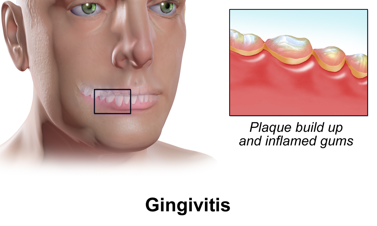
Gingivitis is a non-destructive disease that causes inflammation of the gums. The most common form of gingivitis, and the most common form of periodontal disease overall, is in response to bacterial biofilms (also called plaque) that is attached to tooth surfaces, termed ''plaque-induced gingivitis''. Most forms of gingivitis are plaque-induced. While some cases of gingivitis never progress to periodontitis, periodontitis is always preceded by gingivitis. Gingivitis is reversible with good oral hygiene; however, without treatment, gingivitis can progress to periodontitis, in which the inflammation of the gums results in tissue destruction and bone resorption around the teeth. Periodontitis can ultimately lead to tooth loss. Signs and symptoms The symptoms of gingivitis are somewhat non-specific and manifest in the gum tissue as the classic signs of inflammation: *Swollen gums *Bright red gums *Gums that are tender or painful to the touch *Bleeding gums or bleeding after bru ... [...More Info...] [...Related Items...] OR: [Wikipedia] [Google] [Baidu] |
Dental Plaque
Dental plaque is a biofilm of microorganisms (mostly bacteria, but also fungi) that grows on surfaces within the mouth. It is a sticky colorless deposit at first, but when it forms tartar, it is often brown or pale yellow. It is commonly found between the teeth, on the front of teeth, behind teeth, on chewing surfaces, along the gumline (supragingival), or below the gumline cervical margins (subgingival). Dental plaque is also known as microbial plaque, oral biofilm, dental biofilm, dental plaque biofilm or bacterial plaque biofilm. Bacterial plaque is one of the major causes for dental decay and gum disease. Progression and build-up of dental plaque can give rise to tooth decay – the localised destruction of the tissues of the tooth by acid produced from the bacterial degradation of fermentable sugar – and periodontal problems such as gingivitis and periodontitis; hence it is important to disrupt the mass of bacteria and remove it. Plaque control and removal can be achiev ... [...More Info...] [...Related Items...] OR: [Wikipedia] [Google] [Baidu] |
Eagle Syndrome
Eagle syndrome (also termed stylohyoid syndrome, styloid syndrome, styloid-stylohyoid syndrome, or styloid–carotid artery syndrome) is a rare condition commonly characterized but not limited to sudden, sharp nerve-like pain in the jaw bone and joint, back of the throat, and base of the tongue, triggered by swallowing, moving the jaw, or turning the neck. Since the brain to body's nerve connections pass through the neck, many seemingly random symptoms can be triggered by impingement or entanglement. First described by American otorhinolaryngologist Watt Weems Eagle in 1937, the condition is caused by an elongated or misshapen styloid process (the slender, pointed piece of bone just below the ear) and/or calcification of the stylohyoid ligament, either of which interferes with the functioning of neighboring regions in the body, giving rise to pain. Signs and symptoms Possible symptoms include: Classic eagle syndrome is present on only one side, however, rarely, it may be presen ... [...More Info...] [...Related Items...] OR: [Wikipedia] [Google] [Baidu] |
Torus Mandibularis
Torus mandibularis is a bony growth in the mandible along the surface nearest to the tongue. Mandibular tori are usually present near the premolars and above the location of the mylohyoid muscle's attachment to the mandible. In 90% of cases, there is a torus on both the left and right sides. The prevalence of mandibular tori ranges from 5-40%. It is less common than bony growths occurring on the palate, known as torus palatinus. Mandibular tori are more common in Asian and Inuit populations, and slightly more common in males. In the United States, the prevalence is 7-10% of the population. It is believed that mandibular tori are caused by several factors. They are more common in early adulthood and are associated with bruxism. The size of the tori may fluctuate throughout life, and in some cases the tori can be large enough to touch each other in the midline of mouth. Consequently, it is believed that mandibular tori are the result of local stresses and not due solely to genetic ... [...More Info...] [...Related Items...] OR: [Wikipedia] [Google] [Baidu] |
Torus Palatinus
A torus palatinus (pl. tori palatini), or palatal torus (pl. palatal tori), is a bony protrusion on the palate. Palatal tori are usually present on the midline of the hard palate.Neville, B.W., D. Damm, C. Allen, J. Bouquot. ''Oral & Maxillofacial Pathology''. Second edition. 2002. Page 20. . Most palatal tori are less than 2 cm in diameter, but their size can change throughout life. Types Sometimes, the tori are categorized by their appearance. Arising as a broad base and a smooth surface, flat tori are located on the midline of the palate and extend symmetrically to either side. Spindle tori have a ridge located at their midline. Nodular tori have multiple bony growths that each have their own base. Lobular tori have multiple bony growths with a common base. Cause Although some research suggest palatal tori to be an autosomal dominant trait, it is generally believed that palatal tori are caused by several factors. They are more common in early adult life and can increase ... [...More Info...] [...Related Items...] OR: [Wikipedia] [Google] [Baidu] |
Stafne Defect
The Stafne defect (also termed Stafne's idiopathic bone cavity, Stafne bone cavity, Stafne bone cyst (misnomer), lingual mandibular salivary gland depression, lingual mandibular cortical defect, latent bone cyst, or static bone cyst) is a depression of the mandible, most commonly located on the lingual surface (the side nearest the tongue). The Stafne defect is thought to be a normal anatomical variant, as the depression is created by ectopic salivary gland tissue associated with the submandibular gland and does not represent a pathologic lesion as such. Classification It is a classed as a pseudocyst, since there is no epithelial lining or fluid content. This defect is usually considered with other cysts of the jaws, since it can be mistaken for such on a radiograph. Signs and symptoms There are no symptoms, and no signs can be elicited on examination. Medical imaging such as traditional radiography or computed tomography is required to demonstrate the defect. Usually the de ... [...More Info...] [...Related Items...] OR: [Wikipedia] [Google] [Baidu] |


