|
Obliquus Capitis Superior
The obliquus capitis superior muscle () is a small muscle in the upper back part of the neck and is one of the suboccipital muscles and part of the suboccipital triangle. It arises from the lateral mass of the atlas bone. It passes superiorly and posteriorly to insert into the lateral half of the inferior nuchal line on the external surface of the occipital bone. The muscle is innervated by the suboccipital nerve, the dorsal ramus of the first spinal nerve. It acts at the atlanto-occipital joint to extend the head and flex the head to the ipsilateral side. Additional images File:Obliquus capitis superior muscle - animation04.gif, Position of obliquus capitis superior (shown in red). Animation. File:Obliquus capitis superior muscle05.png, Still image. Posterior view. File:Obliquus capitis superior.png, Deep muscles of the back (obliquus capitis superior labeled at upper left) File:Gray129.png, Occipital bone. Outer surface. Muscle attachments are shown as red circles. ... [...More Info...] [...Related Items...] OR: [Wikipedia] [Google] [Baidu] |
Human Skull
The skull is a bone protective cavity for the brain. The skull is composed of four types of bone i.e., cranial bones, facial bones, ear ossicles and hyoid bone. However two parts are more prominent: the cranium and the mandible. In humans, these two parts are the neurocranium and the viscerocranium ( facial skeleton) that includes the mandible as its largest bone. The skull forms the anterior-most portion of the skeleton and is a product of cephalisation—housing the brain, and several sensory structures such as the eyes, ears, nose, and mouth. In humans these sensory structures are part of the facial skeleton. Functions of the skull include protection of the brain, fixing the distance between the eyes to allow stereoscopic vision, and fixing the position of the ears to enable sound localisation of the direction and distance of sounds. In some animals, such as horned ungulates (mammals with hooves), the skull also has a defensive function by providing the mount (on the front ... [...More Info...] [...Related Items...] OR: [Wikipedia] [Google] [Baidu] |
Occipital Bone
The occipital bone () is a neurocranium, cranial dermal bone and the main bone of the occiput (back and lower part of the skull). It is trapezoidal in shape and curved on itself like a shallow dish. The occipital bone overlies the occipital lobes of the cerebrum. At the base of skull in the occipital bone, there is a large oval opening called the foramen magnum, which allows the passage of the spinal cord. Like the other cranial bones, it is classed as a flat bone. Due to its many attachments and features, the occipital bone is described in terms of separate parts. From its front to the back is the basilar part of occipital bone, basilar part, also called the basioccipital, at the sides of the foramen magnum are the lateral parts of occipital bone, lateral parts, also called the exoccipitals, and the back is named as the squamous part of occipital bone, squamous part. The basilar part is a thick, somewhat quadrilateral piece in front of the foramen magnum and directed towards the ... [...More Info...] [...Related Items...] OR: [Wikipedia] [Google] [Baidu] |
Back Muscles
The human back, also called the dorsum, is the large posterior area of the human body, rising from the top of the buttocks to the back of the neck. It is the surface of the body opposite from the chest and the abdomen. The vertebral column runs the length of the back and creates a central area of recession. The breadth of the back is created by the shoulders at the top and the pelvis at the bottom. Back pain is a common medical condition, generally benign in origin. Structure The central feature of the human back is the vertebral column, specifically the length from the top of the thoracic vertebrae to the bottom of the lumbar vertebrae, which houses the spinal cord in its spinal canal, and which generally has some curvature that gives shape to the back. The ribcage extends from the spine at the top of the back (with the top of the ribcage corresponding to the T1 vertebra), more than halfway down the length of the back, leaving an area with less protection between the bottom ... [...More Info...] [...Related Items...] OR: [Wikipedia] [Google] [Baidu] |
Atlanto-occipital Joint
The atlanto-occipital joint (''Capsula articularis atlantooccipitalis'') is an articulation between the atlas bone and the occipital bone. It consists of a pair of condyloid joints. It is a synovial joint. Structure The atlanto-occipital joint is an articulation between the atlas bone and the occipital bone. It consists of a pair of condyloid joints. It is a synovial joint. Ligaments The ligaments connecting the bones are: * Two articular capsules * Posterior atlanto-occipital membrane * Anterior atlanto-occipital membrane Capsule The capsules of the atlantooccipital articulation surround the condyles of the occipital bone, and connect them with the articular processes of the atlas: they are thin and loose. Function The movements permitted in this joint are: * (a) flexion and extension around the mediolateral axis, which give rise to the ordinary forward and backward nodding of the head. * (b) slight lateral motion, lateroflexion, to one or other side around the anteroposter ... [...More Info...] [...Related Items...] OR: [Wikipedia] [Google] [Baidu] |
Spinal Nerve
A spinal nerve is a mixed nerve, which carries motor, sensory, and autonomic signals between the spinal cord and the body. In the human body there are 31 pairs of spinal nerves, one on each side of the vertebral column. These are grouped into the corresponding cervical, thoracic, lumbar, sacral and coccygeal regions of the spine. There are eight pairs of cervical nerves, twelve pairs of thoracic nerves, five pairs of lumbar nerves, five pairs of sacral nerves, and one pair of coccygeal nerves. The spinal nerves are part of the peripheral nervous system. Structure Each spinal nerve is a mixed nerve, formed from the combination of nerve fibers from its dorsal and ventral roots. The dorsal root is the afferent sensory root and carries sensory information to the brain. The ventral root is the efferent motor root and carries motor information from the brain. The spinal nerve emerges from the spinal column through an opening (intervertebral foramen) between adjacent vertebrae. ... [...More Info...] [...Related Items...] OR: [Wikipedia] [Google] [Baidu] |
Cervical Spinal Nerve 1
The cervical spinal nerve 1 (C1) is a spinal nerve of the cervical segment. from spinalcordinjuryzone.com. Published February 23, 2004 Archived Dec 23, 2011. Retrieved June 12, 2018. C1 carries predominantly motor fibres, but also a small meningeal branch that supplies sensation to parts of the dura around the foramen magnum (via dorsal rami). It originates from the spinal column from above the (C1). The dorsal root and gang ... [...More Info...] [...Related Items...] OR: [Wikipedia] [Google] [Baidu] |
Dorsal Ramus
The dorsal ramus of spinal nerve (or posterior ramus of spinal nerve, or posterior primary division) is the posterior division of a spinal nerve. The dorsal ramus (Latin for branch, plural ''rami'' ) is the dorsal branch of a spinal nerve that forms from the dorsal root of the nerve after it emerges from the spinal cord. The spinal nerve is formed from the dorsal and ventral rami. The dorsal ramus carries information that supplies muscles and skin sensation to the human back. Structure Ventral root axons join with dorsal root ganglia to form mixed spinal nerves (below). These then merge to form peripheral nerves. Shortly after this spinal nerve forms, it then branches into the dorsal ramus and ventral ramus. Spinal nerves are mixed nerves that carry both sensory and motor information. It also branches to form the grey and the white rami communicantes which make connections with the sympathetic ganglia. After it is formed, the dorsal ramus of each spinal nerve travels backward, e ... [...More Info...] [...Related Items...] OR: [Wikipedia] [Google] [Baidu] |
Suboccipital Nerve
The suboccipital nerve (first cervical dorsal ramus) is the dorsal primary ramus of the first cervical nerve (C1). It exits the spinal cord between the skull and the first cervical vertebra In tetrapods, cervical vertebrae (singular: vertebra) are the vertebrae of the neck, immediately below the skull. Truncal vertebrae (divided into thoracic and lumbar vertebrae in mammals) lie caudal (toward the tail) of cervical vertebrae. In sau ..., the Atlas (anatomy), atlas. It lies within the suboccipital triangle along with the vertebral artery, where the artery enters the foramen magnum. It supplies muscles of the suboccipital triangle, the rectus capitis posterior major, obliquus capitis superior, and obliquus capitis inferior. The suboccipital nerve also innervates rectus capitis posterior minor. See also * Vertebral artery Additional images File:Gray792.png, Upper part of medulla spinalis and hind- and mid-brains; posterior aspect, exposed in situ. File:Suboccipital_triangle.P ... [...More Info...] [...Related Items...] OR: [Wikipedia] [Google] [Baidu] |
Atlas Bone
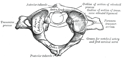
In anatomy, the atlas (C1) is the most superior (first) cervical vertebra of the spine and is located in the neck. It is named for Atlas of Greek mythology because, just as Atlas supported the globe, it supports the entire head. The atlas is the topmost vertebra and, with the axis (the vertebra below it), forms the joint connecting the skull and spine. The atlas and axis are specialized to allow a greater range of motion than normal vertebrae. They are responsible for the nodding and rotation movements of the head. The atlanto-occipital joint allows the head to nod up and down on the vertebral column. The dens acts as a pivot that allows the atlas and attached head to rotate on the axis, side to side. The atlas's chief peculiarity is that it has no body. It is ring-like and consists of an anterior and a posterior arch and two lateral masses. The atlas and axis are important neurologically because the brainstem extends down to the axis. Structure Anterior arch The anterio ... [...More Info...] [...Related Items...] OR: [Wikipedia] [Google] [Baidu] |
Lateral Mass Of Atlas
In anatomy, the atlas (C1) is the most superior (first) cervical vertebra of the spine and is located in the neck. It is named for Atlas of Greek mythology because, just as Atlas supported the globe, it supports the entire head. The atlas is the topmost vertebra and, with the axis (the vertebra below it), forms the joint connecting the skull and spine. The atlas and axis are specialized to allow a greater range of motion than normal vertebrae. They are responsible for the nodding and rotation movements of the head. The atlanto-occipital joint allows the head to nod up and down on the vertebral column. The dens acts as a pivot that allows the atlas and attached head to rotate on the axis, side to side. The atlas's chief peculiarity is that it has no body. It is ring-like and consists of an anterior and a posterior arch and two lateral masses. The atlas and axis are important neurologically because the brainstem extends down to the axis. Structure Anterior arch The ant ... [...More Info...] [...Related Items...] OR: [Wikipedia] [Google] [Baidu] |
Suboccipital Triangle
The suboccipital triangle is a region of the neck bounded by the following three muscles of the suboccipital group of muscles: * Rectus capitis posterior major - above and medially * Obliquus capitis superior - above and laterally * Obliquus capitis inferior - below and laterally (Rectus capitis posterior minor is also in this region but does not form part of the triangle) It is covered by a layer of dense fibro-fatty tissue, situated beneath the semispinalis capitis. The floor is formed by the posterior atlantooccipital membrane, and the posterior arch of the atlas. In the deep groove on the upper surface of the posterior arch of the atlas are the vertebral artery and the first cervical or suboccipital nerve. In the past, the vertebral artery was accessed here in order to conduct angiography of the circle of Willis. Presently, formal angiography of the circle of Willis is performed via catheter angiography, with access usually being acquired at the common femoral artery. Alter ... [...More Info...] [...Related Items...] OR: [Wikipedia] [Google] [Baidu] |


