|
Nondisjunction Diagrams
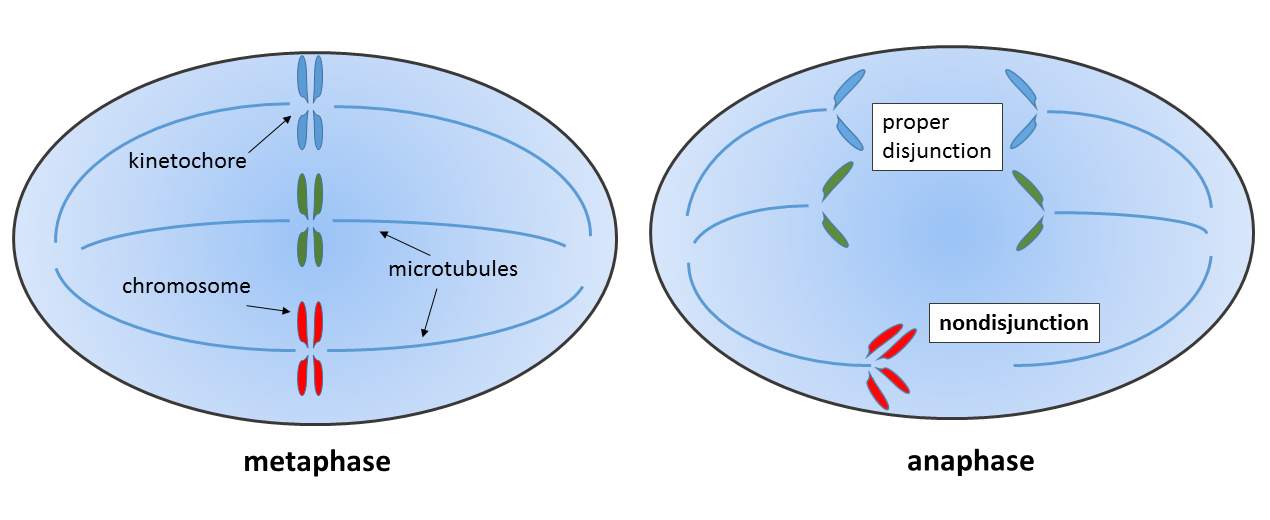
Nondisjunction is the failure of homologous chromosomes or sister chromatids to separate properly during cell division (mitosis/meiosis). There are three forms of nondisjunction: failure of a pair of homologous chromosomes to separate in meiosis I, failure of sister chromatids to separate during meiosis II, and failure of sister chromatids to separate during mitosis. Nondisjunction results in daughter cells with abnormal chromosome numbers (aneuploidy). Calvin Bridges and Thomas Hunt Morgan are credited with discovering nondisjunction in ''Drosophila melanogaster'' sex chromosomes in the spring of 1910, while working in the Zoological Laboratory of Columbia University. Types In general, nondisjunction can occur in any form of cell division that involves ordered distribution of chromosomal material. Higher animals have three distinct forms of such cell divisions: Meiosis I and meiosis II are specialized forms of cell division occurring during generation of gametes (eggs and sperm) ... [...More Info...] [...Related Items...] OR: [Wikipedia] [Google] [Baidu] |
Spermatogenesis
Spermatogenesis is the process by which haploid spermatozoa develop from germ cells in the seminiferous tubules of the testis. This process starts with the mitotic division of the stem cells located close to the basement membrane of the tubules. These cells are called spermatogonial stem cells. The mitotic division of these produces two types of cells. Type A cells replenish the stem cells, and type B cells differentiate into primary spermatocytes. The primary spermatocyte divides meiotically (Meiosis I) into two secondary spermatocytes; each secondary spermatocyte divides into two equal haploid spermatids by Meiosis II. The spermatids are transformed into spermatozoa (sperm) by the process of spermiogenesis. These develop into mature spermatozoa, also known as sperm cells. Thus, the primary spermatocyte gives rise to two cells, the secondary spermatocytes, and the two secondary spermatocytes by their subdivision produce four spermatozoa and four haploid cells. Spermatozoa are ... [...More Info...] [...Related Items...] OR: [Wikipedia] [Google] [Baidu] |
Topoisomerase II
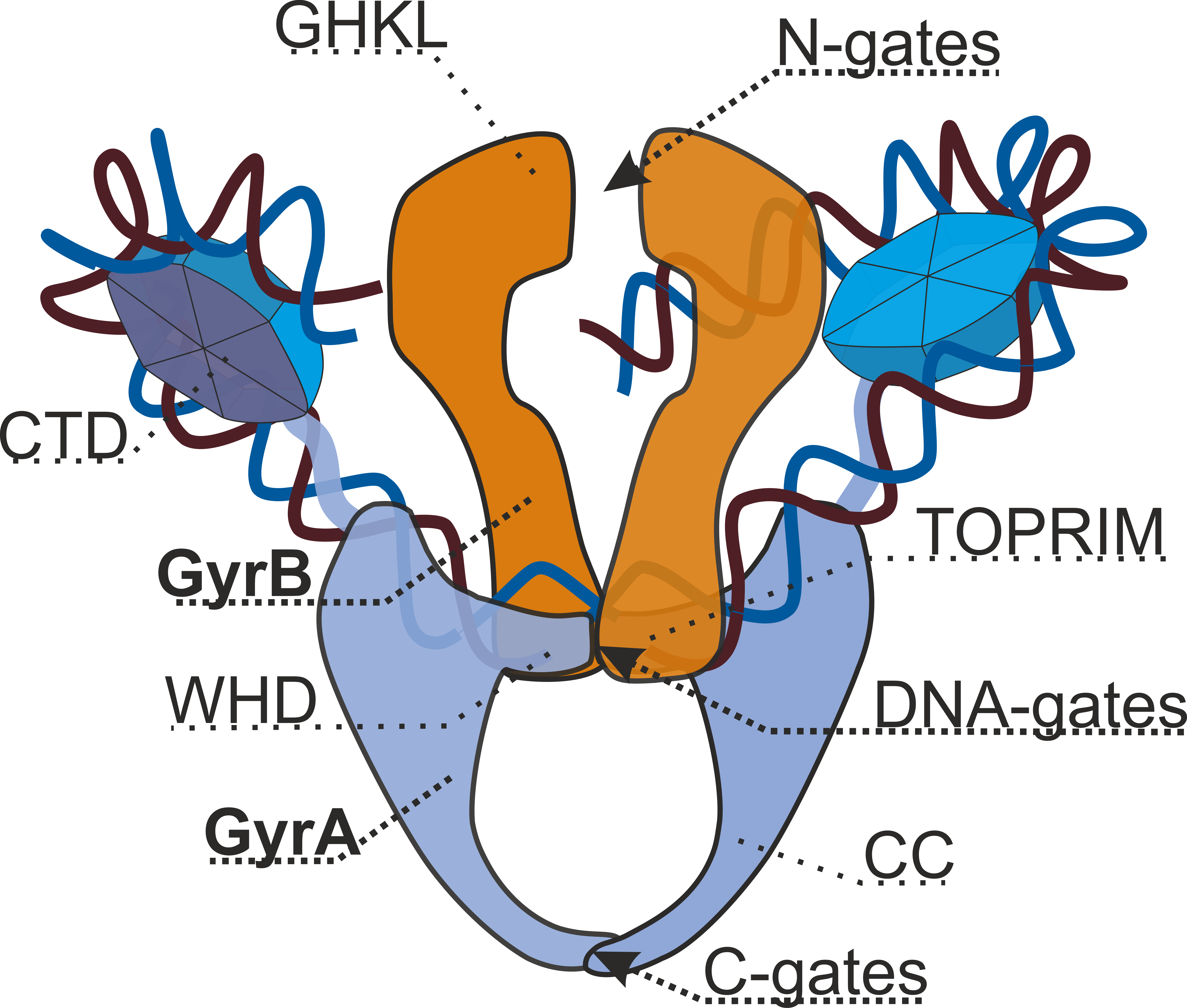
Type II topoisomerases are topoisomerases that cut both strands of the DNA helix simultaneously in order to manage DNA tangles and supercoils. They use the hydrolysis of ATP, unlike Type I topoisomerase. In this process, these enzymes change the linking number of circular DNA by ±2. Topoisomerases are ubiquitous enzymes, found in all living organisms. In animals, topoisomerase II is a chemotherapy target. In prokaryotes, gyrase is an antibacterial target. Indeed, these enzymes are of interest for a wide range of effects. Function Type II topoisomerases increase or decrease the linking number of a DNA loop by 2 units, and it promotes chromosome disentanglement. For example, DNA gyrase, a type II topoisomerase observed in ''E. coli'' and most other prokaryotes, introduces negative supercoils and decreases the linking number by 2. Gyrase is also able to remove knots from the bacterial chromosome. Along with gyrase, most prokaryotes also contain a second type IIA topoisomerase, ter ... [...More Info...] [...Related Items...] OR: [Wikipedia] [Google] [Baidu] |
Retinoblastoma
Retinoblastoma (Rb) is a rare form of cancer that rapidly develops from the immature cells of a retina, the light-detecting tissue of the eye. It is the most common primary malignant intraocular cancer in children, and it is almost exclusively found in young children. Though most children survive this cancer, they may lose their vision in the affected eye(s) or need to have the eye removed. Almost half of children with retinoblastoma have a hereditary genetic defect associated with retinoblastoma. In other cases, it is caused by a congenital mutation in the chromosome 13 gene 13q14 (retinoblastoma protein). Signs and symptoms Retinoblastoma is universally known as the most intrusive intraocular cancer among children. The chance of survival and preservation of the eye depends fully on the severity. Retinoblastoma is extremely rare as there are only about 200 to 300 cases every year in the United States. Looking at retinoblastoma globally, only 1 in about 15,000 children have ... [...More Info...] [...Related Items...] OR: [Wikipedia] [Google] [Baidu] |
Cancer
Cancer is a group of diseases involving abnormal cell growth with the potential to invade or spread to other parts of the body. These contrast with benign tumors, which do not spread. Possible signs and symptoms include a lump, abnormal bleeding, prolonged cough, unexplained weight loss, and a change in bowel movements. While these symptoms may indicate cancer, they can also have other causes. Over 100 types of cancers affect humans. Tobacco use is the cause of about 22% of cancer deaths. Another 10% are due to obesity, poor diet, lack of physical activity or excessive drinking of alcohol. Other factors include certain infections, exposure to ionizing radiation, and environmental pollutants. In the developing world, 15% of cancers are due to infections such as ''Helicobacter pylori'', hepatitis B, hepatitis C, human papillomavirus infection, Epstein–Barr virus and human immunodeficiency virus (HIV). These factors act, at least partly, by changing the genes of ... [...More Info...] [...Related Items...] OR: [Wikipedia] [Google] [Baidu] |
Chromosomes
A chromosome is a long DNA molecule with part or all of the genetic material of an organism. In most chromosomes the very long thin DNA fibers are coated with packaging proteins; in eukaryotic cells the most important of these proteins are the histones. These proteins, aided by chaperone proteins, bind to and condense the DNA molecule to maintain its integrity. These chromosomes display a complex three-dimensional structure, which plays a significant role in transcriptional regulation. Chromosomes are normally visible under a light microscope only during the metaphase of cell division (where all chromosomes are aligned in the center of the cell in their condensed form). Before this happens, each chromosome is duplicated (S phase), and both copies are joined by a centromere, resulting either in an X-shaped structure (pictured above), if the centromere is located equatorially, or a two-arm structure, if the centromere is located distally. The joined copies are now called sis ... [...More Info...] [...Related Items...] OR: [Wikipedia] [Google] [Baidu] |
Mosaicism
Mosaicism or genetic mosaicism is a condition in multicellular organisms in which a single organism possesses more than one genetic line as the result of genetic mutation. This means that various genetic lines resulted from a single fertilized egg. Genetic mosaics may often be confused with chimerism, in which two or more genotypes arise in one individual similarly to mosaicism. In chimerism, though, the two genotypes arise from the fusion of more than one fertilized zygote in the early stages of embryonic development, rather than from a mutation or chromosome loss. Genetic mosaicism can result from many different mechanisms including chromosome nondisjunction, anaphase lag, and endoreplication. Anaphase lagging is the most common way by which mosaicism arises in the preimplantation embryo. Mosaicism can also result from a mutation in one cell during development, in which case the mutation will be passed on only to its daughter cells (and will be present only in certain adult cel ... [...More Info...] [...Related Items...] OR: [Wikipedia] [Google] [Baidu] |
Chromatin Bridge
Chromatin bridge is a mitotic occurrence that forms when telomeres of sister chromatids fuse together and fail to completely segregate into their respective daughter cells. Because this event is most prevalent during anaphase, the term anaphase bridge is often used as a substitute. After the formation of individual daughter cells, the DNA bridge connecting homologous chromosomes remains fixed. As the daughter cells exit mitosis and re-enter interphase, the chromatin bridge becomes known as an interphase bridge. These phenomena are usually visualized using the laboratory techniques of staining and fluorescence microscopy. Background The faithful inheritance of genetic information from one cellular generation to the next heavily relies on the duplication of deoxyribonucleic acid (DNA), as well as the formation of two identical daughter cells. This complicated cellular process, known as mitosis, depends on a multitude of cellular checkpoints, signals, interactions and signal cascades ... [...More Info...] [...Related Items...] OR: [Wikipedia] [Google] [Baidu] |
Centromere
The centromere links a pair of sister chromatids together during cell division. This constricted region of chromosome connects the sister chromatids, creating a short arm (p) and a long arm (q) on the chromatids. During mitosis, spindle fibers attach to the centromere via the kinetochore. The physical role of the centromere is to act as the site of assembly of the kinetochores – a highly complex multiprotein structure that is responsible for the actual events of chromosome segregation – i.e. binding microtubules and signaling to the cell cycle machinery when all chromosomes have adopted correct attachments to the spindle, so that it is safe for cell division to proceed to completion and for cells to enter anaphase. There are, broadly speaking, two types of centromeres. "Point centromeres" bind to specific proteins that recognize particular DNA sequences with high efficiency. Any piece of DNA with the point centromere DNA sequence on it will typically form a centromere if pr ... [...More Info...] [...Related Items...] OR: [Wikipedia] [Google] [Baidu] |
S Phase
S phase (Synthesis Phase) is the phase of the cell cycle in which DNA is replicated, occurring between G1 phase and G2 phase. Since accurate duplication of the genome is critical to successful cell division, the processes that occur during S-phase are tightly regulated and widely conserved. Regulation Entry into S-phase is controlled by the G1 restriction point (R), which commits cells to the remainder of the cell-cycle if there is adequate nutrients and growth signaling. This transition is essentially irreversible; after passing the restriction point, the cell will progress through S-phase even if environmental conditions become unfavorable. Accordingly, entry into S-phase is controlled by molecular pathways that facilitate a rapid, unidirectional shift in cell state. In yeast, for instance, cell growth induces accumulation of Cln3 cyclin, which complexes with the cyclin dependent kinase CDK2. The Cln3-CDK2 complex promotes transcription of S-phase genes by inactivating ... [...More Info...] [...Related Items...] OR: [Wikipedia] [Google] [Baidu] |
Somatic (biology)
The term somatic - etymologically from the Ancient Greek words of "σωματικός" (sōmatikós, “bodily”) and σῶμα (sôma, “body”) - is often used in biology to refer to the cells of the body in contrast to the reproductive (germline) cells, which usually give rise to the egg or sperm (or other gametes in other organisms). These somatic cells are diploid, containing two copies of each chromosome, whereas germ cells are haploid, as they only contain one copy of each chromosome (in preparation for fertilisation). Although under normal circumstances all somatic cells in an organism contain identical DNA, they develop a variety of tissue-specific characteristics. This process is called differentiation, through epigenetic and regulatory alterations. The grouping of similar cells and tissues creates the foundation for organs. Somatic mutations are changes to the genetics of a multicellular organism that are not passed on to its offspring through the germline. Most canc ... [...More Info...] [...Related Items...] OR: [Wikipedia] [Google] [Baidu] |
Mitotic Nondisjunction
Nondisjunction is the failure of homologous chromosomes or sister chromatids to separate properly during cell division (mitosis/meiosis). There are three forms of nondisjunction: failure of a pair of homologous chromosomes to separate in meiosis I, failure of sister chromatids to separate during meiosis II, and failure of sister chromatids to separate during mitosis. Nondisjunction results in daughter cells with abnormal chromosome numbers (aneuploidy). Calvin Bridges and Thomas Hunt Morgan are credited with discovering nondisjunction in ''Drosophila melanogaster'' sex chromosomes in the spring of 1910, while working in the Zoological Laboratory of Columbia University. Types In general, nondisjunction can occur in any form of cell division that involves ordered distribution of chromosomal material. Higher animals have three distinct forms of such cell divisions: Meiosis I and meiosis II are specialized forms of cell division occurring during generation of gametes (eggs and sperm) ... [...More Info...] [...Related Items...] OR: [Wikipedia] [Google] [Baidu] |


