|
Neuropeptide B
Neuropeptide B is a short biologically active peptide whose precursor in humans is encoded by the ''NBP'' gene. Neuropeptide B acts via two G protein-coupled receptors, neuropeptide B/W receptors, called NPBW1 and NPBW2 encoded by the genes ''NPBWR1'' and ''NPBWR2'', respectively. Neuropeptide B is thought to be associated with the regulation of feeding, neuroendocrine system, memory, learning and in the afferent pain pathway. It is expressed throughout the CNS with high levels in the substantia nigra, hypothalamus, hippocampus, and spinal cord The spinal cord is a long, thin, tubular structure made up of nervous tissue, which extends from the medulla oblongata in the brainstem to the lumbar region of the vertebral column (backbone). The backbone encloses the central canal of the spi .... References External linksUniprot: Neuropeptide B precursor {{Portal bar, Biology, border=no G proteins Neuropeptides ... [...More Info...] [...Related Items...] OR: [Wikipedia] [Google] [Baidu] |
Neuropeptide B/W Receptor
The neuropeptide B/W receptors are members of the G-protein coupled receptor superfamily of integral membrane proteins which bind the neuropeptide Neuropeptides are chemical messengers made up of small chains of amino acids that are synthesized and released by neurons. Neuropeptides typically bind to G protein-coupled receptors (GPCRs) to modulate neural activity and other tissues like the ...s B and W. These receptors are predominantly expressed in the CNS and have a number of functions including regulation of the secretion of cortisol. References External links * * * {{DEFAULTSORT:Neuropeptide B W receptor G protein-coupled receptors ... [...More Info...] [...Related Items...] OR: [Wikipedia] [Google] [Baidu] |
NPBWR1
Neuropeptides B/W receptor 1, also known as NPBW1 and GPR7, is a human protein encoded by the NPBWR1 gene. As implied by its name, it and related gene NPBW2 (with which it shares 70% nucleotide identity) are transmembranes protein that bind Neuropeptide B (NPB) and Neuropeptide W (NPW), both proteins expressed strongly in parts of the brain that regulate stress and fear including the extended amygdala and stria terminalis. When originally discovered in 1995, these receptors had no known ligands ("orphan receptors") and were called GPR7 and GPR8, but at least three groups in the early 2000s independently identified their endogenous ligands, triggering the name change in 2005. Structure NPBW1 has seven transmembrane domains, which it unsurprisingly shares with NPBWR2, but also a family of somatostatin and opioid receptors, and like these proteins couple to Gi-class G proteins. Functions In rodent models, NPBWR1 is over-expressed in Schwann cells associated with neuropathic pai ... [...More Info...] [...Related Items...] OR: [Wikipedia] [Google] [Baidu] |
Neuropeptides B/W Receptor 2
Neuropeptides B/W receptor 2, also known as NPBW2, is a human protein encoded by the NPBWR2 gene. The protein encoded by this gene is an integral membrane protein and G protein-coupled receptor. The encoded protein is similar in sequence to another G protein-coupled receptor (GPR7), and it is structurally similar to opioid and somatostatin receptors. This protein binds neuropeptides B and W. This gene is intronless and is expressed primarily in the frontal cortex of the brain. See also * Neuropeptide B/W receptor The neuropeptide B/W receptors are members of the G-protein coupled receptor superfamily of integral membrane proteins which bind the neuropeptide Neuropeptides are chemical messengers made up of small chains of amino acids that are synthesi ... References Further reading * * * * * * * External links * G protein-coupled receptors {{transmembranereceptor-stub ... [...More Info...] [...Related Items...] OR: [Wikipedia] [Google] [Baidu] |
Neuroendocrine System
Neuroendocrinology is the branch of biology (specifically of physiology) which studies the interaction between the nervous system and the endocrine system; i.e. how the brain regulates the hormonal activity in the body. The nervous and endocrine systems often act together in a process called neuroendocrine integration, to regulate the physiological processes of the human body. Neuroendocrinology arose from the recognition that the brain, especially the hypothalamus, controls secretion of pituitary gland hormones, and has subsequently expanded to investigate numerous interconnections of the endocrine and nervous systems. The endocrine system consists of numerous glands throughout the body that produce and secrete hormones of diverse chemical structure, including peptides, steroids, and neuroamines. Collectively, hormones regulate many physiological processes. The neuroendocrine system is the mechanism by which the hypothalamus maintains homeostasis, regulating reproduction, metab ... [...More Info...] [...Related Items...] OR: [Wikipedia] [Google] [Baidu] |
Substantia Nigra
The substantia nigra (SN) is a basal ganglia structure located in the midbrain that plays an important role in reward and movement. ''Substantia nigra'' is Latin for "black substance", reflecting the fact that parts of the substantia nigra appear darker than neighboring areas due to high levels of neuromelanin in dopaminergic neurons. Parkinson's disease is characterized by the loss of dopaminergic neurons in the substantia nigra pars compacta. Although the substantia nigra appears as a continuous band in brain sections, anatomical studies have found that it actually consists of two parts with very different connections and functions: the pars compacta (SNpc) and the pars reticulata (SNpr). The pars compacta serves mainly as a projection to the basal ganglia circuit, supplying the striatum with dopamine. The pars reticulata conveys signals from the basal ganglia to numerous other brain structures. Structure The substantia nigra, along with four other nuclei, is part ... [...More Info...] [...Related Items...] OR: [Wikipedia] [Google] [Baidu] |
Hypothalamus
The hypothalamus () is a part of the brain that contains a number of small nuclei with a variety of functions. One of the most important functions is to link the nervous system to the endocrine system via the pituitary gland. The hypothalamus is located below the thalamus and is part of the limbic system. In the terminology of neuroanatomy, it forms the ventral part of the diencephalon. All vertebrate brains contain a hypothalamus. In humans, it is the size of an almond. The hypothalamus is responsible for regulating certain metabolic processes and other activities of the autonomic nervous system. It synthesizes and secretes certain neurohormones, called releasing hormones or hypothalamic hormones, and these in turn stimulate or inhibit the secretion of hormones from the pituitary gland. The hypothalamus controls body temperature, hunger, important aspects of parenting and maternal attachment behaviours, thirst, fatigue, sleep, and circadian rhythms. Structure T ... [...More Info...] [...Related Items...] OR: [Wikipedia] [Google] [Baidu] |
Hippocampus
The hippocampus (via Latin from Greek , 'seahorse') is a major component of the brain of humans and other vertebrates. Humans and other mammals have two hippocampi, one in each side of the brain. The hippocampus is part of the limbic system, and plays important roles in the consolidation of information from short-term memory to long-term memory, and in spatial memory that enables navigation. The hippocampus is located in the allocortex, with neural projections into the neocortex in humans, as well as primates. The hippocampus, as the medial pallium, is a structure found in all vertebrates. In humans, it contains two main interlocking parts: the hippocampus proper (also called ''Ammon's horn''), and the dentate gyrus. In Alzheimer's disease (and other forms of dementia), the hippocampus is one of the first regions of the brain to suffer damage; short-term memory loss and disorientation are included among the early symptoms. Damage to the hippocampus can also result from ... [...More Info...] [...Related Items...] OR: [Wikipedia] [Google] [Baidu] |
Spinal Cord
The spinal cord is a long, thin, tubular structure made up of nervous tissue, which extends from the medulla oblongata in the brainstem to the lumbar region of the vertebral column (backbone). The backbone encloses the central canal of the spinal cord, which contains cerebrospinal fluid. The brain and spinal cord together make up the central nervous system (CNS). In humans, the spinal cord begins at the occipital bone, passing through the foramen magnum and then enters the spinal canal at the beginning of the cervical vertebrae. The spinal cord extends down to between the first and second lumbar vertebrae, where it ends. The enclosing bony vertebral column protects the relatively shorter spinal cord. It is around long in adult men and around long in adult women. The diameter of the spinal cord ranges from in the cervical and lumbar regions to in the thoracic area. The spinal cord functions primarily in the transmission of nerve signals from the motor cortex to the body, ... [...More Info...] [...Related Items...] OR: [Wikipedia] [Google] [Baidu] |
G Proteins
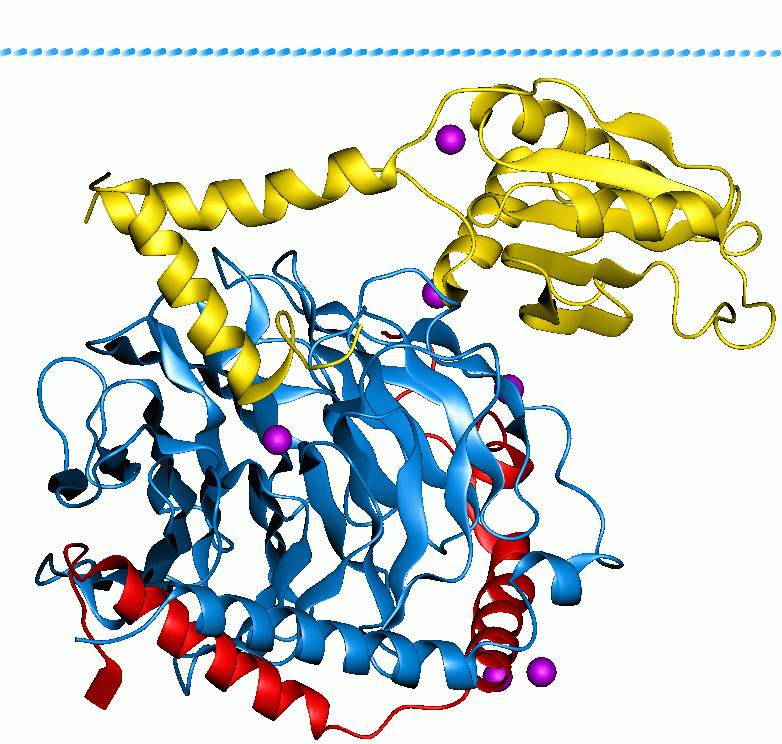
G proteins, also known as guanine nucleotide-binding proteins, are a family of proteins that act as molecular switches inside cells, and are involved in transmitting signals from a variety of stimuli outside a cell to its interior. Their activity is regulated by factors that control their ability to bind to and hydrolyze guanosine triphosphate (GTP) to guanosine diphosphate (GDP). When they are bound to GTP, they are 'on', and, when they are bound to GDP, they are 'off'. G proteins belong to the larger group of enzymes called GTPases. There are two classes of G proteins. The first function as monomeric small GTPases (small G-proteins), while the second function as heterotrimeric G protein complexes. The latter class of complexes is made up of '' alpha'' (α), ''beta'' (β) and ''gamma'' (γ) subunits. In addition, the beta and gamma subunits can form a stable dimeric complex referred to as the beta-gamma complex . Heterotrimeric G proteins located within the cell are activ ... [...More Info...] [...Related Items...] OR: [Wikipedia] [Google] [Baidu] |
.jpg)


