|
Neuronal Tracing
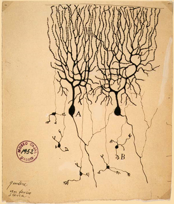
Neuronal tracing, or neuron reconstruction is a technique used in neuroscience to determine the pathway of the neurites or neuronal processes, the axons and dendrites, of a neuron. From a sample preparation point of view, it may refer to some of the following as well as other genetic neuron labeling techniques, * Anterograde tracing, for labeling from the cell body to synapse; * Retrograde tracing, for labeling from the synapse to cell body; * Viral neuronal tracing, for a technique which can be used to label in either direction; * Manual tracing of neuronal imagery. In broad sense, neuron tracing is more often related to digital reconstruction of a neuron's morphology from imaging data of above samples. Digital neuronal reconstruction and neuronal tracing Digital reconstruction or tracing of neuron morphology is a fundamental task in computational neuroscience. It is also critical for mapping neuronal circuits based on advanced microscope images, usually based on light micros ... [...More Info...] [...Related Items...] OR: [Wikipedia] [Google] [Baidu] |
Neuroscience
Neuroscience is the scientific study of the nervous system (the brain, spinal cord, and peripheral nervous system), its functions and disorders. It is a multidisciplinary science that combines physiology, anatomy, molecular biology, developmental biology, cytology, psychology, physics, computer science, chemistry, medicine, statistics, and Mathematical Modeling, mathematical modeling to understand the fundamental and emergent properties of neurons, glia and neural circuits. The understanding of the biological basis of learning, memory, behavior, perception, and consciousness has been described by Eric Kandel as the "epic challenge" of the Biology, biological sciences. The scope of neuroscience has broadened over time to include different approaches used to study the nervous system at different scales. The techniques used by neuroscientists have expanded enormously, from molecular biology, molecular and cell biology, cellular studies of individual neurons to neuroimaging, imaging ... [...More Info...] [...Related Items...] OR: [Wikipedia] [Google] [Baidu] |
Chandelier Cells
Chandelier neurons or chandelier cells are a subset of GABAergic cortical interneurons. They are described as parvalbumin-containing and fast- spiking to distinguish them from other subtypes of GABAergic neurons, although more recent work has suggested that only a subset of chandelier cells test positive for parvalbumin by immunostaining. The name comes from the specific shape of their axon arbors, with the terminals forming distinct arrays called "''cartridges''". The cartridges are immunoreactive to an isoform of the GABA membrane transporter, GAT-1, and this serves as their identifying feature. GAT-1 is involved in the process of GABA reuptake into nerve terminals, thus helping to terminate its synaptic activity. Chandelier neurons synapse exclusively to the axon initial segment of pyramidal neurons, near the site where action potential is generated. It is believed that they provide inhibitory input to the pyramidal neurons, but there is data showing that in some circumstanc ... [...More Info...] [...Related Items...] OR: [Wikipedia] [Google] [Baidu] |
List Of Neuroscience Databases
A number of online neuroscience databases are available which provide information regarding gene expression, neurons, macroscopic brain structure, and neurological or psychiatric disorders. Some databases contain descriptive and numerical data, some to brain function, others offer access to 'raw' imaging data, such as postmortem brain sections or 3D MRI and fMRI images. Some focus on the human brain, others on non-human. As the number of databases that seek to disseminate information about the structure, development and function of the brain has grown, so has the need to collate these resources themselves. As a result, there now exist databases of neuroscience databases, some of which reach over 3000 entries. __TOC__ Neuroscience databases Databases of neuroscience databases Neuroscience article aggregators Neuroscience feed at RightRelevance. See also *Neuroinformatics *Budapest Reference Connectome The Budapest Reference Connectome server computes the frequentl ... [...More Info...] [...Related Items...] OR: [Wikipedia] [Google] [Baidu] |
Janelia Research Campus
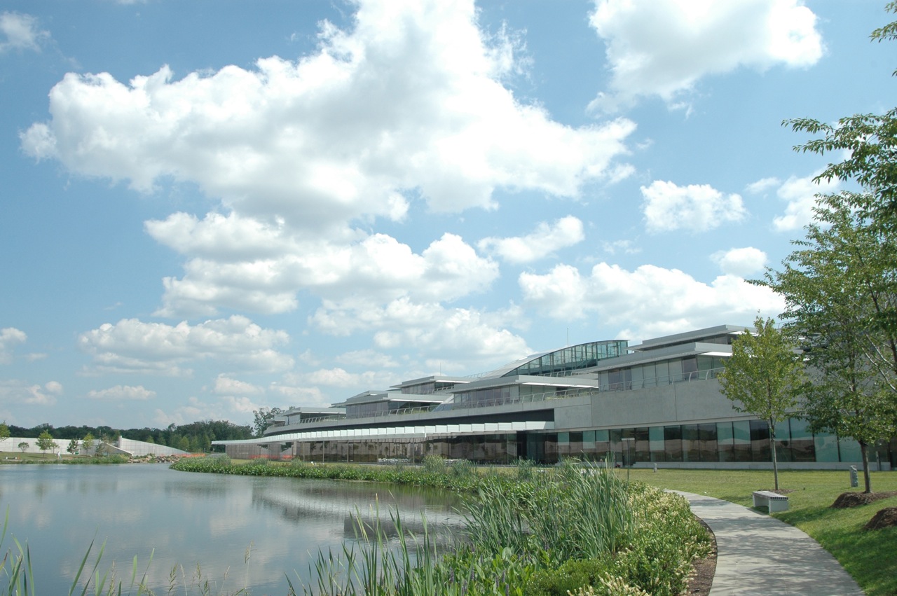
Janelia Research Campus is a scientific research campus of the Howard Hughes Medical Institute that opened in October 2006. The campus is located in Loudoun County, Virginia, near the town of Ashburn. It is known for its scientific research and modern architecture. The current Executive Director of the laboratory is Ronald Vale, who is also a vice-president of HHMI. He succeeded Gerald M. Rubin in 2020. The campus was known as "Janelia Farm Research Campus" until 2014. Research Most HHMI-funded research supports investigators working at their home institution. However, some interdisciplinary problems are difficult to address in existing research settings, and Janelia was built as a separate institution to address such problems in neurobiology. As of November 2011, it has 424 employees and room for 150 more. They specifically address the identification of general principles governing information processing by neuronal circuits, and the development of imaging technologies ... [...More Info...] [...Related Items...] OR: [Wikipedia] [Google] [Baidu] |
Allen Institute For Brain Science
The Allen Institute for Brain Science is a division of the Allen Institute, based in Seattle, Washington, that focuses on bioscience research. Founded in 2003, it is dedicated to accelerating the understanding of how the human brain works. With the intent of catalyzing brain research in different areas, the Allen Institute provides free data and tools to scientists. Started with $100 million in seed money from Microsoft co-founder and philanthropist Paul Allen in 2003, the institute tackles projects at the leading edge of science—far-reaching projects at the intersection of biology and technology. The resulting data create free, publicly available resources that fuel discovery for countless researchers. Hongkui Zeng is the director of the institute.Allen Institute For ... [...More Info...] [...Related Items...] OR: [Wikipedia] [Google] [Baidu] |
NeuronStudio
NeuronStudio was a non-commercial program created at Icahn School of Medicine at Mount Sinai by the Computational Neurobiology and Imaging Center. This program performed automatic tracing and reconstruction of neuron structures from confocal image stacks. The resulting models were then exported to file using standard formats for further processing, modeling, or for statistical analyses. NeuronStudio handled morphologic details on scales spanning local Dendritic spine geometry through complex tree topology to the gross spatial arrangement of multi-neuron networks. Its capability for automated digitization avoided the subjective errors inherent in manual tracing. The program ceased to be supported in 2012 and the project pages were eventually removed from the ISMMS Website. Its documentation and the #External_links, Windows source code however are still available via the Internet Archive. Deconvolution Deconvolution of imaged data is essential for accurate 3D reconstructions. De ... [...More Info...] [...Related Items...] OR: [Wikipedia] [Google] [Baidu] |
Vaa3D
Vaa3D (in Chinese ‘挖三维’) is an Open Source visualization and analysis software suite created mainly by Hanchuan Peng and his team at Janelia Research Campus, HHMI and Allen Institute for Brain Science. The software performs 3D, 4D and 5D rendering and analysis of very large image data sets, especially those generated using various modern microscopy methods, and associated 3D surface objects. This software has been used in several large neuroscience initiatives and a number of applications in other domains. In a recent ''Nature Methods'' review article, it has been viewed as one of the leading open-source software suites in the related research fields. In addition, research using this software was awarded the 2012 Cozzarelli Prize from the National Academy of Sciences. Creation Vaa3D was created in 2007 to tackle the large-scale brain mapping project at Janelia Farm of the Howard Hughes Medical Institute. The initial goal was to quickly visualize any of the tens of thous ... [...More Info...] [...Related Items...] OR: [Wikipedia] [Google] [Baidu] |
Neuron Reconstruction And Tracing Illustration
A neuron, neurone, or nerve cell is an electrically excitable cell that communicates with other cells via specialized connections called synapses. The neuron is the main component of nervous tissue in all animals except sponges and placozoa. Non-animals like plants and fungi do not have nerve cells. Neurons are typically classified into three types based on their function. Sensory neurons respond to stimuli such as touch, sound, or light that affect the cells of the sensory organs, and they send signals to the spinal cord or brain. Motor neurons receive signals from the brain and spinal cord to control everything from muscle contractions to glandular output. Interneurons connect neurons to other neurons within the same region of the brain or spinal cord. When multiple neurons are connected together, they form what is called a neural circuit. A typical neuron consists of a cell body (soma), dendrites, and a single axon. The soma is a compact structure, and the axon and dendri ... [...More Info...] [...Related Items...] OR: [Wikipedia] [Google] [Baidu] |
Electron Microscope
An electron microscope is a microscope that uses a beam of accelerated electrons as a source of illumination. As the wavelength of an electron can be up to 100,000 times shorter than that of visible light photons, electron microscopes have a higher resolving power than light microscopes and can reveal the structure of smaller objects. A scanning transmission electron microscope has achieved better than 50 pm resolution in annular dark-field imaging mode and magnifications of up to about 10,000,000× whereas most light microscopes are limited by diffraction to about 200 nm resolution and useful magnifications below 2000×. Electron microscopes use shaped magnetic fields to form electron optical lens systems that are analogous to the glass lenses of an optical light microscope. Electron microscopes are used to investigate the ultrastructure of a wide range of biological and inorganic specimens including microorganisms, cells, large molecules, biopsy samples, ... [...More Info...] [...Related Items...] OR: [Wikipedia] [Google] [Baidu] |
Fluorescence Microscope
A fluorescence microscope is an optical microscope that uses fluorescence instead of, or in addition to, scattering, reflection, and attenuation or absorption, to study the properties of organic or inorganic substances. "Fluorescence microscope" refers to any microscope that uses fluorescence to generate an image, whether it is a simple set up like an epifluorescence microscope or a more complicated design such as a confocal microscope, which uses optical sectioning to get better resolution of the fluorescence image. Principle The specimen is illuminated with light of a specific wavelength (or wavelengths) which is absorbed by the fluorophores, causing them to emit light of longer wavelengths (i.e., of a different color than the absorbed light). The illumination light is separated from the much weaker emitted fluorescence through the use of a spectral emission filter. Typical components of a fluorescence microscope are a light source (xenon arc lamp or mercury-vapor lamp are ... [...More Info...] [...Related Items...] OR: [Wikipedia] [Google] [Baidu] |
Pyramidal Neurons
Pyramidal cells, or pyramidal neurons, are a type of multipolar neuron found in areas of the brain including the cerebral cortex, the hippocampus, and the amygdala. Pyramidal neurons are the primary excitation units of the mammalian prefrontal cortex and the corticospinal tract. Pyramidal neurons are also one of two cell types where the characteristic sign, Negri bodies, are found in post-mortem rabies infection. Pyramidal neurons were first discovered and studied by Santiago Ramón y Cajal. Since then, studies on pyramidal neurons have focused on topics ranging from neuroplasticity to cognition. Structure File:GFPneuron.png, Pyramidal neuron visualized by green fluorescent protein (gfp) File:Hippocampal-pyramidal-cell.png, A hippocampal pyramidal cell One of the main structural features of the pyramidal neuron is the conic shaped soma, or cell body, after which the neuron is named. Other key structural features of the pyramidal cell are a single axon, a large apical dendrite, ... [...More Info...] [...Related Items...] OR: [Wikipedia] [Google] [Baidu] |
Neural Pathway
In neuroanatomy, a neural pathway is the connection formed by axons that project from neurons to make synapses onto neurons in another location, to enable neurotransmission (the sending of a signal from one region of the nervous system to another). Neurons are connected by a single axon, or by a bundle of axons known as a nerve tract, or fasciculus. Shorter neural pathways are found within grey matter in the brain, whereas longer projections, made up of myelinated axons, constitute white matter. In the hippocampus there are neural pathways involved in its circuitry including the perforant pathway, that provides a connectional route from the entorhinal cortex to all fields of the hippocampal formation, including the dentate gyrus, all CA fields (including CA1), and the subiculum. Descending motor pathways of the pyramidal tracts travel from the cerebral cortex to the brainstem or lower spinal cord. Ascending sensory tracts in the dorsal column–medial lemniscus pathway (DC ... [...More Info...] [...Related Items...] OR: [Wikipedia] [Google] [Baidu] |


