|
Neural Decoding
Neural decoding is a neuroscience field concerned with the hypothetical reconstruction of sensory and other stimuli from information that has already been encoded and represented in the brain by networks of neurons. Reconstruction refers to the ability of the researcher to predict what sensory stimuli the subject is receiving based purely on neuron action potentials. Therefore, the main goal of neural decoding is to characterize how the electrical activity of neurons elicit activity and responses in the brain. This article specifically refers to neural decoding as it pertains to the mammalian neocortex. Overview When looking at a picture, people's brains are constantly making decisions about what object they are looking at, where they need to move their eyes next, and what they find to be the most salient aspects of the input stimulus. As these images hit the back of the retina, these stimuli are converted from varying wavelengths to a series of neural spikes called action potent ... [...More Info...] [...Related Items...] OR: [Wikipedia] [Google] [Baidu] |
Neuroscience
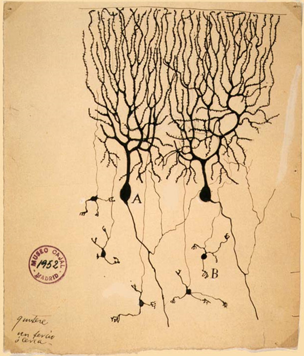
Neuroscience is the scientific study of the nervous system (the brain, spinal cord, and peripheral nervous system), its functions and disorders. It is a multidisciplinary science that combines physiology, anatomy, molecular biology, developmental biology, cytology, psychology, physics, computer science, chemistry, medicine, statistics, and Mathematical Modeling, mathematical modeling to understand the fundamental and emergent properties of neurons, glia and neural circuits. The understanding of the biological basis of learning, memory, behavior, perception, and consciousness has been described by Eric Kandel as the "epic challenge" of the Biology, biological sciences. The scope of neuroscience has broadened over time to include different approaches used to study the nervous system at different scales. The techniques used by neuroscientists have expanded enormously, from molecular biology, molecular and cell biology, cellular studies of individual neurons to neuroimaging, imaging ... [...More Info...] [...Related Items...] OR: [Wikipedia] [Google] [Baidu] |
Rods And Cones
A photoreceptor cell is a specialized type of neuroepithelial cell found in the retina that is capable of visual phototransduction. The great biological importance of photoreceptors is that they convert light (visible electromagnetic radiation) into signals that can stimulate biological processes. To be more specific, photoreceptor proteins in the cell absorb photons, triggering a change in the cell's membrane potential. There are currently three known types of photoreceptor cells in mammalian eyes: rods, cones, and intrinsically photosensitive retinal ganglion cells. The two classic photoreceptor cells are rods and cones, each contributing information used by the visual system to form an image of the environment, sight. Rods primarily mediate scotopic vision (dim conditions) whereas cones primarily mediate to photopic vision (bright conditions), but the processes in each that supports phototransduction is similar. A third class of mammalian photoreceptor cell was discovere ... [...More Info...] [...Related Items...] OR: [Wikipedia] [Google] [Baidu] |
Lateral Geniculate Nucleus
In neuroanatomy, the lateral geniculate nucleus (LGN; also called the lateral geniculate body or lateral geniculate complex) is a structure in the thalamus and a key component of the mammalian visual pathway. It is a small, ovoid, ventral projection of the thalamus where the thalamus connects with the optic nerve. There are two LGNs, one on the left and another on the right side of the thalamus. In humans, both LGNs have six layers of neurons (grey matter) alternating with optic fibers (white matter). The LGN receives information directly from the ascending retinal ganglion cells via the optic tract and from the reticular activating system. Neurons of the LGN send their axons through the optic radiation, a direct pathway to the primary visual cortex. In addition, the LGN receives many strong feedback connections from the primary visual cortex. In humans as well as other mammals, the two strongest pathways linking the eye to the brain are those projecting to the dorsal part of th ... [...More Info...] [...Related Items...] OR: [Wikipedia] [Google] [Baidu] |
Retina
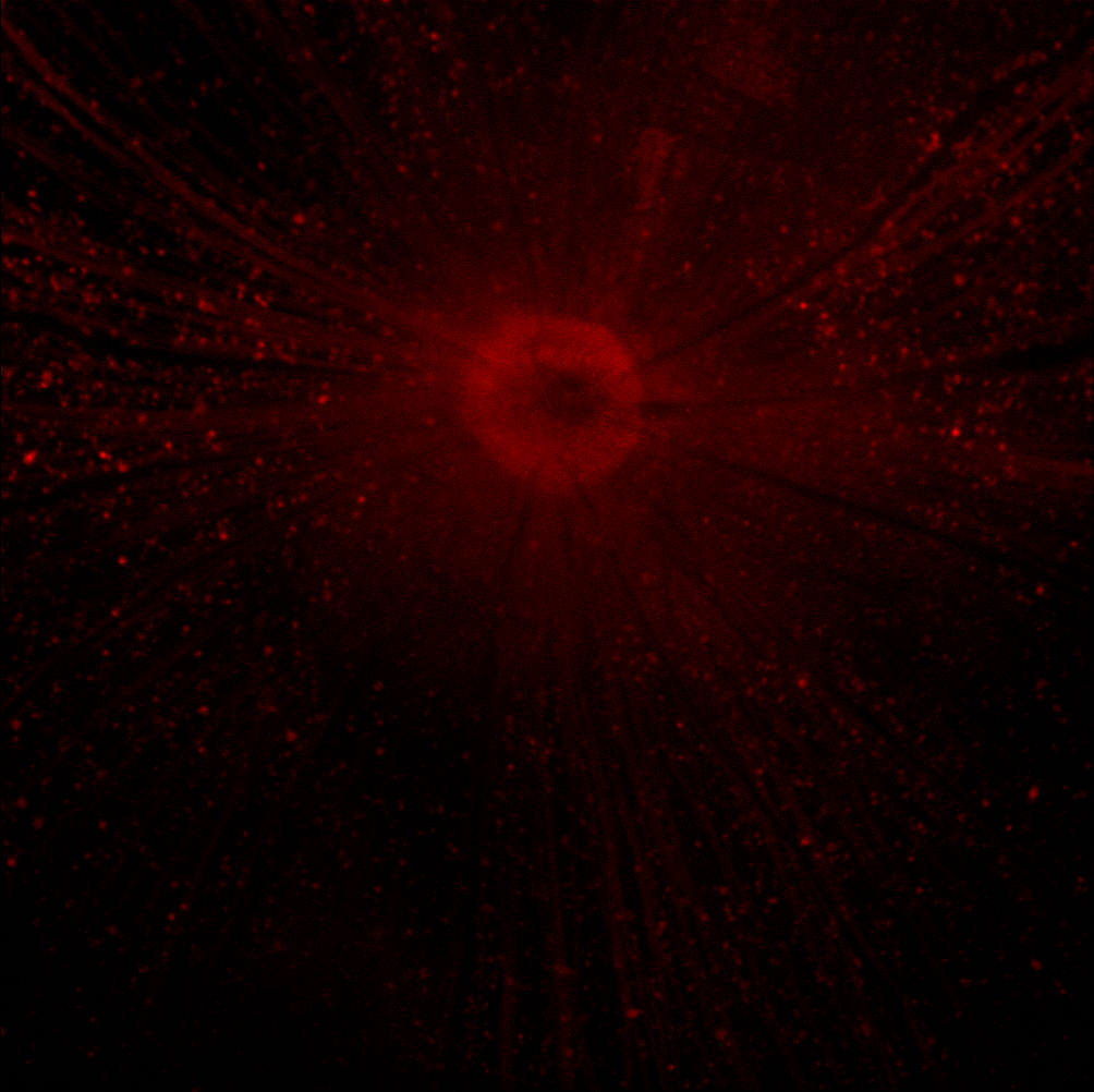
The retina (from la, rete "net") is the innermost, light-sensitive layer of tissue of the eye of most vertebrates and some molluscs. The optics of the eye create a focused two-dimensional image of the visual world on the retina, which then processes that image within the retina and sends nerve impulses along the optic nerve to the visual cortex to create visual perception. The retina serves a function which is in many ways analogous to that of the film or image sensor in a camera. The neural retina consists of several layers of neurons interconnected by synapses and is supported by an outer layer of pigmented epithelial cells. The primary light-sensing cells in the retina are the photoreceptor cells, which are of two types: rods and cones. Rods function mainly in dim light and provide monochromatic vision. Cones function in well-lit conditions and are responsible for the perception of colour through the use of a range of opsins, as well as high-acuity vision used for task ... [...More Info...] [...Related Items...] OR: [Wikipedia] [Google] [Baidu] |
Voltage-gated Potassium Channel
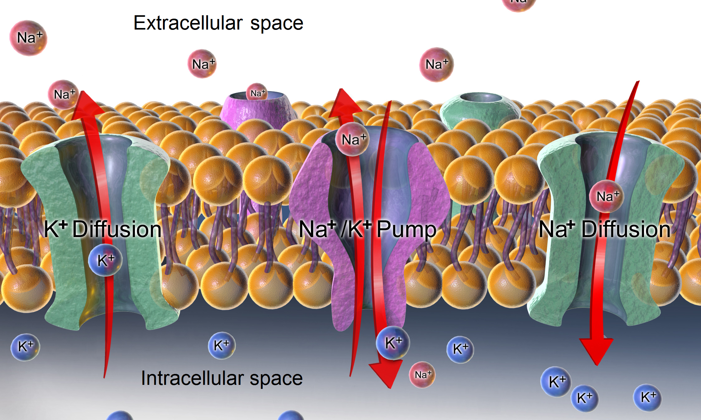
Voltage-gated potassium channels (VGKCs) are transmembrane channels specific for potassium and sensitive to voltage changes in the cell's membrane potential. During action potentials, they play a crucial role in returning the depolarized cell to a resting state. Classification Alpha subunits Alpha subunits form the actual conductance pore. Based on sequence homology of the hydrophobic transmembrane cores, the alpha subunits of voltage-gated potassium channels are grouped into 12 classes. These are labeled Kvα1-12. The following is a list of the 40 known human voltage-gated potassium channel alpha subunits grouped first according to function and then subgrouped according to the Kv sequence homology classification scheme: Delayed rectifier slowly inactivating or non-inactivating *Kvα1.x - Shaker-related: Kv1.1 (KCNA1), Kv1.2 (KCNA2), Kv1.3 (KCNA3), Kv1.5 (KCNA5), Kv1.6 (KCNA6), Kv1.7 ( KCNA7), Kv1.8 (KCNA10) *Kvα2.x - Shab-related: Kv2.1 (KCNB1), Kv2.2 (KCNB2) *Kvα3.x - ... [...More Info...] [...Related Items...] OR: [Wikipedia] [Google] [Baidu] |
Voltage-gated Sodium Channel
Sodium channels are integral membrane proteins that form ion channels, conducting sodium ions (Na+) through a cell's membrane. They belong to the superfamily of cation channels and can be classified according to the trigger that opens the channel for such ions, i.e. either a voltage-change ("voltage-gated", "voltage-sensitive", or "voltage-dependent" sodium channel; also called "VGSCs" or "Nav channel") or a binding of a substance (a ligand) to the channel (ligand-gated sodium channels). In excitable cells such as neurons, myocytes, and certain types of glia, sodium channels are responsible for the rising phase of action potentials. These channels go through three different states called resting, active and inactive states. Even though the resting and inactive states would not allow the ions to flow through the channels the difference exists with respect to their structural conformation. Selectivity Sodium channels are highly selective for the transport of ions across cell membr ... [...More Info...] [...Related Items...] OR: [Wikipedia] [Google] [Baidu] |
Resting Potential
A relatively static membrane potential which is usually referred to as the ground value for trans-membrane voltage. The relatively static membrane potential of quiescent cells is called the resting membrane potential (or resting voltage), as opposed to the specific dynamic electrochemical phenomena called action potential and graded membrane potential. Apart from the latter two, which occur in excitable cells (neurons, muscles, and some secretory cells in glands), membrane voltage in the majority of non-excitable cells can also undergo changes in response to environmental or intracellular stimuli. The resting potential exists due to the differences in membrane permeabilities for potassium, sodium, calcium, and chloride ions, which in turn result from functional activity of various ion channels, ion transporters, and exchangers. Conventionally, resting membrane potential can be defined as a relatively stable, ground value of transmembrane voltage in animal and plant cells. Beca ... [...More Info...] [...Related Items...] OR: [Wikipedia] [Google] [Baidu] |
Electric Potential
The electric potential (also called the ''electric field potential'', potential drop, the electrostatic potential) is defined as the amount of work energy needed to move a unit of electric charge from a reference point to the specific point in an electric field. More precisely, it is the energy per unit charge for a test charge that is so small that the disturbance of the field under consideration is negligible. Furthermore, the motion across the field is supposed to proceed with negligible acceleration, so as to avoid the test charge acquiring kinetic energy or producing radiation. By definition, the electric potential at the reference point is zero units. Typically, the reference point is earth or a point at infinity, although any point can be used. In classical electrostatics, the electrostatic field is a vector quantity expressed as the gradient of the electrostatic potential, which is a scalar quantity denoted by or occasionally , equal to the electric potential energy o ... [...More Info...] [...Related Items...] OR: [Wikipedia] [Google] [Baidu] |
Retinotopy
Retinotopy (from Greek τόπος, place) is the mapping of visual input from the retina to neurons, particularly those neurons within the visual stream. For clarity, 'retinotopy' can be replaced with 'retinal mapping', and 'retinotopic' with 'retinally mapped'. Visual field maps (retinotopic maps) are found in many amphibian and mammalian species, though the specific size, number, and spatial arrangement of these maps can differ considerably. Sensory topographies can be found throughout the brain and are critical to the understanding of one's external environment. Moreover, the study of sensory topographies and retinotopy in particular has furthered our understanding of how neurons encode and organize sensory signals. Retinal mapping of the visual field is maintained through various points of the visual pathway including but not limited to the retina, the dorsal lateral geniculate nucleus, the optic tectum, the primary visual cortex (V1), and higher visual areas (V2-V4). Retino ... [...More Info...] [...Related Items...] OR: [Wikipedia] [Google] [Baidu] |
Feedforward Neural Network
A feedforward neural network (FNN) is an artificial neural network wherein connections between the nodes do ''not'' form a cycle. As such, it is different from its descendant: recurrent neural networks. The feedforward neural network was the first and simplest type of artificial neural network devised. In this network, the information moves in only one direction—forward—from the input nodes, through the hidden nodes (if any) and to the output nodes. There are no cycles or loops in the network. Single-layer perceptron The simplest kind of neural network is a ''single-layer perceptron'' network, which consists of a single layer of output nodes; the inputs are fed directly to the outputs via a series of weights. The sum of the products of the weights and the inputs is calculated in each node, and if the value is above some threshold (typically 0) the neuron fires and takes the activated value (typically 1); otherwise it takes the deactivated value (typically -1). Ne ... [...More Info...] [...Related Items...] OR: [Wikipedia] [Google] [Baidu] |
Retinal Ganglion Cell
A retinal ganglion cell (RGC) is a type of neuron located near the inner surface (the ganglion cell layer) of the retina of the human eye, eye. It receives visual information from photoreceptor cell, photoreceptors via two intermediate neuron types: Bipolar cell of the retina, bipolar cells and retina amacrine cells. Retina amacrine cells, particularly narrow field cells, are important for creating functional subunits within the ganglion cell layer and making it so that ganglion cells can observe a small dot moving a small distance. Retinal ganglion cells collectively transmit image-forming and non-image forming visual information from the retina in the form of action potential to several regions in the thalamus, hypothalamus, and mesencephalon, or midbrain. Retinal ganglion cells vary significantly in terms of their size, connections, and responses to visual stimulation but they all share the defining property of having a long axon that extends into the brain. These axons form th ... [...More Info...] [...Related Items...] OR: [Wikipedia] [Google] [Baidu] |
Two-photon Excitation Microscopy
Two-photon excitation microscopy (TPEF or 2PEF) is a fluorescence imaging technique that allows imaging of living tissue up to about one millimeter in thickness, with 0.64 μm lateral and 3.35 μm axial spatial resolution. Unlike traditional fluorescence microscopy, in which the excitation wavelength is shorter than the emission wavelength, two-photon excitation requires simultaneous excitation by two photons with longer wavelength than the emitted light. Two-photon excitation microscopy typically uses near-infrared (NIR) excitation light which can also excite fluorescent dyes. However, for each excitation, two photons of NIR light are absorbed. Using infrared light minimizes scattering in the tissue. Due to the multiphoton absorption, the background signal is strongly suppressed. Both effects lead to an increased penetration depth for this technique. Two-photon excitation can be a superior alternative to confocal microscopy due to its deeper tissue penetration, efficient light d ... [...More Info...] [...Related Items...] OR: [Wikipedia] [Google] [Baidu] |


