|
NRAP
Nebulin-related-anchoring protein (N-RAP) is a protein that in humans is encoded by the ''NRAP'' gene. N-RAP is a muscle-specific isoform belonging to the nebulin family of proteins. This family is composed of 5 members: N-RAP, nebulin, nebulette, LASP-1 and LASP-2. N-RAP is involved in both myofibrillar myogenesis during development and cell-cell connections in mature muscle. Structure N-RAP is a 197 kDa protein composed of 1730 amino acids. As a member of the nebulin family of proteins, N-RAP is characterized by 35 amino acid stretches of ‘‘nebulin repeats’’, which are actin binding domains containing a conserved S Dxx Y K motif. Like nebulin, groups of seven single repeats within N-RAP form “super repeats”, which incorporate a single conserved motif W L K G I G W at the end of the third repeat. A unique feature of NRAP relative to nebulin is its N-terminal cysteine-rich LIM domain, a feature shared with LASP-1 and LASP-2. Function An important role has ... [...More Info...] [...Related Items...] OR: [Wikipedia] [Google] [Baidu] |
Nebulette
Nebulette is a cardiac-specific isoform belonging to the nebulin family of proteins. It is encoded by the ''NEBL'' gene. This family is composed of 5 members: nebulette, nebulin, N-RAP, LASP-1 and LASP-2. Nebulette localizes to Z-discs of cardiac muscle and appears to regulate the length of actin thin filaments. Structure Nebulette is a 116.4 kDa protein composed of 1014 amino acids. As a member of the nebulin family of proteins, nebulette is characterized by 35 amino acid stretches of ‘‘nebulin repeats’’, which are actin binding domains containing a conserved S Dxx Y K motif. Like nebulin, nebulette has an acidic region with unknown structure at its N-terminus, and a serine-rich region adjacent to an SH3 domain at its C-terminus. Though nebulette shares structural features with nebulin, nebulin is expressed preferentially in skeletal muscle and has an enormous size (600-900 kDa), while nebulette is expressed in cardiac muscle at Z-disc regions and is significantly ... [...More Info...] [...Related Items...] OR: [Wikipedia] [Google] [Baidu] |
Protein
Proteins are large biomolecules and macromolecules that comprise one or more long chains of amino acid residues. Proteins perform a vast array of functions within organisms, including catalysing metabolic reactions, DNA replication, responding to stimuli, providing structure to cells and organisms, and transporting molecules from one location to another. Proteins differ from one another primarily in their sequence of amino acids, which is dictated by the nucleotide sequence of their genes, and which usually results in protein folding into a specific 3D structure that determines its activity. A linear chain of amino acid residues is called a polypeptide. A protein contains at least one long polypeptide. Short polypeptides, containing less than 20–30 residues, are rarely considered to be proteins and are commonly called peptides. The individual amino acid residues are bonded together by peptide bonds and adjacent amino acid residues. The sequence of amino acid residue ... [...More Info...] [...Related Items...] OR: [Wikipedia] [Google] [Baidu] |
Glycine
Glycine (symbol Gly or G; ) is an amino acid that has a single hydrogen atom as its side chain. It is the simplest stable amino acid (carbamic acid is unstable), with the chemical formula NH2‐ CH2‐ COOH. Glycine is one of the proteinogenic amino acids. It is encoded by all the codons starting with GG (GGU, GGC, GGA, GGG). Glycine is integral to the formation of alpha-helices in secondary protein structure due to its compact form. For the same reason, it is the most abundant amino acid in collagen triple-helices. Glycine is also an inhibitory neurotransmitter – interference with its release within the spinal cord (such as during a ''Clostridium tetani'' infection) can cause spastic paralysis due to uninhibited muscle contraction. It is the only achiral proteinogenic amino acid. It can fit into hydrophilic or hydrophobic environments, due to its minimal side chain of only one hydrogen atom. History and etymology Glycine was discovered in 1820 by the French chemist He ... [...More Info...] [...Related Items...] OR: [Wikipedia] [Google] [Baidu] |
Dilated Cardiomyopathy
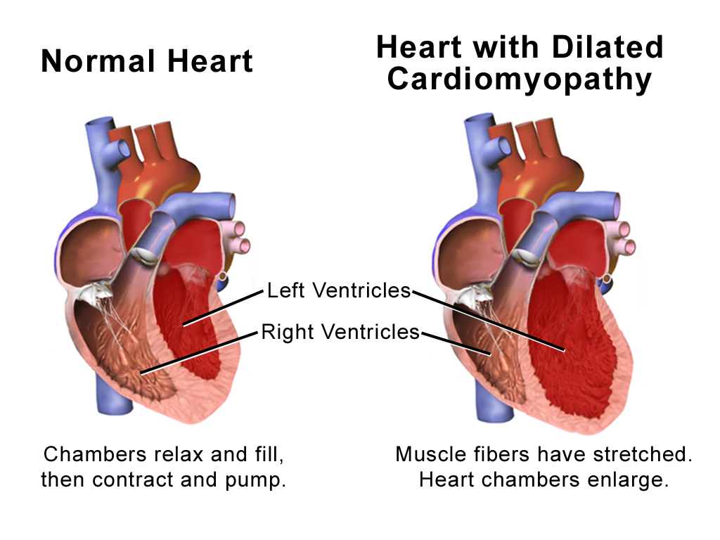
Dilated cardiomyopathy (DCM) is a condition in which the heart becomes enlarged and cannot pump blood effectively. Symptoms vary from none to feeling tired, leg swelling, and shortness of breath. It may also result in chest pain or fainting. Complications can include heart failure, heart valve disease, or an irregular heartbeat. Causes include genetics, alcohol, cocaine, certain toxins, complications of pregnancy, and certain infections. Coronary artery disease and high blood pressure may play a role, but are not the primary cause. In many cases the cause remains unclear. It is a type of cardiomyopathy, a group of diseases that primarily affects the heart muscle. The diagnosis may be supported by an electrocardiogram, chest X-ray, or echocardiogram. In those with heart failure, treatment may include medications in the ACE inhibitor, beta blocker, and diuretic families. A low salt diet may also be helpful. In those with certain types of irregular heartbeat, blood thinners or ... [...More Info...] [...Related Items...] OR: [Wikipedia] [Google] [Baidu] |
Sarcolemma
The sarcolemma (''sarco'' (from ''sarx'') from Greek; flesh, and ''lemma'' from Greek; sheath) also called the myolemma, is the cell membrane surrounding a skeletal muscle fiber or a cardiomyocyte. It consists of a lipid bilayer and a thin outer coat of polysaccharide material (glycocalyx) that contacts the basement membrane. The basement membrane contains numerous thin collagen fibrils and specialized proteins such as laminin that provide a scaffold to which the muscle fiber can adhere. Through transmembrane proteins in the plasma membrane, the actin skeleton inside the cell is connected to the basement membrane and the cell's exterior. At each end of the muscle fiber, the surface layer of the sarcolemma fuses with a tendon fiber, and the tendon fibers, in turn, collect into bundles to form the muscle tendons that adhere to bones. The sarcolemma generally maintains the same function in muscle cells as the plasma membrane does in other eukaryote cells. It acts as a barrier betwe ... [...More Info...] [...Related Items...] OR: [Wikipedia] [Google] [Baidu] |
Intercalated Discs
Intercalated discs or lines of Eberth are microscopic identifying features of cardiac muscle. Cardiac muscle consists of individual heart muscle cells (cardiomyocytes) connected by intercalated discs to work as a single functional syncytium. By contrast, skeletal muscle consists of multinucleated muscle fibers and exhibits no intercalated discs. Intercalated discs support synchronized contraction of cardiac tissue. They occur at the Z line of the sarcomere and can be visualized easily when observing a longitudinal section of the tissue. Structure Intercalated discs are complex structures that connect adjacent cardiac muscle cells. The three types of cell junction recognised as making up an intercalated disc are desmosomes, fascia adherens junctions, and gap junctions. * Fascia adherens are anchoring sites for actin, and connect to the closest sarcomere. * Desmosomes prevent separation during contraction by binding intermediate filaments, anchoring the cell membrane to the intermed ... [...More Info...] [...Related Items...] OR: [Wikipedia] [Google] [Baidu] |
Sarcomere
A sarcomere (Greek σάρξ ''sarx'' "flesh", μέρος ''meros'' "part") is the smallest functional unit of striated muscle tissue. It is the repeating unit between two Z-lines. Skeletal muscles are composed of tubular muscle cells (called muscle fibers or myofibers) which are formed during embryonic myogenesis. Muscle fibers contain numerous tubular myofibrils. Myofibrils are composed of repeating sections of sarcomeres, which appear under the microscope as alternating dark and light bands. Sarcomeres are composed of long, fibrous proteins as filaments that slide past each other when a muscle contracts or relaxes. The costamere is a different component that connects the sarcomere to the sarcolemma. Two of the important proteins are myosin, which forms the thick filament, and actin, which forms the thin filament. Myosin has a long, fibrous tail and a globular head, which binds to actin. The myosin head also binds to ATP, which is the source of energy for muscle movement. Myos ... [...More Info...] [...Related Items...] OR: [Wikipedia] [Google] [Baidu] |
ACTC1
ACTC1 encodes cardiac muscle alpha actin. This isoform differs from the alpha actin that is expressed in skeletal muscle, ACTA1. Alpha cardiac actin is the major protein of the thin filament in cardiac sarcomeres, which are responsible for muscle contraction and generation of force to support the pump function of the heart. Structure Cardiac alpha actin is a 42.0 kDa protein composed of 377 amino acids. Cardiac alpha actin is a filamentous protein extending from a complex mesh with cardiac alpha-actinin (ACTN2) at Z-lines towards the center of the sarcomere. Polymerization of globular actin (G-actin) leads to a structural filament (F-actin) in the form of a two-stranded helix. Each actin can bind to four others. The atomic structure of monomeric actin was solved by Kabsch et al., and closely thereafter this same group published the structure of the actin filament. Actins are highly conserved proteins; the alpha actins are found in muscle tissues and are a major constituent of ... [...More Info...] [...Related Items...] OR: [Wikipedia] [Google] [Baidu] |
ACTN2
Alpha-actinin-2 is a protein which in humans is encoded by the ''ACTN2'' gene. This gene encodes an alpha-actinin isoform that is expressed in both skeletal and cardiac muscles and functions to anchor myofibrillar actin thin filaments and titin to Z-discs. Structure Alpha-actinin-2 is a 103.8 kDa protein composed of 894 amino acids. Each molecule is rod-shaped (35 nm in length) and it homodimerizes in an anti-parallel fashion. Each monomer has an N-terminal actin-binding region composed of two calponin homology domains, two C-terminal EF hand domains, and four tandem spectrin-like repeats form the rod domain in the central region of the molecule. The high-resolution crystal structure of human alpha-actinin 2 at 3.5 Å was recently resolved. Alpha actinins belong to the spectrin gene superfamily which represents a diverse group of actin-binding cytoskeletal proteins, including spectrin, dystrophin, utrophin and fimbrin. Skeletal, cardiac, and smooth muscle isofor ... [...More Info...] [...Related Items...] OR: [Wikipedia] [Google] [Baidu] |
LIM Domain
LIM domains are protein structural domains, composed of two contiguous zinc fingers, separated by a two-amino acid residue hydrophobic linker. The domain name is an acronym of the three genes in which it was first identified (LIN-11, Isl-1 and MEC-3). LIM is a protein interaction domain that is involved in binding to many structurally and functionally diverse partners. The LIM domain appeared in eukaryotes sometime prior to the most recent common ancestor of plants, fungi, amoeba and animals. In animal cells, LIM domain-containing proteins often shuttle between the cell nucleus where they can regulate gene expression, and the cytoplasm where they are usually associated with actin cytoskeletal structures involved in connecting cells together and to the surrounding matrix, such as stress fibers, focal adhesions and adherens junctions. Discovery LIM domains are named after their initial discovery in the three homeobox proteins that have the following functions: * Lin-11 � ... [...More Info...] [...Related Items...] OR: [Wikipedia] [Google] [Baidu] |
Cysteine
Cysteine (symbol Cys or C; ) is a semiessential proteinogenic amino acid with the formula . The thiol side chain in cysteine often participates in enzymatic reactions as a nucleophile. When present as a deprotonated catalytic residue, sometimes the symbol Cyz is used. The deprotonated form can generally be described by the symbol Cym as well. The thiol is susceptible to oxidation to give the disulfide derivative cystine, which serves an important structural role in many proteins. In this case, the symbol Cyx is sometimes used. When used as a food additive, it has the E number E920. Cysteine is encoded by the codons UGU and UGC. The sulfur-containing amino acids cysteine and methionine are more easily oxidized than the other amino acids. Structure Like other amino acids (not as a residue of a protein), cysteine exists as a zwitterion. Cysteine has chirality in the older / notation based on homology to - and -glyceraldehyde. In the newer ''R''/''S'' system of designating chi ... [...More Info...] [...Related Items...] OR: [Wikipedia] [Google] [Baidu] |
Isoleucine
Isoleucine (symbol Ile or I) is an α-amino acid that is used in the biosynthesis of proteins. It contains an α-amino group (which is in the protonated −NH form under biological conditions), an α-carboxylic acid group (which is in the deprotonated −COO form under biological conditions), and a hydrocarbon side chain with a branch (a central carbon atom bound to three other carbon atoms). It is classified as a non-polar, uncharged (at physiological pH), branched-chain, aliphatic amino acid. It is essential in humans, meaning the body cannot synthesize it, and must be ingested in our diet. Isoleucine is synthesized from pyruvate employing leucine biosynthesis enzymes in other organisms such as bacteria. It is encoded by the codons AUU, AUC, and AUA. Metabolism Biosynthesis As an essential nutrient, it is not synthesized in the body, hence it must be ingested, usually as a component of proteins. In plants and microorganisms, it is synthesized via several steps, startin ... [...More Info...] [...Related Items...] OR: [Wikipedia] [Google] [Baidu] |


