|
Nucleus Cuneatus
In neuroanatomy, the dorsal column nuclei are a pair of nuclei in the dorsal columns in the brainstem. The name refers collectively to the cuneate nucleus and gracile nucleus, which are present at the bottom of the medulla oblongata. Both nuclei contain second-order neurons of the dorsal column–medial lemniscus pathway, which carries fine touch and proprioceptive information from the body to the brain. Fibres reach the thalamus. Structure Nerve pathways The dorsal column nuclei each have an associated nerve tract in the spinal cord, the gracile fasciculus and the cuneate fasciculus. Both dorsal column nuclei contain synapses from afferent nerve fibers that have travelled in the spinal cord. They then send on second-order neurons of the dorsal column–medial lemniscal pathway. Neurons of the dorsal column nuclei eventually reach the midbrain and the thalamus. They send axons that form the internal arcuate fibers. These cross over at the sensory decussation to form ... [...More Info...] [...Related Items...] OR: [Wikipedia] [Google] [Baidu] |
Spinal Cord
The spinal cord is a long, thin, tubular structure made up of nervous tissue, which extends from the medulla oblongata in the brainstem to the lumbar region of the vertebral column (backbone). The backbone encloses the central canal of the spinal cord, which contains cerebrospinal fluid. The brain and spinal cord together make up the central nervous system (CNS). In humans, the spinal cord begins at the occipital bone, passing through the foramen magnum and then enters the spinal canal at the beginning of the cervical vertebrae. The spinal cord extends down to between the first and second lumbar vertebrae, where it ends. The enclosing bony vertebral column protects the relatively shorter spinal cord. It is around long in adult men and around long in adult women. The diameter of the spinal cord ranges from in the cervical and lumbar regions to in the thoracic area. The spinal cord functions primarily in the transmission of nerve signals from the motor cortex to the ... [...More Info...] [...Related Items...] OR: [Wikipedia] [Google] [Baidu] |
Academic Press
Academic Press (AP) is an academic book publisher founded in 1941. It was acquired by Harcourt, Brace & World in 1969. Reed Elsevier bought Harcourt in 2000, and Academic Press is now an imprint of Elsevier. Academic Press publishes reference books, serials and online products in the subject areas of: * Communications engineering * Economics * Environmental science * Finance * Food science and nutrition * Geophysics * Life sciences * Mathematics and statistics * Neuroscience * Physical sciences * Psychology Well-known products include the ''Methods in Enzymology'' series and encyclopedias such as ''The International Encyclopedia of Public Health'' and the ''Encyclopedia of Neuroscience''. See also * Akademische Verlagsgesellschaft (AVG) — the German predecessor, founded in 1906 by Leo Jolowicz (1868–1940), the father of Walter Jolowicz Walter may refer to: People * Walter (name), both a surname and a given name * Little Walter, American blues harmonica player ... [...More Info...] [...Related Items...] OR: [Wikipedia] [Google] [Baidu] |
Kinesthetic
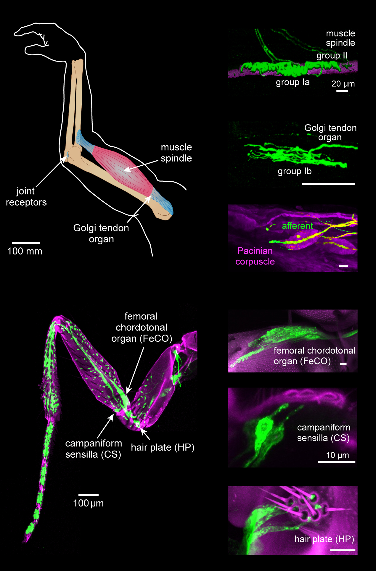
Proprioception ( ), also referred to as kinaesthesia (or kinesthesia), is the sense of self-movement, force, and body position. It is sometimes described as the "sixth sense". Proprioception is mediated by proprioceptors, mechanosensory neurons located within muscles, tendons, and joints. Most animals possess multiple subtypes of proprioceptors, which detect distinct kinematic parameters, such as joint position, movement, and load. Although all mobile animals possess proprioceptors, the structure of the sensory organs can vary across species. Proprioceptive signals are transmitted to the central nervous system, where they are integrated with information from other sensory systems, such as the visual system and the vestibular system, to create an overall representation of body position, movement, and acceleration. In many animals, sensory feedback from proprioceptors is essential for stabilizing body posture and coordinating body movement. System overview In vertebrates, limb ve ... [...More Info...] [...Related Items...] OR: [Wikipedia] [Google] [Baidu] |
Epicritic
Cutaneous innervation refers to the area of the skin which is supplied by a specific cutaneous nerve. Dermatomes are similar; however, a dermatome only specifies the area served by a spinal nerve. In some cases, the dermatome is less specific (when a spinal nerve is the source for more than one cutaneous nerve), and in other cases it is more specific (when a cutaneous nerve is derived from multiple spinal nerves.) Modern texts are in agreement about which areas of the skin are served by which nerves, but there are minor variations in some of the details. The borders designated by the diagrams in the 1918 edition of Gray's Anatomy are similar, but not identical, to those generally accepted today. Importance of the peripheral nervous system The peripheral nervous system (PNS) is divided into the somatic nervous system, the autonomic nervous system, and the enteric nervous system. However, it is the somatic nervous system, responsible for body movement and the reception of externa ... [...More Info...] [...Related Items...] OR: [Wikipedia] [Google] [Baidu] |
Human Leg
The human leg, in the general word sense, is the entire lower limb of the human body, including the foot, thigh or sometimes even the hip or gluteal region. However, the definition in human anatomy refers only to the section of the lower limb extending from the knee to the ankle, also known as the crus or, especially in non-technical use, the shank. Legs are used for standing, and all forms of locomotion including recreational such as dancing, and constitute a significant portion of a person's mass. Female legs generally have greater hip anteversion and tibiofemoral angles, but shorter femur and tibial lengths than those in males. Structure In human anatomy, the lower leg is the part of the lower limb that lies between the knee and the ankle. Anatomists restrict the term ''leg'' to this use, rather than to the entire lower limb. The thigh is between the hip and knee and makes up the rest of the lower limb. The term ''lower limb'' or ''lower extremity'' is commonly u ... [...More Info...] [...Related Items...] OR: [Wikipedia] [Google] [Baidu] |
Torso
The torso or trunk is an anatomical term for the central part, or the core, of the body of many animals (including humans), from which the head, neck, limbs, tail and other appendages extend. The tetrapod torso — including that of a human — is usually divided into the ''thoracic'' segment (also known as the upper torso, where the forelimbs extend), the '' abdominal'' segment (also known as the "mid-section" or "midriff"), and the '' pelvic'' and '' perineal'' segments (sometimes known together with the abdomen as the lower torso, where the hindlimbs extend). Anatomy Major organs In humans, most critical organs, with the notable exception of the brain, are housed within the torso. In the upper chest, the heart and lungs are protected by the rib cage, and the abdomen contains most of the organs responsible for digestion: the stomach, which breaks down partially digested food via gastric acid; the liver, which respectively produces bile necessary for digestion; the la ... [...More Info...] [...Related Items...] OR: [Wikipedia] [Google] [Baidu] |
Sensory Neuron
Sensory neurons, also known as afferent neurons, are neurons in the nervous system, that convert a specific type of stimulus, via their receptors, into action potentials or graded potentials. This process is called sensory transduction. The cell bodies of the sensory neurons are located in the dorsal ganglia of the spinal cord. The sensory information travels on the afferent nerve fibers in a sensory nerve, to the brain via the spinal cord. The stimulus can come from ''exteroreceptors'' outside the body, for example those that detect light and sound, or from ''interoreceptors'' inside the body, for example those that are responsive to blood pressure or the sense of body position. Types and function Different types of sensory neurons have different sensory receptors that respond to different kinds of stimuli. There are at least six external and two internal sensory receptors: External receptors External receptors that respond to stimuli from outside the body are called ... [...More Info...] [...Related Items...] OR: [Wikipedia] [Google] [Baidu] |
Dorsal Root Ganglion
A dorsal root ganglion (or spinal ganglion; also known as a posterior root ganglion) is a cluster of neurons (a ganglion) in a dorsal root of a spinal nerve. The cell bodies of sensory neurons known as first-order neurons are located in the dorsal root ganglia. The axons of dorsal root ganglion neurons are known as afferents. In the peripheral nervous system, afferents refer to the axons that relay sensory information into the central nervous system (i.e. the brain and the spinal cord). Structure The neurons comprising the dorsal root ganglion are of the pseudo-unipolar type, meaning they have a cell body (soma) with two branches that act as a single axon, often referred to as a ''distal process'' and a ''proximal process''. Unlike the majority of neurons found in the central nervous system, an action potential in posterior root ganglion neuron may initiate in the ''distal process'' in the periphery, bypass the cell body, and continue to propagate along the ''proximal pro ... [...More Info...] [...Related Items...] OR: [Wikipedia] [Google] [Baidu] |
Synapse
In the nervous system, a synapse is a structure that permits a neuron (or nerve cell) to pass an electrical or chemical signal to another neuron or to the target effector cell. Synapses are essential to the transmission of nervous impulses from one neuron to another. Neurons are specialized to pass signals to individual target cells, and synapses are the means by which they do so. At a synapse, the plasma membrane of the signal-passing neuron (the ''presynaptic'' neuron) comes into close apposition with the membrane of the target (''postsynaptic'') cell. Both the presynaptic and postsynaptic sites contain extensive arrays of Molecular biology, molecular machinery that link the two membranes together and carry out the signaling process. In many synapses, the presynaptic part is located on an axon and the postsynaptic part is located on a dendrite or soma (biology), soma. Astrocytes also exchange information with the synaptic neurons, responding to synaptic activity and, in turn, r ... [...More Info...] [...Related Items...] OR: [Wikipedia] [Google] [Baidu] |
Medial Lemniscus
In neuroanatomy, the medial lemniscus, also known as Reil's band or Reil's ribbon (for German anatomist Johann Christian Reil), is a large ascending bundle of heavily myelinated axons that decussate (cross) in the brainstem, specifically in the medulla oblongata. The medial lemniscus is formed by the crossings of the internal arcuate fibers. The internal arcuate fibers are composed of axons of nucleus gracilis and nucleus cuneatus. The axons of the nucleus gracilis and nucleus cuneatus in the medial lemniscus have cell bodies that lie contralaterally. The medial lemniscus is part of the dorsal column–medial lemniscus pathway, which ascends from the skin to the thalamus, which is important for somatosensation from the skin and joints, therefore, lesion of the medial lemnisci causes an impairment of vibratory and touch-pressure sense. Etymology Lemniscus means "ribbon", so named because the medial lemniscus "spirals" or "turns" as it ascends. Path After neurons carrying pro ... [...More Info...] [...Related Items...] OR: [Wikipedia] [Google] [Baidu] |
Sensory Decussation
In neuroanatomy, the sensory decussation or decussation of the lemnisci is a decussation (i.e. crossover) of axons from the gracile nucleus and cuneate nucleus, which are responsible for fine touch, vibration, proprioception and two-point discrimination of the body. The fibres of this decussation are called the internal arcuate fibres and are found at the superior aspect of the closed medulla superior to the motor decussation. It is part of the second neuron in the posterior column–medial lemniscus pathway. Structure At the level of the closed medulla in the posterior white column, two large nuclei namely the gracile nucleus and the cuneate nucleus can be found. The two nuclei receive the impulse from the two ascending tracts: fasciculus gracilis and fasciculus cuneatus. After the two tracts terminate upon these nuclei, the heavily myelinated fibres arise and ascend anteromedially around the periaqueductal gray as internal arcuate fibres. These fibres decussate (cross) ... [...More Info...] [...Related Items...] OR: [Wikipedia] [Google] [Baidu] |
Internal Arcuate Fiber
In neuroanatomy, the internal arcuate fibers or internal arcuate tract are the axons of second-order sensory neurons that compose the gracile and cuneate nuclei of the medulla oblongata. These second-order neurons begin in the gracile and cuneate nuclei in the medulla. They receive input from first-order sensory neurons, which provide sensation to many areas of the body and have cell bodies in the dorsal root ganglia of the dorsal root of the spinal nerves. Upon decussation (crossing over) from one side of the medulla to the other, also known as the sensory decussation, they are then called the medial lemniscus. The internal arcuate fibers are part of the second-order neurons of the posterior column-medial lemniscus system, and are important for relaying the sensation of fine touch and proprioception to the thalamus and ultimately to the cerebral cortex The cerebral cortex, also known as the cerebral mantle, is the outer layer of neural tissue of the cerebrum of the brain ... [...More Info...] [...Related Items...] OR: [Wikipedia] [Google] [Baidu] |


