|
Nodes Of Ranvier
In neuroscience and anatomy, nodes of Ranvier ( ), also known as myelin-sheath gaps, occur along a myelinated axon where the axolemma is exposed to the extracellular space. Nodes of Ranvier are uninsulated and highly enriched in ion channels, allowing them to participate in the exchange of ions required to regenerate the action potential. Nerve conduction in myelinated axons is referred to as saltatory conduction () due to the manner in which the action potential seems to "jump" from one node to the next along the axon. This results in faster conduction of the action potential. Overview Many vertebrate axons are surrounded by a myelin sheath, allowing rapid and efficient saltatory ("jumping") propagation of action potentials. The contacts between neurons and glial cells display a very high level of spatial and temporal organization in myelinated fibers. The myelinating glial cells - oligodendrocytes in the central nervous system (CNS), and Schwann cells in the peripheral ne ... [...More Info...] [...Related Items...] OR: [Wikipedia] [Google] [Baidu] |
Nervous System
In Biology, biology, the nervous system is the Complex system, highly complex part of an animal that coordinates its Behavior, actions and Sense, sensory information by transmitting action potential, signals to and from different parts of its body. The nervous system detects environmental changes that impact the body, then works in tandem with the endocrine system to respond to such events. Nervous tissue first arose in Ediacara biota, wormlike organisms about 550 to 600 million years ago. In vertebrates it consists of two main parts, the central nervous system (CNS) and the peripheral nervous system (PNS). The CNS consists of the brain and spinal cord. The PNS consists mainly of nerves, which are enclosed bundles of the long fibers or axons, that connect the CNS to every other part of the body. Nerves that transmit signals from the brain are called motor nerves or ''Efferent nerve fiber, efferent'' nerves, while those nerves that transmit information from the body to the CNS a ... [...More Info...] [...Related Items...] OR: [Wikipedia] [Google] [Baidu] |
Central Nervous System
The central nervous system (CNS) is the part of the nervous system consisting primarily of the brain and spinal cord. The CNS is so named because the brain integrates the received information and coordinates and influences the activity of all parts of the bodies of bilaterally symmetric and triploblastic animals—that is, all multicellular animals except sponges and diploblasts. It is a structure composed of nervous tissue positioned along the rostral (nose end) to caudal (tail end) axis of the body and may have an enlarged section at the rostral end which is a brain. Only arthropods, cephalopods and vertebrates have a true brain (precursor structures exist in onychophorans, gastropods and lancelets). The rest of this article exclusively discusses the vertebrate central nervous system, which is radically distinct from all other animals. Overview In vertebrates, the brain and spinal cord are both enclosed in the meninges. The meninges provide a barrier to chemicals d ... [...More Info...] [...Related Items...] OR: [Wikipedia] [Google] [Baidu] |
Phosphacan
Receptor-type tyrosine-protein phosphatase zeta also known as phosphacan is an enzyme that in humans is encoded by the ''PTPRZ1'' gene. Function This gene is a member of the receptor tyrosine phosphatase family and encodes a single-pass type I membrane protein with two cytoplasmic tyrosine-protein phosphatase domains, an alpha-carbonic anhydrase domain and a fibronectin type III domain. Alternative splice variants that encode different protein isoforms have been described but their full-length nature has not been determined. Clinical significance Expression of this gene is induced in gastric cancer cells, in the remyelinating oligodendrocytes of multiple sclerosis lesions, and in human embryonic kidney cells under hypoxic conditions. Both the protein and transcript are overexpressed in glioblastoma Glioblastoma, previously known as glioblastoma multiforme (GBM), is one of the most aggressive types of cancer that begin within the brain. Initially, signs and symptoms of gl ... [...More Info...] [...Related Items...] OR: [Wikipedia] [Google] [Baidu] |
HAPLN2
Hyaluronan and proteoglycan link protein 2 (HAPLN2) also known as brain link protein 1 (BRAL1) is a protein that in humans is encoded by the ''HAPLN2'' gene. HAPLN1 codes for a related link protein that is expressed in cartilage while Bral1 is expressed in brain. Function Bral1 interacts with versican and brevican in nodes of Ranvier. In mice with reduced Bralp1 expression the extracellular matrix at nodes of Ranvier is disrupted and action potential An action potential occurs when the membrane potential of a specific cell location rapidly rises and falls. This depolarization then causes adjacent locations to similarly depolarize. Action potentials occur in several types of animal cells, ... conduction is abnormal. References Further reading * * * * * * * {{gene-1-stub Human proteins ... [...More Info...] [...Related Items...] OR: [Wikipedia] [Google] [Baidu] |
Tenascin-R
Tenascin-R is a protein that in humans is encoded by the ''TNR'' gene. Function Tenascin-R (TNR) is an extracellular matrix protein expressed primarily in the central nervous system. It is a member of the tenascin (TN) gene family, which includes 4 genes in mammals: TNC (or hexabrachion), TNX is a Japanese holding company for various entertainment companies. Its subsidiaries include the talent agency Up-Front Promotion and Up-Front Works, a music production and sales company that manages such record labels as Zetima, Piccolo Town, ... (TNXB), TNW (also known as TNN) and TNR. The genes are expressed in distinct tissues at different times during embryonic development and are present in adult tissues. upplied by OMIMref name="entrez"/> References Further reading * * * * * * * * * * * Tenascins {{gene-1-stub ... [...More Info...] [...Related Items...] OR: [Wikipedia] [Google] [Baidu] |
Ankyrin
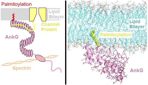
Ankyrins are a family of proteins that mediate the attachment of integral membrane proteins to the spectrin-actin based membrane cytoskeleton. Ankyrins have binding sites for the beta subunit of spectrin and at least 12 families of integral membrane proteins. This linkage is required to maintain the integrity of the plasma membranes and to anchor specific ion channels, ion exchangers and ion transporters in the plasma membrane. The name is derived from the Greek word ἄγκυρα (''ankyra'') for "anchor". Structure Ankyrins contain four functional domains: an N-terminal domain that contains 24 tandem ankyrin repeats, a central domain that binds to spectrin, a death domain that binds to proteins involved in apoptosis, and a C-terminal regulatory domain that is highly variable between different ankyrin proteins. Membrane protein recognition The 24 tandem ankyrin repeats are responsible for the recognition of a wide range of membrane proteins. These 24 repeats contain 3 struc ... [...More Info...] [...Related Items...] OR: [Wikipedia] [Google] [Baidu] |
Neurofascin
Neurofascin is a protein that in humans is encoded by the ''NFASC'' gene. Function Neurofascin is an L1 family immunoglobulin cell adhesion molecule (see L1CAM) involved in axon subcellular targeting and synapse formation during neural development The development of the nervous system, or neural development (neurodevelopment), refers to the processes that generate, shape, and reshape the nervous system of animals, from the earliest stages of embryonic development to adulthood. The fiel .... Clinical importance A homozygous mutation causing loss of Nfasc155 causes severe congenital hypotonia, contractures of fingers and toes and no reaction to touch or pain. References Further reading * * * * * * * * * * * * * Human proteins {{gene-1-stub ... [...More Info...] [...Related Items...] OR: [Wikipedia] [Google] [Baidu] |
Neurofilaments
Neurofilaments (NF) are classed as type IV intermediate filaments found in the cytoplasm of neurons. They are protein polymers measuring 10 nm in diameter and many micrometers in length. Together with microtubules (~25 nm) and microfilaments (7 nm), they form the neuronal cytoskeleton. They are believed to function primarily to provide structural support for axons and to regulate axon diameter, which influences nerve conduction velocity. The proteins that form neurofilaments are members of the intermediate filament protein family, which is divided into six types based on their gene organization and protein structure. Types I and II are the keratins which are expressed in epithelia. Type III contains the proteins vimentin, desmin, peripherin and glial fibrillary acidic protein (GFAP). Type IV consists of the neurofilament proteins L, M, H and internexin. Type V consists of the nuclear lamins, and type VI consists of the protein nestin. The type IV inter ... [...More Info...] [...Related Items...] OR: [Wikipedia] [Google] [Baidu] |
Neuron (journal)
''Neuron'' is a biweekly peer-reviewed scientific journal published by Cell Press, and imprint of Elsevier. It was established in 1988, and covers neuroscience and related biological processes. The current editor in chief is Mariela Zirlinger. The founding editors were Lily Jan, A. James Hudspeth A. James Hudspeth is the F.M. Kirby Professor at Rockefeller University in New York City, where he is director of the F.M. Kirby Center for Sensory Neuroscience. His laboratory studies the physiological basis of hearing. Early life and education ..., Louis Reichardt, Roger Nicoll, and Zach Hall. A past Editor in Chief was Katja Brose. Transcript and video available. Click on "Transcript" for text. * See alsoA Career in Science Editing: Katja BroseEditor in Chief, Neuron References External links * Neuroscience journals Cell Press academic journals Publications established in 1988 English-language journals Biweekly journals {{neuroscience-journal-stub ... [...More Info...] [...Related Items...] OR: [Wikipedia] [Google] [Baidu] |
Astrocytes
Astrocytes (from Ancient Greek , , "star" + , , "cavity", "cell"), also known collectively as astroglia, are characteristic star-shaped glial cells in the brain and spinal cord. They perform many functions, including biochemical control of endothelial cells that form the blood–brain barrier, provision of nutrients to the nervous tissue, maintenance of extracellular ion balance, regulation of cerebral blood flow, and a role in the repair and scarring process of the brain and spinal cord following infection and traumatic injuries. The proportion of astrocytes in the brain is not well defined; depending on the counting technique used, studies have found that the astrocyte proportion varies by region and ranges from 20% to 40% of all glia. Another study reports that astrocytes are the most numerous cell type in the brain. Astrocytes are the major source of cholesterol in the central nervous system. Apolipoprotein E transports cholesterol from astrocytes to neurons and other glial ... [...More Info...] [...Related Items...] OR: [Wikipedia] [Google] [Baidu] |
Schwann Cell
Schwann cells or neurolemmocytes (named after German physiologist Theodor Schwann) are the principal glia of the peripheral nervous system (PNS). Glial cells function to support neurons and in the PNS, also include satellite cells, olfactory ensheathing cells, enteric glia and glia that reside at sensory nerve endings, such as the Pacinian corpuscle. The two types of Schwann cells are myelinating and nonmyelinating. Myelinating Schwann cells wrap around axons of motor and sensory neurons to form the myelin sheath. The Schwann cell promoter is present in the downstream region of the human dystrophin gene that gives shortened transcript that are again synthesized in a tissue-specific manner. During the development of the PNS, the regulatory mechanisms of myelination are controlled by feedforward interaction of specific genes, influencing transcriptional cascades and shaping the morphology of the myelinated nerve fibers. Schwann cells are involved in many important aspect ... [...More Info...] [...Related Items...] OR: [Wikipedia] [Google] [Baidu] |
Microvillus
Microvilli (singular: microvillus) are microscopic cellular membrane protrusions that increase the surface area for diffusion and minimize any increase in volume, and are involved in a wide variety of functions, including absorption, secretion, cellular adhesion, and mechanotransduction. Structure Microvilli are covered in plasma membrane, which encloses cytoplasm and microfilaments. Though these are cellular extensions, there are little or no cellular organelles present in the microvilli. Each microvillus has a dense bundle of cross-linked actin filaments, which serves as its structural core. 20 to 30 tightly bundled actin filaments are cross-linked by bundling proteins fimbrin (or plastin-1), villin and espin to form the core of the microvilli. In the enterocyte microvillus, the structural core is attached to the plasma membrane along its length by lateral arms made of myosin 1a and Ca2+ binding protein calmodulin. Myosin 1a functions through a binding site for filamentous ... [...More Info...] [...Related Items...] OR: [Wikipedia] [Google] [Baidu] |


