|
Nevilleiella Marchantii
''Nevilleiella marchantii'' is a species of terricolous (ground-dwelling), crustose lichen in the family Teloschistaceae. Found in Australia, it was formally described as a new species in 2007. The thallus of ''Nevilleiella marchantii'' spreads 1–3 cm wide, with distinctive, almost spherical, -like formations that give it an appearance resembling a bunch of grapes. These formations vary in shape and colour from yellow-brown to orange-brown. Taxonomy Sergey Kondratyuk and Ingvar Kärnefelt formally described this lichen as a new species in 2007; they initially classified it in the genus ''Caloplaca''. The type specimen was gathered in January 2004, approximately 5 km away from the town of Lake King, situated on the eastern fringe of a lake with the same name in Western Australia. It was found growing in a chenopod heath habitat, situated on clay and sandy soil. The species epithet honours Western Australian botanist Neville Graeme Marchant, who assisted the au ... [...More Info...] [...Related Items...] OR: [Wikipedia] [Google] [Baidu] |
Ingvar Kärnefelt
Jan Eric Ingvar Kärnefelt (born 1944) is a Swedish lichenologist. Early life and education Kärnefelt was born in Gothenburg, Sweden in 1944. His initial goal in his higher-level studies at University of Cologne in 1966–1967 was to become a dentist. He changed courses in 1968, turning instead to biology at the University of Gothenburg in 1968. Gunnar Degelius, his first teacher during undergraduate studies in botany in 1968, inspired him and others. After Degelius' retirement in 1969, Ingvar continued his studies at Lund University, where Hans Runemark held a position in systematic botany. In 1971 he met Ove Almborn, who became his supervisor. In 1979, he defended his thesis titled "The brown fruticose species of ''Cetraria''". The thesis was later awarded a prize for the best doctoral dissertation in botany at Lund University during a 5-year period by the Royal Physiographic Society in Lund. Career Kärnefelt became associate professor at the Department of Systematic Bo ... [...More Info...] [...Related Items...] OR: [Wikipedia] [Google] [Baidu] |
Taxon
In biology, a taxon (back-formation from ''taxonomy''; plural taxa) is a group of one or more populations of an organism or organisms seen by taxonomists to form a unit. Although neither is required, a taxon is usually known by a particular name and given a particular ranking, especially if and when it is accepted or becomes established. It is very common, however, for taxonomists to remain at odds over what belongs to a taxon and the criteria used for inclusion. If a taxon is given a formal scientific name, its use is then governed by one of the nomenclature codes specifying which scientific name is correct for a particular grouping. Initial attempts at classifying and ordering organisms (plants and animals) were set forth in Carl Linnaeus's Linnaean taxonomy, system in ''Systema Naturae'', 10th edition (1758), as well as an unpublished work by Bernard de Jussieu, Bernard and Antoine Laurent de Jussieu. The idea of a unit-based system of biological classification was first mad ... [...More Info...] [...Related Items...] OR: [Wikipedia] [Google] [Baidu] |
Parietin
Parietin is the predominant cortical pigment of lichens in the genus '' Caloplaca'', a secondary product of the lichen ''Xanthoria parietina'', and a pigment found in the roots of Curled Dock (''Rumex crispus''). It has an orangy-yellow color and absorbs blue light. It is also known as physcion. It has also been shown to protect lichens against UV-B light, at high altitudes in Alpine regions. The UV-B light stimulates production of parietin and the parietin protects the lichens from damage. Lichens in arctic regions such as Svarlbard retain this capability though they do not encounter damaging levels of UV-B, a capability that could help protect the lichens in case of Ozone layer thinning. It has also shown anti-fungal activity against barley powdery mildew and cucumber powdery mildew, more efficiently in the latter case than treatments with fenarimol and polyoxin B. It reacts with KOH to form a deep, reddish-magenta compound. Effect on human cancer cells Also found in rhu ... [...More Info...] [...Related Items...] OR: [Wikipedia] [Google] [Baidu] |
Spot Test (lichen)
A spot test in lichenology is a spot analysis used to help identify lichens. It is performed by placing a drop of a chemical on different parts of the lichen and noting the colour change (or lack thereof) associated with application of the chemical. The tests are routinely encountered in dichotomous keys for lichen species, and they take advantage of the wide array of lichen products produced by lichens and their uniqueness among taxa. As such, spot tests reveal the presence or absence of chemicals in various parts of a lichen. They were first proposed by the botanist William Nylander in 1866. Three common spot tests use either 10% aqueous KOH solution (K test), saturated aqueous solution of bleaching powder or calcium hypochlorite (C test), or 5% alcoholic ''p''-phenylenediamine solution (P test). The colour changes occur due to presence of particular secondary metabolites in the lichen. There are several other less frequently used spot tests of more limited use that are employed ... [...More Info...] [...Related Items...] OR: [Wikipedia] [Google] [Baidu] |
Conidiomata
Conidiomata (singular: Conidioma) are blister-like fruiting structures produced by a specific type of fungus called a coelomycete. They are formed as a means of dispersing asexual spores call conidia, which they accomplish by creating the blister-like formations which then rupture to release the contained spores. Structure Conidiomata mainly consist of a mass of densely packed hypha which develops below the surface of the host cuticle, and the fungus may or may not use some of the host’s own tissue to construct the structure. Development of these structures can either occur just below the cuticle or below the epidermal layer of the tissue. Formation on the differing levels is dependent upon the type of conidiomata being formed. Five types of conidioma have been found and are classified as acervuli, pycnidia, sporodochia, synnemata and corenima. Types Acervuli Acervuli is one of the two major groups of conidiomata (the other being pycnidia). Conidiomata of this type form ju ... [...More Info...] [...Related Items...] OR: [Wikipedia] [Google] [Baidu] |
Staining
Staining is a technique used to enhance contrast in samples, generally at the microscopic level. Stains and dyes are frequently used in histology (microscopic study of biological tissues), in cytology (microscopic study of cells), and in the medical fields of histopathology, hematology, and cytopathology that focus on the study and diagnoses of diseases at the microscopic level. Stains may be used to define biological tissues (highlighting, for example, muscle fibers or connective tissue), cell populations (classifying different blood cells), or organelles within individual cells. In biochemistry, it involves adding a class-specific ( DNA, proteins, lipids, carbohydrates) dye to a substrate to qualify or quantify the presence of a specific compound. Staining and fluorescent tagging can serve similar purposes. Biological staining is also used to mark cells in flow cytometry, and to flag proteins or nucleic acids in gel electrophoresis. Light microscopes are used for viewin ... [...More Info...] [...Related Items...] OR: [Wikipedia] [Google] [Baidu] |
Septum
In biology, a septum (Latin for ''something that encloses''; plural septa) is a wall, dividing a cavity or structure into smaller ones. A cavity or structure divided in this way may be referred to as septate. Examples Human anatomy * Interatrial septum, the wall of tissue that is a sectional part of the left and right atria of the heart * Interventricular septum, the wall separating the left and right ventricles of the heart * Lingual septum, a vertical layer of fibrous tissue that separates the halves of the tongue. *Nasal septum: the cartilage wall separating the nostrils of the nose * Alveolar septum: the thin wall which separates the alveoli from each other in the lungs * Orbital septum, a palpebral ligament in the upper and lower eyelids * Septum pellucidum or septum lucidum, a thin structure separating two fluid pockets in the brain * Uterine septum, a malformation of the uterus * Vaginal septum, a lateral or transverse partition inside the vagina * Intermuscular sep ... [...More Info...] [...Related Items...] OR: [Wikipedia] [Google] [Baidu] |
Paraphyses
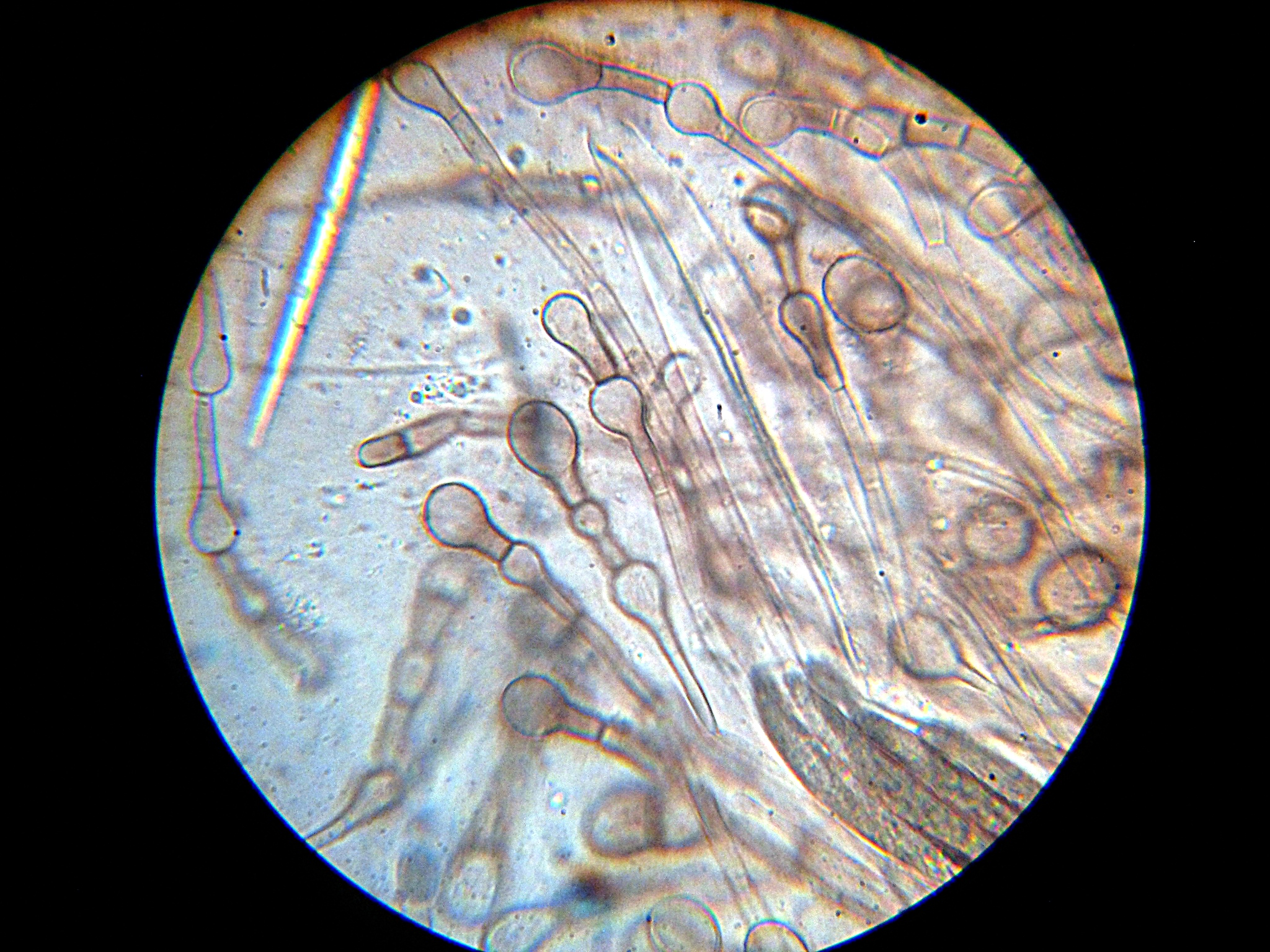
Paraphyses are erect sterile filament-like support structures occurring among the reproductive apparatuses of fungi, ferns, bryophytes and some thallophytes. The singular form of the word is paraphysis. In certain fungi, they are part of the fertile spore-bearing layer. More specifically, paraphyses are sterile filamentous hyphal end cells composing part of the hymenium of Ascomycota and Basidiomycota interspersed among either the asci or basidia respectively, and not sufficiently differentiated to be called cystidia A cystidium (plural cystidia) is a relatively large cell found on the sporocarp of a basidiomycete (for example, on the surface of a mushroom gill), often between clusters of basidia. Since cystidia have highly varied and distinct shapes that ar ..., which are specialized, swollen, often protruding cells. The tips of paraphyses may contain the pigments which colour the hymenium. In ferns and mosses, they are filament-like structures that are found on sporangia ... [...More Info...] [...Related Items...] OR: [Wikipedia] [Google] [Baidu] |
Ascus
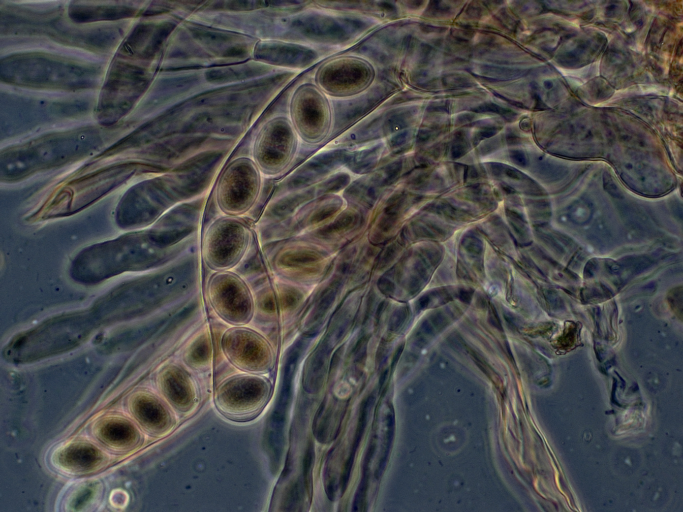
An ascus (; ) is the sexual spore-bearing cell produced in ascomycete fungi. Each ascus usually contains eight ascospores (or octad), produced by meiosis followed, in most species, by a mitotic cell division. However, asci in some genera or species can occur in numbers of one (e.g. ''Monosporascus cannonballus''), two, four, or multiples of four. In a few cases, the ascospores can bud off conidia that may fill the asci (e.g. ''Tympanis'') with hundreds of conidia, or the ascospores may fragment, e.g. some ''Cordyceps'', also filling the asci with smaller cells. Ascospores are nonmotile, usually single celled, but not infrequently may be coenocytic (lacking a septum), and in some cases coenocytic in multiple planes. Mitotic divisions within the developing spores populate each resulting cell in septate ascospores with nuclei. The term ocular chamber, or oculus, refers to the epiplasm (the portion of cytoplasm not used in ascospore formation) that is surrounded by the "bourrelet ... [...More Info...] [...Related Items...] OR: [Wikipedia] [Google] [Baidu] |
Hymenium
The hymenium is the tissue layer on the hymenophore of a fungal fruiting body where the cells develop into basidia or asci, which produce spores. In some species all of the cells of the hymenium develop into basidia or asci, while in others some cells develop into sterile cells called cystidia (basidiomycetes) or paraphyses (ascomycetes). Cystidia are often important for microscopic identification. The subhymenium consists of the supportive hyphae from which the cells of the hymenium grow, beneath which is the hymenophoral trama, the hyphae that make up the mass of the hymenophore. The position of the hymenium is traditionally the first characteristic used in the classification and identification of mushrooms. Below are some examples of the diverse types which exist among the macroscopic Basidiomycota and Ascomycota. * In agarics, the hymenium is on the vertical faces of the gills. * In boletes and polypores, it is in a spongy mass of downward-pointing tubes. * In puffballs, ... [...More Info...] [...Related Items...] OR: [Wikipedia] [Google] [Baidu] |
Apothecia
An ascocarp, or ascoma (), is the fruiting body ( sporocarp) of an ascomycete phylum fungus. It consists of very tightly interwoven hyphae and millions of embedded asci, each of which typically contains four to eight ascospores. Ascocarps are most commonly bowl-shaped (apothecia) but may take on a spherical or flask-like form that has a pore opening to release spores (perithecia) or no opening (cleistothecia). Classification The ascocarp is classified according to its placement (in ways not fundamental to the basic taxonomy). It is called ''epigeous'' if it grows above ground, as with the morels, while underground ascocarps, such as truffles, are termed ''hypogeous''. The structure enclosing the hymenium is divided into the types described below (apothecium, cleistothecium, etc.) and this character ''is'' important for the taxonomic classification of the fungus. Apothecia can be relatively large and fleshy, whereas the others are microscopic—about the size of flecks of ... [...More Info...] [...Related Items...] OR: [Wikipedia] [Google] [Baidu] |
Micrometre
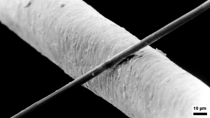
The micrometre ( international spelling as used by the International Bureau of Weights and Measures; SI symbol: μm) or micrometer (American spelling), also commonly known as a micron, is a unit of length in the International System of Units (SI) equalling (SI standard prefix "micro-" = ); that is, one millionth of a metre (or one thousandth of a millimetre, , or about ). The nearest smaller common SI unit is the nanometre, equivalent to one thousandth of a micrometre, one millionth of a millimetre or one billionth of a metre (). The micrometre is a common unit of measurement for wavelengths of infrared radiation as well as sizes of biological cells and bacteria, and for grading wool by the diameter of the fibres. The width of a single human hair ranges from approximately 20 to . The longest human chromosome, chromosome 1, is approximately in length. Examples Between 1 μm and 10 μm: * 1–10 μm – length of a typical bacterium * 3–8 μm – width of ... [...More Info...] [...Related Items...] OR: [Wikipedia] [Google] [Baidu] |


