|
NG2 Proteoglycan
Neural/glial antigen 2, or NG2, is a rat integral membrane proteoglycan found in the plasma membrane of many diverse cell types.Nishiyama A, Dahlin KJ, Prince JT, Johnstone SR, Stallcup WB. "The primary structure of NG2: a novel membrane-spanning proteoglycan." ''J Cell Biol''. 1991 Jul;114(2):359-71. . Homologous proteins in other species include human CSPG4, also known as melanoma-associated chondroitin sulfate proteoglycan (MCSP), Mouse AN2, and Sea urchin ECM3.Asher RA, Morgenstern DA, Fawcett JW. "Chondroitin sulphate proteoglycans: inhibitory components of the glial scar." Prog Brain Res. 2001;132:611-9. . This single-pass transmembrane molecule may be plasma membrane-bound or secreted and associated with the extracellular matrix.Nishiyama A, Lin ZH, Stallcup WB. "Generation of truncated forms of the NG2 proteoglycan by cell surface proteolysis." ''Mol Biol Cell''. 1995 Dec;6(12):1819-32. It is believed to play a role in functions such as cell adhesion, cell-cell and cell-ECM ... [...More Info...] [...Related Items...] OR: [Wikipedia] [Google] [Baidu] |
CSPG4
Chondroitin sulfate proteoglycan 4, also known as melanoma-associated chondroitin sulfate proteoglycan (MCSP) or neuron-glial antigen 2 (NG2), is a chondroitin sulfate proteoglycan that in humans is encoded by the ''CSPG4'' gene. Function CSPG4 plays a role in stabilizing cell-substratum interactions during early events of melanoma cell spreading on endothelial basement membranes. It represents an integral membrane chondroitin sulfate proteoglycan expressed by human malignant melanoma cells. Implications in disease CSPG4/NG2 is also a hallmark protein of oligodendrocyte progenitor cells (OPCs) and OPC dysfunction has been implicated as a candidate pathophysiological mechanism of familial schizophrenia. A research group investigating the role of genetics in schizophrenia, reported, two rare missense mutations in ''CSPG4'' gene'','' segregating within families (''CSPG4A131T'' and ''CSPG4V901G'' mutations). The researchers also demonstrate that the induced pluripotent stem cells ... [...More Info...] [...Related Items...] OR: [Wikipedia] [Google] [Baidu] |
Oligodendrocyte Progenitor
Oligodendrocyte progenitor cells (OPCs), also known as oligodendrocyte precursor cells, NG2-glia, O2A cells, or polydendrocytes, are a subtype of glia in the central nervous system named for their essential role as precursors to oligodendrocytes. They are typically identified by coexpression of PDGFRA and NG2. OPCs play a critical role in developmental and adult myelinogenesis by giving rise to oligodendrocytes, which then ensheath axons and provide electrical insulation in the form of a myelin sheath, enabling faster action potential propagation and high fidelity transmission without a need for an increase in axonal diameter. The loss or lack of OPCs, and consequent lack of differentiated oligodendrocytes, is associated with a loss of myelination and subsequent impairment of neurological functions. In addition, OPCs express receptors for various neurotransmitters and undergo membrane depolarization when they receive synaptic inputs from neurons. Structure OPCs are glial cells ... [...More Info...] [...Related Items...] OR: [Wikipedia] [Google] [Baidu] |
Chondroblasts
Chondroblasts, or perichondrial cells, is the name given to mesenchymal progenitor cells in situ which, from endochondral ossification, will form chondrocytes in the growing cartilage matrix. Another name for them is subchondral cortico-spongious progenitors. They have euchromatic nuclei and stain by basic dyes. These cells are extremely important in chondrogenesis due to their role in forming both the chondrocytes and cartilage matrix which will eventually form cartilage. Use of the term is technically inaccurate since mesenchymal progenitors can also technically differentiate into osteoblasts or fat. Chondroblasts are called chondrocytes when they embed themselves in the cartilage matrix, consisting of proteoglycan and collagen fibers, until they lie in the matrix lacunae. Once they embed themselves into the cartilage matrix, they grow the cartilage matrix by growing more cartilage extracellular matrix rather than by dividing further. Structure Within adults and developing ... [...More Info...] [...Related Items...] OR: [Wikipedia] [Google] [Baidu] |
Myoblasts
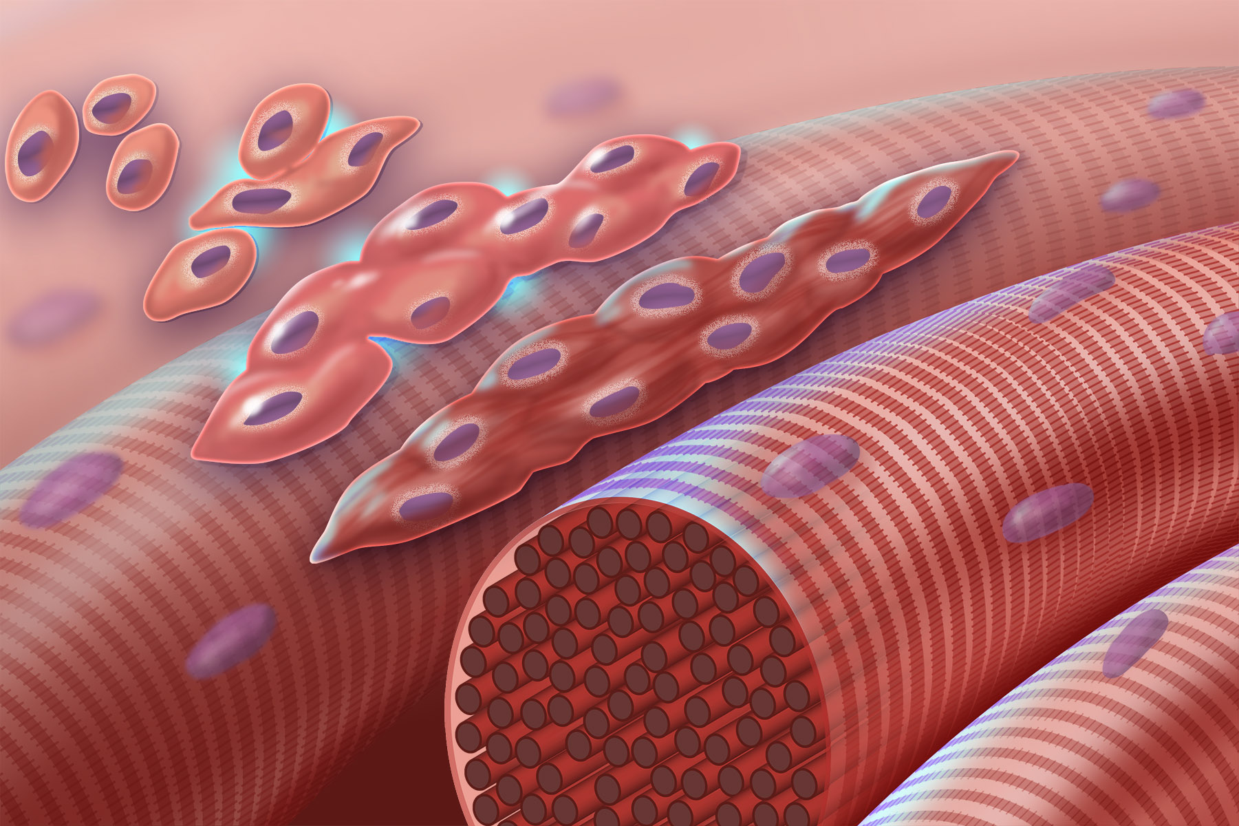
Myogenesis is the formation of skeletal muscular tissue, particularly during embryonic development. Muscle fibers generally form through the fusion of precursor myoblasts into multinucleated fibers called ''myotubes''. In the early development of an embryo, myoblasts can either proliferate, or differentiate into a myotube. What controls this choice in vivo is generally unclear. If placed in cell culture, most myoblasts will proliferate if enough fibroblast growth factor (FGF) or another growth factor is present in the medium surrounding the cells. When the growth factor runs out, the myoblasts cease division and undergo terminal differentiation into myotubes. Myoblast differentiation proceeds in stages. The first stage, involves cell cycle exit and the commencement of expression of certain genes. The second stage of differentiation involves the alignment of the myoblasts with one another. Studies have shown that even rat and chick myoblasts can recognise and align with one ... [...More Info...] [...Related Items...] OR: [Wikipedia] [Google] [Baidu] |
Pericytes
Pericytes (previously known as Rouget cells) are multi-functional mural cells of the microcirculation that wrap around the endothelial cells that line the capillaries throughout the body. Pericytes are embedded in the basement membrane of blood capillaries, where they communicate with endothelial cells by means of both direct physical contact and paracrine signaling. The morphology, distribution, density and molecular fingerprints of pericytes vary between organs and vascular beds. Pericytes help to maintain homeostatic and hemostatic functions in the brain, one of the organs with higher pericyte coverage, and also sustain the blood–brain barrier. These cells are also a key component of the neurovascular unit, which includes endothelial cells, astrocytes, and neurons. Pericytes have been postulated to regulate capillary blood flow and the clearance and phagocytosis of cellular debris ''in vitro.'' Pericytes stabilize and monitor the maturation of endothelial cells by means of ... [...More Info...] [...Related Items...] OR: [Wikipedia] [Google] [Baidu] |
Glioblastoma Multiforme
Glioblastoma, previously known as glioblastoma multiforme (GBM), is one of the most aggressive types of cancer that begin within the brain. Initially, signs and symptoms of glioblastoma are nonspecific. They may include headaches, personality changes, nausea, and symptoms similar to those of a stroke. Symptoms often worsen rapidly and may progress to unconsciousness. The cause of most cases of glioblastoma is not known. Uncommon risk factors include genetic disorders, such as neurofibromatosis and Li–Fraumeni syndrome, and previous radiation therapy. Glioblastomas represent 15% of all brain tumors. They can either start from normal brain cells or develop from an existing low-grade astrocytoma. The diagnosis typically is made by a combination of a CT scan, MRI scan, and tissue biopsy. There is no known method of preventing the cancer. Treatment usually involves surgery, after which chemotherapy and radiation therapy are used. The medication temozolomide is frequently used as ... [...More Info...] [...Related Items...] OR: [Wikipedia] [Google] [Baidu] |
Melanoma
Melanoma, also redundantly known as malignant melanoma, is a type of skin cancer that develops from the pigment-producing cells known as melanocytes. Melanomas typically occur in the skin, but may rarely occur in the mouth, intestines, or eye (uveal melanoma). In women, they most commonly occur on the legs, while in men, they most commonly occur on the back. About 25% of melanomas develop from moles. Changes in a mole that can indicate melanoma include an increase in size, irregular edges, change in color, itchiness, or skin breakdown. The primary cause of melanoma is ultraviolet light (UV) exposure in those with low levels of the skin pigment melanin. The UV light may be from the sun or other sources, such as tanning devices. Those with many moles, a history of affected family members, and poor immune function are at greater risk. A number of rare genetic conditions, such as xeroderma pigmentosum, also increase the risk. Diagnosis is by biopsy and analysis of any skin lesio ... [...More Info...] [...Related Items...] OR: [Wikipedia] [Google] [Baidu] |
Chondroitin Sulfate
Chondroitin sulfate is a sulfated glycosaminoglycan (GAG) composed of a chain of alternating sugars ( N-acetylgalactosamine and glucuronic acid). It is usually found attached to proteins as part of a proteoglycan. A chondroitin chain can have over 100 individual sugars, each of which can be sulfated in variable positions and quantities. Chondroitin sulfate is an important structural component of cartilage, and provides much of its resistance to compression. Along with glucosamine, chondroitin sulfate has become a widely used dietary supplement for treatment of osteoarthritis, although large clinical trials failed to demonstrate any symptomatic benefit of chondroitin. Medical use Chondroitin is used in dietary supplements as an alternative medicine to treat osteoarthritis. It is also approved and regulated as a symptomatic slow-acting drug for this disease (SYSADOA) in Europe and some other countries. It is commonly sold together with glucosamine. A 2015 Cochrane review of ... [...More Info...] [...Related Items...] OR: [Wikipedia] [Google] [Baidu] |

