|
Myeloid Sarcoma
A myeloid sarcoma (chloroma, granulocytic sarcoma, extramedullary myeloid tumor) is a solid tumor composed of immature white blood cells called myeloblasts. A chloroma is an extramedullary manifestation of acute myeloid leukemia; in other words, it is a solid collection of leukemic cells occurring outside of the bone marrow. Types In acute leukemia Chloromas are rare; exact estimates of their prevalence are lacking, but they are uncommonly seen even by physicians specializing in the treatment of leukemia. Chloromas may be somewhat more common in patients with the following disease features: * French–American–British (FAB) classification class M2 * WHO Classification (2016 revision) is a separate entity under the "Acute myeloid leukemia (AML) and related neoplasms" * those with specific cytogenetic abnormalities (e.g. t(8;21) or inv(16)) * those whose myeloblasts express T-cell surface markers, CD13, or CD14 * those with high peripheral white blood cell counts However ... [...More Info...] [...Related Items...] OR: [Wikipedia] [Google] [Baidu] |
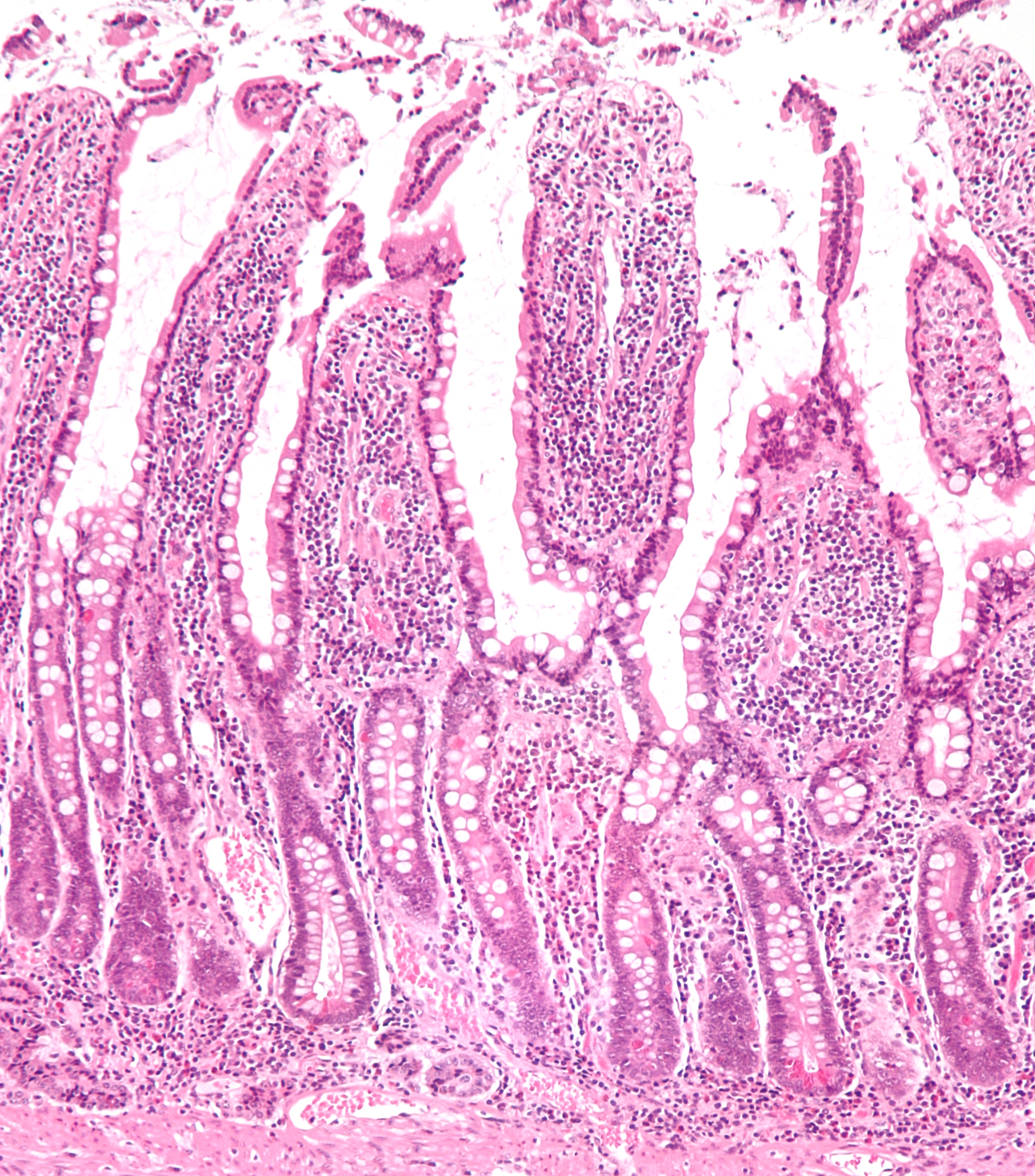
Micrograph
A micrograph or photomicrograph is a photograph or digital image taken through a microscope or similar device to show a magnified image of an object. This is opposed to a macrograph or photomacrograph, an image which is also taken on a microscope but is only slightly magnified, usually less than 10 times. Micrography is the practice or art of using microscopes to make photographs. A micrograph contains extensive details of microstructure. A wealth of information can be obtained from a simple micrograph like behavior of the material under different conditions, the phases found in the system, failure analysis, grain size estimation, elemental analysis and so on. Micrographs are widely used in all fields of microscopy. Types Photomicrograph A light micrograph or photomicrograph is a micrograph prepared using an optical microscope, a process referred to as ''photomicroscopy''. At a basic level, photomicroscopy may be performed simply by connecting a camera to a microscope, th ... [...More Info...] [...Related Items...] OR: [Wikipedia] [Google] [Baidu] |
Chronic Myelogenous Leukemia
Chronic myelogenous leukemia (CML), also known as chronic myeloid leukemia, is a cancer of the white blood cells. It is a form of leukemia characterized by the increased and unregulated growth of myeloid cells in the bone marrow and the accumulation of these cells in the blood. CML is a clonal bone marrow stem cell disorder in which a proliferation of mature granulocytes (neutrophils, eosinophils and basophils) and their precursors is found. It is a type of myeloproliferative neoplasm associated with a characteristic chromosomal translocation called the Philadelphia chromosome. CML is largely treated with targeted drugs called tyrosine-kinase inhibitors (TKIs) which have led to dramatically improved long-term survival rates since 2001. These drugs have revolutionized treatment of this disease and allow most patients to have a good quality of life when compared to the former chemotherapy drugs. In Western countries, CML accounts for 15–25% of all adult leukemias and 14% of leuke ... [...More Info...] [...Related Items...] OR: [Wikipedia] [Google] [Baidu] |
Mediastinum
The mediastinum (from ) is the central compartment of the thoracic cavity. Surrounded by loose connective tissue, it is an undelineated region that contains a group of structures within the thorax, namely the heart and its vessels, the esophagus, the trachea, the phrenic nerve, phrenic and cardiac nerves, the thoracic duct, the thymus and the lymph nodes of the central chest. Anatomy The mediastinum lies within the thorax and is enclosed on the right and left by pulmonary pleurae, pleurae. It is surrounded by the chest wall in front, the lungs to the sides and the Spine (anatomy), spine at the back. It extends from the sternum in front to the vertebral column behind. It contains all the organs of the thorax except the lungs. It is continuous with the loose connective tissue of the neck. The mediastinum can be divided into an upper (or superior) and lower (or inferior) part: * The superior mediastinum starts at the superior thoracic aperture and ends at the #Thoracic plane, t ... [...More Info...] [...Related Items...] OR: [Wikipedia] [Google] [Baidu] |
Small Intestine
The small intestine or small bowel is an organ in the gastrointestinal tract where most of the absorption of nutrients from food takes place. It lies between the stomach and large intestine, and receives bile and pancreatic juice through the pancreatic duct to aid in digestion. The small intestine is about long and folds many times to fit in the abdomen. Although it is longer than the large intestine, it is called the small intestine because it is narrower in diameter. The small intestine has three distinct regions – the duodenum, jejunum, and ileum. The duodenum, the shortest, is where preparation for absorption through small finger-like protrusions called villi begins. The jejunum is specialized for the absorption through its lining by enterocytes: small nutrient particles which have been previously digested by enzymes in the duodenum. The main function of the ileum is to absorb vitamin B12, bile salts, and whatever products of digestion that were not absorbed by the ... [...More Info...] [...Related Items...] OR: [Wikipedia] [Google] [Baidu] |
Lymph Node
A lymph node, or lymph gland, is a kidney-shaped organ of the lymphatic system and the adaptive immune system. A large number of lymph nodes are linked throughout the body by the lymphatic vessels. They are major sites of lymphocytes that include B and T cells. Lymph nodes are important for the proper functioning of the immune system, acting as filters for foreign particles including cancer cells, but have no detoxification function. In the lymphatic system a lymph node is a secondary lymphoid organ. A lymph node is enclosed in a fibrous capsule and is made up of an outer cortex and an inner medulla. Lymph nodes become inflamed or enlarged in various diseases, which may range from trivial throat infections to life-threatening cancers. The condition of lymph nodes is very important in cancer staging, which decides the treatment to be used and determines the prognosis. Lymphadenopathy refers to glands that are enlarged or swollen. When inflamed or enlarged, lymph nodes can be ... [...More Info...] [...Related Items...] OR: [Wikipedia] [Google] [Baidu] |
Gingival Enlargement
Gingival enlargement is an increase in the size of the gingiva (gums). It is a common feature of gingival disease. Gingival enlargement can be caused by a number of factors, including inflammatory conditions and the side effects of certain medications. The treatment is based on the cause. A closely related term is epulis, denoting a localized tumor (i.e. lump) on the gingiva. Classification The terms gingival hyperplasia and gingival hypertrophy have been used to describe this topic in the past. These are not precise descriptions of gingival enlargement because these terms are strictly histologic diagnoses, and such diagnoses require microscopic analysis of a tissue sample. Hyperplasia refers to an increased number of cells, and hypertrophy refers to an increase in the size of individual cells. As these identifications cannot be performed with a clinical examination and evaluation of the tissue, the term ''gingival enlargement'' is more properly applied. Gingival enlargement has ... [...More Info...] [...Related Items...] OR: [Wikipedia] [Google] [Baidu] |
Paraneoplastic
A paraneoplastic syndrome is a syndrome (a set of signs and symptoms) that is the consequence of a tumor in the body (usually a cancerous one), specifically due to the production of chemical signaling molecules (such as hormones or cytokines) by tumor cells or by an immune response against the tumor. Unlike a mass effect, it is not due to the local presence of cancer cells. Paraneoplastic syndromes are typical among middle-aged to older patients, and they most commonly present with cancers of the lung, breast, ovaries or lymphatic system (a lymphoma). Sometimes, the symptoms of paraneoplastic syndromes show before the diagnosis of a malignancy, which has been hypothesized to relate to the disease pathogenesis. In this paradigm, tumor cells express tissue-restricted antigens (e.g., neuronal proteins), triggering an anti-tumor immune response which may be partially or, rarely, completely effective in suppressing tumor growth and symptoms. Patients then come to clinical attention whe ... [...More Info...] [...Related Items...] OR: [Wikipedia] [Google] [Baidu] |
Sweet Syndrome
Sweet syndrome (SS), or acute febrile neutrophilic dermatosis, is a skin disease characterized by the sudden onset of fever, an elevated white blood cell count, and tender, red, well-demarcated papules and plaques that show dense infiltrates by neutrophil granulocytes on histologic examination. The syndrome was first described in 1964 by Robert Douglas Sweet. It was also known as Gomm–Button disease in honour of the first two patients Sweet diagnosed with the condition. Signs and symptoms Acute, tender, erythematous plaques, nodes, pseudovesicles and, occasionally, blisters with an annular or arciform pattern occur on the head, neck, legs, and arms, particularly the back of the hands and fingers. The trunk is rarely involved. Fever (50%); arthralgia or arthritis (62%); eye involvement, most frequently conjunctivitis or iridocyclitis (38%); and oral aphthae (13%) are associated features. Cause SS can be classified based upon the clinical setting in which it occurs: classica ... [...More Info...] [...Related Items...] OR: [Wikipedia] [Google] [Baidu] |
Biopsy
A biopsy is a medical test commonly performed by a surgeon, interventional radiologist, or an interventional cardiologist. The process involves extraction of sample cells or tissues for examination to determine the presence or extent of a disease. The tissue is then fixed, dehydrated, embedded, sectioned, stained and mounted before it is generally examined under a microscope by a pathologist; it may also be analyzed chemically. When an entire lump or suspicious area is removed, the procedure is called an excisional biopsy. An incisional biopsy or core biopsy samples a portion of the abnormal tissue without attempting to remove the entire lesion or tumor. When a sample of tissue or fluid is removed with a needle in such a way that cells are removed without preserving the histological architecture of the tissue cells, the procedure is called a needle aspiration biopsy. Biopsies are most commonly performed for insight into possible cancerous or inflammatory conditions. History T ... [...More Info...] [...Related Items...] OR: [Wikipedia] [Google] [Baidu] |
Acute Promyelocytic Leukemia
Acute promyelocytic leukemia (APML, APL) is a subtype of acute myeloid leukemia (AML), a cancer of the white blood cells. In APL, there is an abnormal accumulation of immature granulocytes called promyelocytes. The disease is characterized by a chromosomal translocation involving the retinoic acid receptor alpha (RARA) gene and is distinguished from other forms of AML by its responsiveness to all-''trans'' retinoic acid (ATRA; also known as tretinoin) therapy. Acute promyelocytic leukemia was first characterized in 1957 by French and Norwegian physicians as a hyperacute fatal illness, with a median survival time of less than a week. Today, prognoses have drastically improved; 10-year survival rates are estimated to be approximately 80-90% according to one study. Signs and symptoms The symptoms tend to be similar to AML in general with the following being possible symptoms: * Anemia * Fatigue * Weakness * Chills * Depression * Difficulty breathing (dyspnea) * Low platelets (thr ... [...More Info...] [...Related Items...] OR: [Wikipedia] [Google] [Baidu] |
Imatinib
Imatinib, sold under the brand names Gleevec and Glivec (both marketed worldwide by Novartis) among others, is an oral chemotherapy medication used to treat cancer. Imatinib is a small molecule inhibitor targeting multiple receptor tyrosine kinases such as CSF1R, ABL, c-KIT, FLT3, and PDGFR-β. Specifically, it is used for chronic myelogenous leukemia (CML) and acute lymphocytic leukemia (ALL) that are Philadelphia chromosome-positive (Ph+), certain types of gastrointestinal stromal tumors (GIST), hypereosinophilic syndrome (HES), chronic eosinophilic leukemia (CEL), systemic mastocytosis, and myelodysplastic syndrome. Common side effects include vomiting, diarrhea, muscle pain, headache, and rash. Severe side effects may include fluid retention, gastrointestinal bleeding, bone marrow suppression, liver problems, and heart failure. Use during pregnancy may result in harm to the baby. Imatinib works by stopping the Bcr-Abl tyrosine-kinase. This can slow growth or result in p ... [...More Info...] [...Related Items...] OR: [Wikipedia] [Google] [Baidu] |
FIP1L1
Factor interacting with PAPOLA and CPSF1 (i.e, FIP1L1; also termed Pre-mRNA 3'-end-processing factor FIP1) is a protein that in humans is encoded by the ''FIP1L1'' gene (also known as Rhe, FIP1, and hFip1). A medically important aspect of the ''FIP1L1'' gene is its fusion with other genes to form fusion genes which cause clonal hypereosinophilia and leukemic diseases in humans. Gene The human ''FIP1L1'' gene is located on chromosome 4 at position q12 (4q12), contains 19 exons, and codes for a complete protein consisting of 594 amino acids. However, alternative splicing of its Precursor mRNA results in multiple transcript variants encoding distinct FIP1L1 protein isoforms. The ''FIP1L1'' gene is found in a wide range of species, being designated as FIP1 in Saccharomyces cerevisiae (yeast) and fip1l1 in coho salmon as well as mice and numerous other mammalian species. In humans, an interstitial chromosomal deletion of about 800 kilobases at 4q12 deletes the ''CHIC2'' gene (i.e.c ... [...More Info...] [...Related Items...] OR: [Wikipedia] [Google] [Baidu] |


.jpg)

