|
Muscular Triangle
The inferior carotid triangle (or muscular triangle), is bounded, in front, by the median line of the neck from the hyoid bone to the sternum; behind, by the anterior margin of the sternocleidomastoid; above, by the superior belly of the omohyoid. It is covered by the integument, superficial fascia, platysma, and deep fascia, ramifying in which are some of the branches of the supraclavicular nerves. Beneath these superficial structures are the sternohyoid and sternothyroid, which, together with the anterior margin of the sternocleidomastoid, conceal the lower part of the common carotid artery. This vessel is enclosed within its sheath, together with the internal jugular vein and vagus nerve; the vein lies lateral to the artery on the right side of the neck, but overlaps it below on the left side; the nerve lies between the artery and vein, on a plane posterior to both. In front of the sheath are a few descending filaments from the ansa cervicalis; behind the sheath are the i ... [...More Info...] [...Related Items...] OR: [Wikipedia] [Google] [Baidu] [Amazon] |
Hyoid Bone
The hyoid-bone (lingual-bone or tongue-bone) () is a horseshoe-shaped bone situated in the anterior midline of the neck between the chin and the thyroid-cartilage. At rest, it lies between the base of the mandible and the third cervical vertebra. Unlike other bones, the hyoid is only distantly articulated to other bones by muscles or ligaments. It is the only bone in the human body that is not connected to any other bones. The hyoid is anchored by muscles from the anterior, posterior and inferior directions, and aids in tongue movement and swallowing. The hyoid bone provides attachment to the muscles of the floor of the mouth and the tongue above, the larynx below, and the epiglottis and pharynx behind. Its name is derived . Structure The hyoid bone is classed as an irregular bone and consists of a central part called the body, and two pairs of horns, the greater and lesser horns. Body The body of the hyoid bone is the central part of the hyoid bone. *At the fron ... [...More Info...] [...Related Items...] OR: [Wikipedia] [Google] [Baidu] [Amazon] |
Inferior Thyroid Artery
The inferior thyroid artery is an artery in the neck. It arises from the thyrocervical trunk and passes upward, in front of the vertebral artery and longus colli muscle. It then turns medially behind the carotid sheath and its contents, and also behind the sympathetic trunk, the middle cervical ganglion resting upon the vessel. Reaching the lower border of the thyroid gland it divides into two branches, which supply the postero-inferior parts of the gland, and anastomose with the superior thyroid artery, and with the corresponding artery of the opposite side. Structure The branches of the inferior thyroid artery are the inferior laryngeal, the oesophageal, the tracheal, the ascending cervical and the pharyngeal arteries. Branches Inferior laryngeal artery The ''inferior laryngeal artery'' - accompanied by the recurrent laryngeal nerve - passes superior-ward upon the trachea deep to the inferior pharyngeal constrictor muscle to reach the posterior surface of the larynx. ... [...More Info...] [...Related Items...] OR: [Wikipedia] [Google] [Baidu] [Amazon] |
Anterior Triangle Of The Neck
The anterior triangle is a region of the neck. Structure The triangle is inverted with its apex inferior to its base which is under the chin. Investing fascia covers the roof of the triangle while visceral fascia covers the floor. Anatomy Muscles: * Suprahyoid muscles - Digastric (Ant and post belly), mylohyoid, geniohyoid and stylohyoid. * Infrahyoid muscles - Omohyoid, sternohyoid, sternothyroid, and thyrohyoid. Nerve supply 2 Bellies of digastric * Anterior: Mylohyoid nerve * Posterior: Facial nerve Stylohyoid: by the facial nerve, by a branch from that to the posterior belly of digastric. Mylohyoid: by its own nerve, a branch of the inferior alveolar (from the mandibular division of trigeminal nerve), which arises just before the parent nerve enters the mandibular foramen, pierces the sphenomandibular ligament, and runs forward on the inferior surface of the mylohyoid, supplying it and the anterior belly of the digastric. Geniohyoid: by a branch from the hypoglossa ... [...More Info...] [...Related Items...] OR: [Wikipedia] [Google] [Baidu] [Amazon] |
Sternocleidomastoid Muscle
The sternocleidomastoid muscle is one of the largest and most superficial cervical muscles. The primary actions of the muscle are rotation of the head to the opposite side and Anatomical terms of motion#Flexion and extension, flexion of the neck. The sternocleidomastoid is innervated by the accessory nerve. Etymology and location It is given the name ''sternocleidomastoid'' because it originates at the manubrium of the Human sternum, sternum (''sterno-'') and the clavicle (''cleido-'') and has an Insertion (anatomy), insertion at the mastoid process of the temporal bone of the human skull, skull. Structure The sternocleidomastoid muscle originates from two locations: the manubrium of the Human sternum, sternum and the clavicle, hence it is said to have two heads: sternal head and clavicular head. It travels obliquely across the side of the neck and inserts at the mastoid process of the temporal bone of the human skull, skull by a thin aponeurosis. The sternocleidomastoid is thick ... [...More Info...] [...Related Items...] OR: [Wikipedia] [Google] [Baidu] [Amazon] |
Larynx
The larynx (), commonly called the voice box, is an organ (anatomy), organ in the top of the neck involved in breathing, producing sound and protecting the trachea against food aspiration. The opening of larynx into pharynx known as the laryngeal inlet is about 4–5 centimeters in diameter. The larynx houses the vocal cords, and manipulates pitch (music), pitch and sound pressure, volume, which is essential for phonation. It is situated just below where the tract of the pharynx splits into the trachea and the esophagus. The word 'larynx' (: larynges) comes from the Ancient Greek word ''lárunx'' ʻlarynx, gullet, throatʼ. Structure The triangle-shaped larynx consists largely of cartilages that are attached to one another, and to surrounding structures, by muscles or by fibrous and elastic tissue components. The larynx is lined by a respiratory epithelium, ciliated columnar epithelium except for the vocal folds. The laryngeal cavity, cavity of the larynx extends from its tria ... [...More Info...] [...Related Items...] OR: [Wikipedia] [Google] [Baidu] [Amazon] |
Thyroid Gland
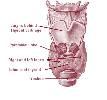
The thyroid, or thyroid gland, is an endocrine gland in vertebrates. In humans, it is a butterfly-shaped gland located in the neck below the Adam's apple. It consists of two connected lobes. The lower two thirds of the lobes are connected by a thin band of tissue called the isthmus (: isthmi). Microscopically, the functional unit of the thyroid gland is the spherical thyroid follicle, lined with follicular cells (thyrocytes), and occasional parafollicular cells that surround a lumen containing colloid. The thyroid gland secretes three hormones: the two thyroid hormones triiodothyronine (T3) and thyroxine (T4)and a peptide hormone, calcitonin. The thyroid hormones influence the metabolic rate and protein synthesis and growth and development in children. Calcitonin plays a role in calcium homeostasis. Secretion of the two thyroid hormones is regulated by thyroid-stimulating hormone (TSH), which is secreted from the anterior pituitary gland. TSH is regulated by th ... [...More Info...] [...Related Items...] OR: [Wikipedia] [Google] [Baidu] [Amazon] |
Vertebrate Trachea
The trachea (: tracheae or tracheas), also known as the windpipe, is a cartilaginous tube that connects the larynx to the bronchi of the lungs, allowing the passage of air, and so is present in almost all animals' lungs. The trachea extends from the larynx and branches into the two primary bronchi. At the top of the trachea, the cricoid cartilage attaches it to the larynx. The trachea is formed by a number of horseshoe-shaped rings, joined together vertically by overlying ligaments, and by the trachealis muscle at their ends. The epiglottis closes the opening to the larynx during swallowing. The trachea begins to form in the second month of embryo development, becoming longer and more fixed in its position over time. Its epithelium is lined with column-shaped cells that have hair-like extensions called cilia, with scattered goblet cells that produce protective mucins. The trachea can be affected by inflammation or infection, usually as a result of a viral illness affectin ... [...More Info...] [...Related Items...] OR: [Wikipedia] [Google] [Baidu] [Amazon] |
Esophagus
The esophagus (American English), oesophagus (British English), or œsophagus (Œ, archaic spelling) (American and British English spelling differences#ae and oe, see spelling difference) all ; : ((o)e)(œ)sophagi or ((o)e)(œ)sophaguses), colloquially known also as the food pipe, food tube, or gullet, is an Organ (anatomy), organ in vertebrates through which food passes, aided by Peristalsis, peristaltic contractions, from the Human pharynx, pharynx to the stomach. The esophagus is a :wiktionary:fibromuscular, fibromuscular tube, about long in adults, that travels behind the trachea and human heart, heart, passes through the Thoracic diaphragm, diaphragm, and empties into the uppermost region of the stomach. During swallowing, the epiglottis tilts backwards to prevent food from going down the larynx and lungs. The word ''esophagus'' is from Ancient Greek οἰσοφάγος (oisophágos), from οἴσω (oísō), future form of φέρω (phérō, "I carry") + ἔφαγον ( ... [...More Info...] [...Related Items...] OR: [Wikipedia] [Google] [Baidu] [Amazon] |
Sympathetic Trunk
The sympathetic trunk (sympathetic chain, gangliated cord) is a paired bundle of nerve fibers that run from the base of the skull to the coccyx. It is a major component of the sympathetic nervous system. Structure The sympathetic trunk lies just lateral to the vertebral bodies for the entire length of the vertebral column. It interacts with the anterior rami of spinal nerves by way of rami communicantes. The sympathetic trunk permits preganglionic fibers of the sympathetic nervous system to ascend to spinal levels superior to T1 and descend to spinal levels inferior to L2/3.Greenstein B., Greenstein A. (2002): Color atlas of neuroscience – Neuroanatomy and neurophysiology. Thieme, Stuttgart – New York, . The superior end of it is continued upward through the carotid canal into the skull, and forms a plexus on the internal carotid artery; the inferior part travels in front of the coccyx, where it converges with the other trunk at a structure known as the ganglion impar ... [...More Info...] [...Related Items...] OR: [Wikipedia] [Google] [Baidu] [Amazon] |
Recurrent Nerve
The recurrent laryngeal nerve (RLN), also known as nervus recurrens, is a branch of the vagus nerve (cranial nerve X) that supplies all the intrinsic muscles of the larynx, with the exception of the cricothyroid muscles. There are two recurrent laryngeal nerves, right and left. The right and left nerves are not symmetrical, with the left nerve looping under the aortic arch, and the right nerve looping under the right subclavian artery, then traveling upwards. They both travel alongside the trachea. Additionally, the nerves are among the few nerves that follow a ''recurrent'' course, moving in the opposite direction to the nerve they branch from, a fact from which they gain their name. The recurrent laryngeal nerves supply sensation to the larynx below the vocal cords, give cardiac branches to the deep cardiac plexus, and branch to the trachea, esophagus and the inferior constrictor muscles. The posterior cricoarytenoid muscles, the only muscles that can open the vocal fol ... [...More Info...] [...Related Items...] OR: [Wikipedia] [Google] [Baidu] [Amazon] |
Ansa Cervicalis
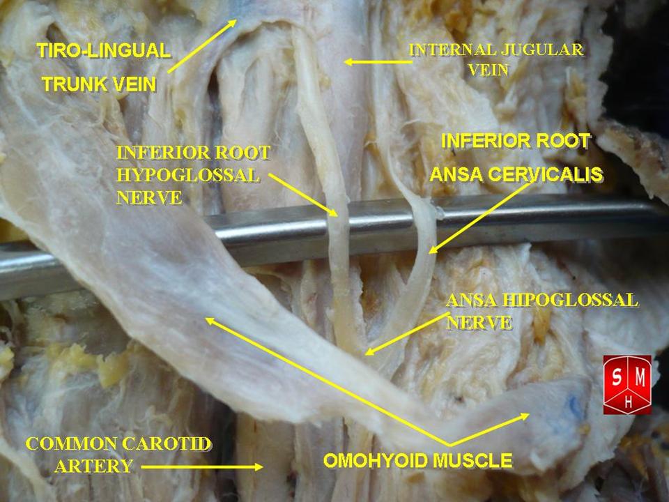
The ansa cervicalis (or ansa hypoglossi in older literature) is a loop formed by muscular branches of the cervical plexus formed by branches of cervical spinal nerves C1-C3. The ansa cervicalis has two roots - a superior root (formed by branch of C1) and an inferior root (formed by union of branches of C2 and C3) - that unite distally, forming a loop. It is situated anterior to the carotid sheath. Branches of the ansa cervicalis innervate three of the four infrahyoid muscles: the sternothyroid, sternohyoid, and omohyoid muscles (note that the thyrohyoid muscle is the one infrahyoid muscle not innervated by the ansa cervicalis - it is instead innervated by cervical spinal nerve 1 via a separate thyrohyoid branch). Its name means "handle of the neck" in Latin. Anatomy The ansa cervicalis is typically embedded within the anterior wall of the carotid sheath anterior to the internal jugular vein. Superior root The superior root of the ansa cervicalis (formerly known as d ... [...More Info...] [...Related Items...] OR: [Wikipedia] [Google] [Baidu] [Amazon] |
Human Sternum
The sternum (: sternums or sterna) or breastbone is a long flat bone located in the central part of the chest. It connects to the ribs via cartilage and forms the front of the rib cage, thus helping to protect the heart, lungs, and major blood vessels from injury. Shaped roughly like a necktie, it is one of the largest and longest flat bones of the body. Its three regions are the manubrium, the body, and the xiphoid process. The word ''sternum'' originates from Ancient Greek στέρνον (''stérnon'') 'chest'. Structure The sternum is a narrow, flat bone, forming the middle portion of the front of the chest. The top of the sternum supports the clavicles (collarbones) and its edges join with the costal cartilages of the first two pairs of ribs. The inner surface of the sternum is also the attachment of the sternopericardial ligaments. Its top is also connected to the sternocleidomastoid muscle. The sternum consists of three main parts, listed from the top: * Manubr ... [...More Info...] [...Related Items...] OR: [Wikipedia] [Google] [Baidu] [Amazon] |


