|
Molecular Neuroscience
Molecular neuroscience is a branch of neuroscience that observes concepts in molecular biology applied to the nervous systems of animals. The scope of this subject covers topics such as molecular neuroanatomy, mechanisms of molecular signaling in the nervous system, the effects of genetics and epigenetics on neuronal development, and the molecular basis for neuroplasticity and neurodegenerative diseases. As with molecular biology, molecular neuroscience is a relatively new field that is considerably dynamic. Locating neurotransmitters In molecular biology, communication between neurons typically occurs by chemical transmission across gaps between the cells called synapses. The transmitted chemicals, known as neurotransmitters, regulate a significant fraction of vital body functions. It is possible to anatomically locate neurotransmitters by labeling techniques. It is possible to chemically identify certain neurotransmitters such as catecholamines by fixing neural tissue sectio ... [...More Info...] [...Related Items...] OR: [Wikipedia] [Google] [Baidu] |
Neuroscience
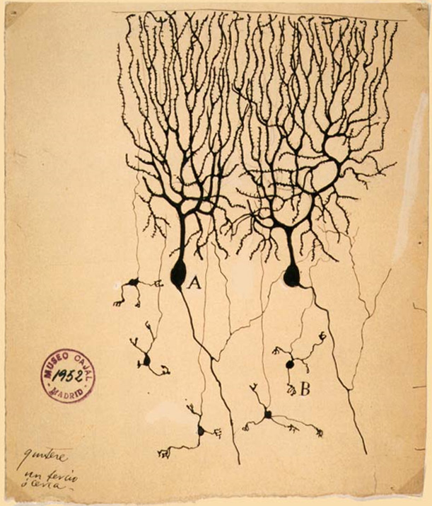
Neuroscience is the scientific study of the nervous system (the brain, spinal cord, and peripheral nervous system), its functions and disorders. It is a multidisciplinary science that combines physiology, anatomy, molecular biology, developmental biology, cytology, psychology, physics, computer science, chemistry, medicine, statistics, and Mathematical Modeling, mathematical modeling to understand the fundamental and emergent properties of neurons, glia and neural circuits. The understanding of the biological basis of learning, memory, behavior, perception, and consciousness has been described by Eric Kandel as the "epic challenge" of the Biology, biological sciences. The scope of neuroscience has broadened over time to include different approaches used to study the nervous system at different scales. The techniques used by neuroscientists have expanded enormously, from molecular biology, molecular and cell biology, cellular studies of individual neurons to neuroimaging, imaging ... [...More Info...] [...Related Items...] OR: [Wikipedia] [Google] [Baidu] |
Dopamine
Dopamine (DA, a contraction of 3,4-dihydroxyphenethylamine) is a neuromodulatory molecule that plays several important roles in cells. It is an organic compound, organic chemical of the catecholamine and phenethylamine families. Dopamine constitutes about 80% of the catecholamine content in the brain. It is an amine synthesized by removing a carboxyl group from a molecule of its precursor (chemistry), precursor chemical, L-DOPA, which is biosynthesis, synthesized in the brain and kidneys. Dopamine is also synthesized in plants and most animals. In the brain, dopamine functions as a neurotransmitter—a chemical released by neurons (nerve cells) to send signals to other nerve cells. Neurotransmitters are synthesized in specific regions of the brain, but affect many regions systemically. The brain includes several distinct dopaminergic pathway, dopamine pathways, one of which plays a major role in the motivational component of reward system, reward-motivated behavior. The anticipa ... [...More Info...] [...Related Items...] OR: [Wikipedia] [Google] [Baidu] |
Andrew Fielding Huxley
Sir Andrew Fielding Huxley (22 November 191730 May 2012) was an English physiologist and biophysicist. He was born into the prominent Huxley family. After leaving Westminster School in central London, he went to Trinity College, Cambridge on a scholarship, after which he joined Alan Lloyd Hodgkin to study nerve impulses. Their eventual discovery of the basis for propagation of nerve impulses (called an action potential) earned them the Nobel Prize in Physiology or Medicine in 1963. They made their discovery from the giant axon of the Atlantic squid. Soon after the outbreak of the Second World War, Huxley was recruited by the British Anti-Aircraft Command and later transferred to the Admiralty. After the war he resumed research at the University of Cambridge, where he developed interference microscopy that would be suitable for studying muscle fibres. In 1952, he was joined by a German physiologist Rolf Niedergerke. Together they discovered in 1954 the mechanism of muscle con ... [...More Info...] [...Related Items...] OR: [Wikipedia] [Google] [Baidu] |
Alan Lloyd Hodgkin
Sir Alan Lloyd Hodgkin (5 February 1914 – 20 December 1998) was an English physiologist and biophysicist who shared the 1963 Nobel Prize in Physiology or Medicine with Andrew Huxley and John Eccles. Early life and education Hodgkin was born in Banbury, Oxfordshire, on 5 February 1914. He was the oldest of three sons of Quakers George Hodgkin and Mary Wilson Hodgkin. His father was the son of Thomas Hodgkin and had read for the Natural Science Tripos at Cambridge where he had befriended electrophysiologist Keith Lucas. Because of poor eyesight he was unable to study medicine and eventually ended up working for a bank in Banbury. As members of the Society of Friends, George and Mary opposed the Military Service Act of 1916 and had to endure a great deal of abuse from their local community, including an attempt to throw George in one of the town canals. In 1916 George Hodgkin travelled to Armenia as part of an investigation of distress. Moved by the misery and suffering of ... [...More Info...] [...Related Items...] OR: [Wikipedia] [Google] [Baidu] |
Voltage-gated Ion Channels
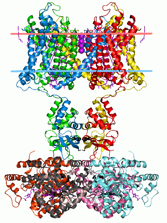
Voltage-gated ion channels are a class of transmembrane proteins that form ion channels that are activated by changes in the electrical membrane potential near the channel. The membrane potential alters the conformation of the channel proteins, regulating their opening and closing. Cell membranes are generally impermeable to ions, thus they must diffuse through the membrane through transmembrane protein channels. They have a crucial role in excitable cells such as neuronal and muscle tissues, allowing a rapid and co-ordinated depolarization in response to triggering voltage change. Found along the axon and at the synapse, voltage-gated ion channels directionally propagate electrical signals. Voltage-gated ion-channels are usually ion-specific, and channels specific to sodium (Na+), potassium (K+), calcium (Ca2+), and chloride (Cl−) ions have been identified. The opening and closing of the channels are triggered by changing ion concentration, and hence charge gradient, between t ... [...More Info...] [...Related Items...] OR: [Wikipedia] [Google] [Baidu] |
Chemiluminescence
Chemiluminescence (also chemoluminescence) is the emission of light (luminescence) as the result of a chemical reaction. There may also be limited emission of heat. Given reactants A and B, with an excited intermediate ◊, : + -> lozenge -> roducts+ light For example, if is luminol and is hydrogen peroxide in the presence of a suitable catalyst we have: :\underset + \underset -> 3-APAlozenge-> + light where: * 3-APA is 3-aminophthalate * 3-APA ''◊is the vibronic excited state fluorescing as it decays to a lower energy level. General description The decay of this excited state ''◊to a lower energy level causes light emission. In theory, one photon of light should be given off for each molecule of reactant. This is equivalent to the Avogadro number of photons per mole of reactant. In actual practice, non-enzymatic reactions seldom exceed 1% QC, quantum efficiency. In a chemical reaction, reactants collide to form a transition state, the enthalpic maximum in a r ... [...More Info...] [...Related Items...] OR: [Wikipedia] [Google] [Baidu] |
Fluorophore
A fluorophore (or fluorochrome, similarly to a chromophore) is a fluorescent chemical compound that can re-emit light upon light excitation. Fluorophores typically contain several combined aromatic groups, or planar or cyclic molecules with several π bonds. Fluorophores are sometimes used alone, as a tracer in fluids, as a dye for staining of certain structures, as a substrate of enzymes, or as a probe or indicator (when its fluorescence is affected by environmental aspects such as polarity or ions). More generally they are covalently bonded to a macromolecule, serving as a marker (or dye, or tag, or reporter) for affine or bioactive reagents (antibodies, peptides, nucleic acids). Fluorophores are notably used to stain tissues, cells, or materials in a variety of analytical methods, i.e., fluorescent imaging and spectroscopy. Fluorescein, via its amine-reactive isothiocyanate derivative fluorescein isothiocyanate (FITC), has been one of the most popular fluorophores. Fro ... [...More Info...] [...Related Items...] OR: [Wikipedia] [Google] [Baidu] |
Precipitate
In an aqueous solution, precipitation is the process of transforming a dissolved substance into an insoluble solid from a super-saturated solution. The solid formed is called the precipitate. In case of an inorganic chemical reaction leading to precipitation, the chemical reagent causing the solid to form is called the ''precipitant''. The clear liquid remaining above the precipitated or the centrifuged solid phase is also called the 'supernate' or 'supernatant'. The notion of precipitation can also be extended to other domains of chemistry (organic chemistry and biochemistry) and even be applied to the solid phases (''e.g.'', metallurgy and alloys) when solid impurities segregate from a solid phase. Supersaturation The precipitation of a compound may occur when its concentration exceeds its solubility. This can be due to temperature changes, solvent evaporation, or by mixing solvents. Precipitation occurs more rapidly from a strongly supersaturated solution. The formati ... [...More Info...] [...Related Items...] OR: [Wikipedia] [Google] [Baidu] |
ELISA
The enzyme-linked immunosorbent assay (ELISA) (, ) is a commonly used analytical biochemistry assay, first described by Eva Engvall and Peter Perlmann in 1971. The assay uses a solid-phase type of enzyme immunoassay (EIA) to detect the presence of a ligand (commonly a protein) in a liquid sample using antibodies directed against the protein to be measured. ELISA has been used as a diagnostic tool in medicine, plant pathology, and biotechnology, as well as a quality control check in various industries. In the most simple form of an ELISA, antigens from the sample to be tested are attached to a surface. Then, a matching antibody is applied over the surface so it can bind the antigen. This antibody is linked to an enzyme and then any unbound antibodies are removed. In the final step, a substance containing the enzyme's substrate is added. If there was binding, the subsequent reaction produces a detectable signal, most commonly a color change. Performing an ELISA involves at least ... [...More Info...] [...Related Items...] OR: [Wikipedia] [Google] [Baidu] |
Autoradiography
An autoradiograph is an image on an X-ray film or nuclear emulsion produced by the pattern of decay emissions (e.g., beta particles or gamma rays) from a distribution of a radioactive substance. Alternatively, the autoradiograph is also available as a digital image (digital autoradiography), due to the recent development of scintillation gas detectors or rare earth phosphorimaging systems. The film or emulsion is apposed to the labeled tissue section to obtain the autoradiograph (also called an autoradiogram). The ''auto-'' prefix indicates that the radioactive substance is within the sample, as distinguished from the case of historadiography or microradiography, in which the sample is marked using an external source. Some autoradiographs can be examined microscopically for localization of silver grains (such as on the interiors or exteriors of cells or organelles) in which the process is termed micro-autoradiography. For example, micro-autoradiography was used to examine whether ... [...More Info...] [...Related Items...] OR: [Wikipedia] [Google] [Baidu] |
Primary And Secondary Antibodies
Primary and secondary antibodies are two groups of antibodies that are classified based on whether they bind to ''antigens or proteins'' directly or target another (primary) antibody that, in turn, is bound to an ''antigen or protein''. Primary A primary antibody can be very useful for the detection of biomarkers for diseases such as cancer, diabetes, Parkinson’s and Alzheimer’s disease and they are used for the study of absorption, distribution, metabolism, and excretion (ADME) and multi-drug resistance (MDR) of therapeutic agents. Secondary Secondary antibodies provide signal detection and amplification along with extending the utility of an antibody through conjugation to proteins. Secondary antibodies are especially efficient in immunolabeling. Secondary antibodies bind to primary antibodies, which are directly bound to the target antigen(s). In immunolabeling, the primary antibody's Fab domain binds to an antigen and exposes its Fc domain to secondary antibody. Then, ... [...More Info...] [...Related Items...] OR: [Wikipedia] [Google] [Baidu] |


