|
Metanephric Dysplastic Hematoma Of The Sacral Region
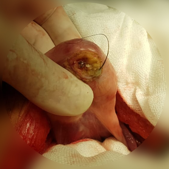
Metanephric dysplastic hematoma of the sacral region (MDHSR) has been described by Cozzutto and Lazzaroni-Fossati in 1980, by Posalaki et al. in 1981 and by Cozzutto et al. in 1982. Three additional cases were seen by Finegold. Case studies The case reported by Cozzutto and Lazzaroni-Fossati involved a premature male newborn with bilateral renal dysplasia and a sacrococcygeal mass featuring a histological picture of renal dysplasia. The case reported by Cozzutto et al. and those studied by Finegold featured changes of renal dysplasia including immature tubules surrounded by a collarette of cellular mesenchyme, glomeruloid figures, tubules and nests of cartilage in a background of adipose tissue and fibrous tissue where muscle fibres, nerve bundles and calcospherites were also seen. The two cases reported by Posalaki et al. showed blastema, glomeruli and tubuli. In several cases the mass was removed during reparative surgery for meningocele or myelomeningocele. Alston et al. ... [...More Info...] [...Related Items...] OR: [Wikipedia] [Google] [Baidu] |
Hematoma
A hematoma, also spelled haematoma, or blood suffusion is a localized bleeding outside of blood vessels, due to either disease or trauma including injury or surgery and may involve blood continuing to seep from broken capillary, capillaries. A hematoma is benign and is initially in liquid form spread among the tissues including in sacs between tissues where it may coagulate and solidify before blood is reabsorbed into blood vessels. An ecchymosis is a hematoma of the skin larger than 10 mm. They may occur among and or within many areas such as skin and other organs, connective tissues, bone, joints and muscle. A collection of blood (or even a hemorrhage) may be aggravated by anticoagulant medication (blood thinner). Blood seepage and collection of blood may occur if heparin is given via an Intramuscular injection, intramuscular route; to avoid this, heparin must be given intravenously or subcutaneous injection, subcutaneously. Signs and symptoms Some hematomas are visible ... [...More Info...] [...Related Items...] OR: [Wikipedia] [Google] [Baidu] |
Meningocele
Spina bifida (Latin for 'split spine'; SB) is a birth defect in which there is incomplete closing of the spine and the membranes around the spinal cord during early development in pregnancy. There are three main types: spina bifida occulta, meningocele and myelomeningocele. Meningocele and myelomeningocele may be grouped as spina bifida cystica. The most common location is the lower back, but in rare cases it may be in the middle back or neck. Occulta has no or only mild signs, which may include a hairy patch, dimple, dark spot or swelling on the back at the site of the gap in the spine. Meningocele typically causes mild problems, with a sac of fluid present at the gap in the spine. Myelomeningocele, also known as open spina bifida, is the most severe form. Problems associated with this form include poor ability to walk, impaired bladder or bowel control, accumulation of fluid in the brain (hydrocephalus), a tethered spinal cord and latex allergy. Learning problems are relat ... [...More Info...] [...Related Items...] OR: [Wikipedia] [Google] [Baidu] |
Nephroblastoma
Wilms' tumor or Wilms tumor, also known as nephroblastoma, is a cancer of the kidneys that typically occurs in children, rarely in adults.; and occurs most commonly as a renal tumor in child patients. It is named after Max Wilms, the German surgeon (1867–1918) who first described it. Approximately 650 cases are diagnosed in the U.S. annually. The majority of cases occur in children with no associated genetic syndromes; however, a minority of children with Wilms' tumor have a congenital abnormality. It is highly responsive to treatment, with about 90 percent of children being cured. Signs and symptoms Typical signs and symptoms of Wilms' tumor include the following: * a painless, palpable abdominal mass * loss of appetite * abdominal pain * fever * nausea and vomiting * blood in the urine (in about 20% of cases) * high blood pressure in some cases (especially if synchronous or metachronous bilateral kidney involvement) * Rarely as varicoceleErginel B, Vural S, Akın M, K ... [...More Info...] [...Related Items...] OR: [Wikipedia] [Google] [Baidu] |
Teratoma
A teratoma is a tumor made up of several different types of tissue, such as hair, muscle, teeth, or bone. Teratomata typically form in the ovary, testicle, or coccyx. Symptoms Symptoms may be minimal if the tumor is small. A testicular teratoma may present as a painless lump. Complications may include ovarian torsion, testicular torsion, or hydrops fetalis. They are a type of germ cell tumor (a tumor that begins in the cells that give rise to sperm or eggs). They are divided into two types: mature and immature. Mature teratomas include dermoid cysts and are generally benign. Immature teratomas may be cancerous. Most ovarian teratomas are mature. In adults, testicular teratomas are generally cancerous. Definitive diagnosis is based on a tissue biopsy. Treatment of coccyx, testicular, and ovarian teratomas is generally by surgery. Testicular and immature ovarian teratomas are also frequently treated with chemotherapy. Teratomas occur in the coccyx in about one in 30,000 n ... [...More Info...] [...Related Items...] OR: [Wikipedia] [Google] [Baidu] |
Neuroglial
Glia, also called glial cells (gliocytes) or neuroglia, are non-neuronal cells in the central nervous system (brain and spinal cord) and the peripheral nervous system that do not produce electrical impulses. They maintain homeostasis, form myelin in the peripheral nervous system, and provide support and protection for neurons. In the central nervous system, glial cells include oligodendrocytes, astrocytes, ependymal cells, and microglia, and in the peripheral nervous system they include Schwann cells and satellite cells. Function They have four main functions: *to surround neurons and hold them in place *to supply nutrients and oxygen to neurons *to insulate one neuron from another *to destroy pathogens and remove dead neurons. They also play a role in neurotransmission and synaptic connections, and in physiological processes such as breathing. While glia were thought to outnumber neurons by a ratio of 10:1, recent studies using newer methods and reappraisal of historical quanti ... [...More Info...] [...Related Items...] OR: [Wikipedia] [Google] [Baidu] |
Spinal Dysraphism
Neural tube defects (NTDs) are a group of birth defects in which an opening in the spine or cranium remains from early in human development. In the third week of pregnancy called gastrulation, specialized cells on the dorsal side of the embryo begin to change shape and form the neural tube. When the neural tube does not close completely, an NTD develops. Specific types include: spina bifida which affects the spine, anencephaly which results in little to no brain, encephalocele which affects the skull, and iniencephaly which results in severe neck problems. NTDs are one of the most common birth defects, affecting over 300,000 births each year worldwide. For example, spina bifida affects approximately 1,500 births annually in the United States, or about 3.5 in every 10,000 (0.035% of US births), which has decreased from around 5 per 10,000 (0.05% of US births) since folate fortification of grain products was started. The number of deaths in the US each year due to neural tube d ... [...More Info...] [...Related Items...] OR: [Wikipedia] [Google] [Baidu] |
Stroma (tissue)
Stroma () is the part of a tissue or organ with a structural or connective role. It is made up of all the parts without specific functions of the organ - for example, connective tissue, blood vessels, ducts, etc. The other part, the parenchyma, consists of the cells that perform the function of the tissue or organ. There are multiple ways of classifying tissues: one classification scheme is based on tissue functions and another analyzes their cellular components. Stromal tissue falls into the "functional" class that contributes to the body's support and movement. The cells which make up stroma tissues serve as a matrix in which the other cells are embedded. Stroma is made of various types of stromal cells. Examples of stroma include: * stroma of iris * stroma of cornea * stroma of ovary * stroma of thyroid gland * stroma of thymus * stroma of bone marrow * lymph node stromal cell * multipotent stromal cell (mesenchymal stem cell) Structure Stromal connective tissues are fou ... [...More Info...] [...Related Items...] OR: [Wikipedia] [Google] [Baidu] |
Lipoma
A lipoma is a benign tumor made of fat tissue. They are generally soft to the touch, movable, and painless. They usually occur just under the skin, but occasionally may be deeper. Most are less than in size. Common locations include upper back, shoulders, and abdomen. It is possible to have a number of lipomas. The cause is generally unclear. Risk factors include family history, obesity, and lack of exercise. Diagnosis is typically based on a physical exam. Occasionally medical imaging or tissue biopsy is used to confirm the diagnosis. Treatment is typically by observation or surgical removal. Rarely, the condition may recur following removal, but this can generally be managed with repeat surgery. They are not generally associated with a future risk of cancer. Lipomas have a prevalence of roughly 2 out of every 100 people. Lipomas typically occur in adults between 40 and 60 years of age. Males are more often affected than females. They are the most common noncancerous soft-t ... [...More Info...] [...Related Items...] OR: [Wikipedia] [Google] [Baidu] |
Myelomeningocele
Spina bifida (Latin for 'split spine'; SB) is a birth defect in which there is incomplete closing of the spine and the membranes around the spinal cord during early development in pregnancy. There are three main types: spina bifida occulta, meningocele and myelomeningocele. Meningocele and myelomeningocele may be grouped as spina bifida cystica. The most common location is the lower back, but in rare cases it may be in the middle back or neck. Occulta has no or only mild signs, which may include a hairy patch, dimple, dark spot or swelling on the back at the site of the gap in the spine. Meningocele typically causes mild problems, with a sac of fluid present at the gap in the spine. Myelomeningocele, also known as open spina bifida, is the most severe form. Problems associated with this form include poor ability to walk, impaired bladder or bowel control, accumulation of fluid in the brain (hydrocephalus), a tethered spinal cord and latex allergy. Learning problems are relat ... [...More Info...] [...Related Items...] OR: [Wikipedia] [Google] [Baidu] |
Glomeruli
''Glomerulus'' () is a common term used in anatomy to describe globular structures of entwined vessels, fibers, or neurons. ''Glomerulus'' is the diminutive of the Latin ''glomus'', meaning "ball of yarn". ''Glomerulus'' may refer to: * the filtering unit of the kidney; see Glomerulus (kidney). * a structure in the olfactory bulb; see Glomerulus (olfaction). * the contact between specific cells in the cerebellum; see Glomerulus (cerebellum) The cerebellar glomerulus is a small, intertwined mass of nerve fiber terminals in the granular layer of the cerebellar cortex. It consists of post-synaptic granule cell dendrites and pre-synaptic Golgi cell axon terminals surrounding the pre- .... See also * Glomerulation, a hemorrhage of the bladder {{SIA ... [...More Info...] [...Related Items...] OR: [Wikipedia] [Google] [Baidu] |
Sacral Region
The sacrum (plural: ''sacra'' or ''sacrums''), in human anatomy, is a large, triangular bone at the base of the spine that forms by the fusing of the sacral vertebrae (S1S5) between ages 18 and 30. The sacrum situates at the upper, back part of the pelvic cavity, between the two wings of the pelvis. It forms joints with four other bones. The two projections at the sides of the sacrum are called the alae (wings), and articulate with the ilium at the L-shaped sacroiliac joints. The upper part of the sacrum connects with the last lumbar vertebra (L5), and its lower part with the coccyx (tailbone) via the sacral and coccygeal cornua. The sacrum has three different surfaces which are shaped to accommodate surrounding pelvic structures. Overall it is concave (curved upon itself). The base of the sacrum, the broadest and uppermost part, is tilted forward as the sacral promontory internally. The central part is curved outward toward the posterior, allowing greater room for the pel ... [...More Info...] [...Related Items...] OR: [Wikipedia] [Google] [Baidu] |
Blastema
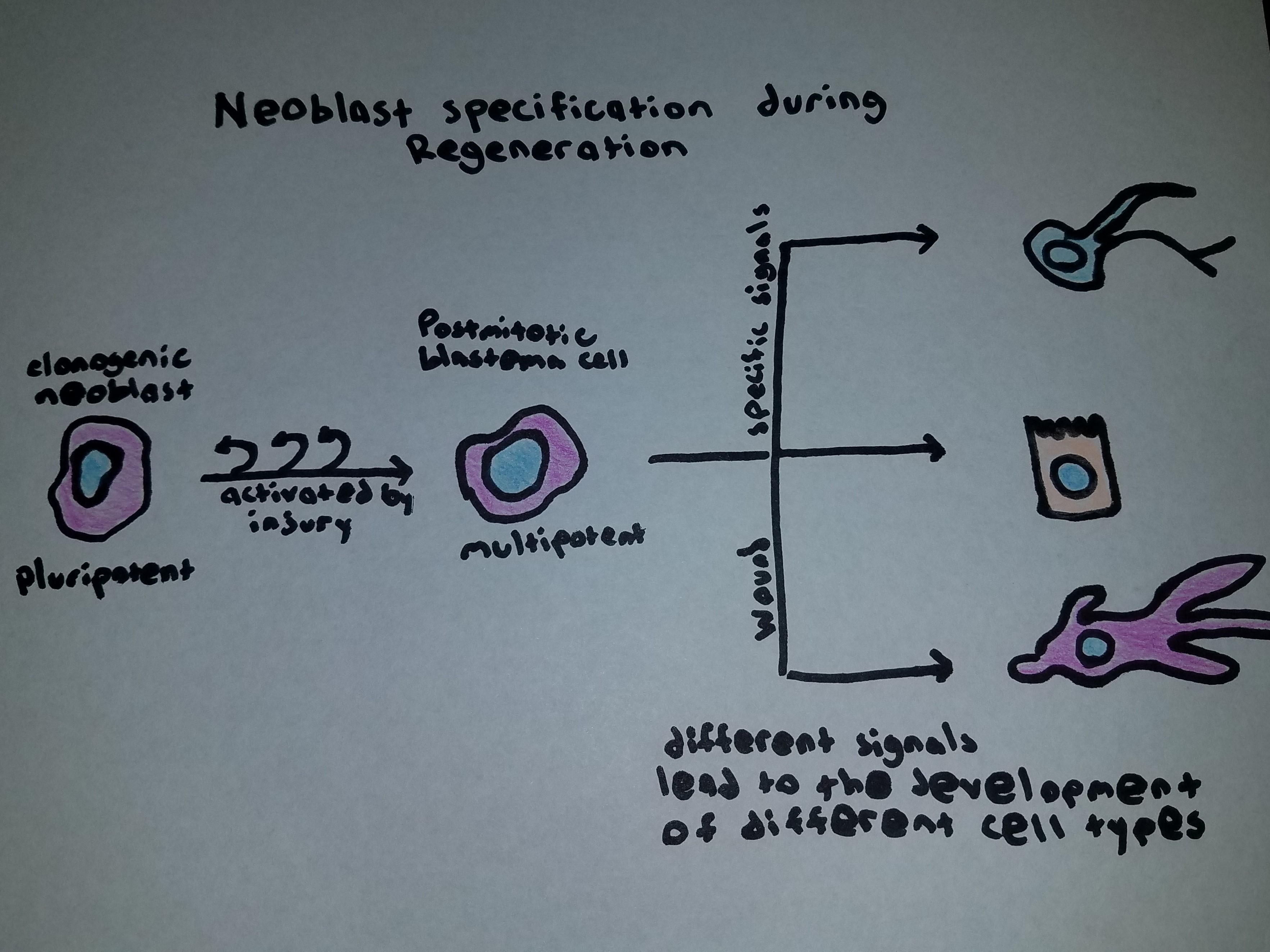
A blastema (Greek ''βλάστημα'', "offspring") is a mass of cells capable of growth and regeneration into organs or body parts. The changing definition of the word "blastema" has been reviewed by Holland (2021). A broad survey of how blastema has been used over time brings to light a somewhat involved history. The word entered the biomedical vocabulary in 1799 to designate a sinister acellular slime that was the starting point for the growth of cancers, themselves, at the time, thought to be acellular, as reviewed by Hajdu (2011, Cancer 118: 1155-1168). Then, during the early nineteenth century, the definition broadened to include growth zones (still considered acellular) in healthy, normally developing plant and animal embryos. Contemporaneously, cancer specialists dropped the term from their vocabulary, perhaps because they felt a term connoting a state of health and normalcy was not appropriate for describing a pathological condition. During the middle decades of the nine ... [...More Info...] [...Related Items...] OR: [Wikipedia] [Google] [Baidu] |


