|
Megalopapilla
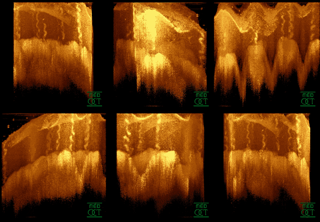
Megalopapilla is a non-progressive human eye condition in which the optic nerve head (optic disc) has an enlarged diameter, exceeding 2.1 mm with no other morphological abnormalities. Clinical features In megalopapilla the optic disc diameter exceeds 2.1 mm (or surface area more than 2.5 mm2) with an increased cup-to-disc ratio. Although the optic disc is looks abnormal, the disc colour, sharpness of disc margin, rim volume, configuration of blood vessels and intraocular pressure will be normal. Visual field is normal except for an enlarged Blind spot (vision), blind spot. Visual acuity may be normal, or occasionally reduced. Types There are two types of megalopapilla. Type 1, which is the most common, is bilateral with a normal configuration of the Optic cup (anatomical), optic cup. Type 2 is unilateral with a superiorly displaced cup that eliminates the adjacent neuro-retinal rim. Diagnosis and differential diagnosis Large disc size can be seen in Fundus (eye), fundus ... [...More Info...] [...Related Items...] OR: [Wikipedia] [Google] [Baidu] |
Optic Nerve Head
The optic disc or optic nerve head is the point of exit for ganglion cell axons leaving the eye. Because there are no rods or cones overlying the optic disc, it corresponds to a small blind spot in each eye. The ganglion cell axons form the optic nerve after they leave the eye. The optic disc represents the beginning of the optic nerve and is the point where the axons of retinal ganglion cells come together. The optic disc is also the entry point for the major blood vessels that supply the retina. The optic disc in a normal human eye carries 1–1.2 million afferent nerve fibers from the eye towards the brain. Structure The optic disc is placed 3 to 4 mm to the nasal side of the fovea. It is a vertical oval, with average dimensions of 1.76mm horizontally by 1.92mm vertically. There is a central depression, of variable size, called the optic cup. This depression can be a variety of shapes from a shallow indentation to a bean pot—this shape can be significant for dia ... [...More Info...] [...Related Items...] OR: [Wikipedia] [Google] [Baidu] |
Optic Disc
The optic disc or optic nerve head is the point of exit for ganglion cell axons leaving the eye. Because there are no rods or cones overlying the optic disc, it corresponds to a small blind spot in each eye. The ganglion cell axons form the optic nerve after they leave the eye. The optic disc represents the beginning of the optic nerve and is the point where the axons of retinal ganglion cells come together. The optic disc is also the entry point for the major blood vessels that supply the retina. The optic disc in a normal human eye carries 1–1.2 million afferent nerve fibers from the eye towards the brain. Structure The optic disc is placed 3 to 4 mm to the nasal side of the fovea. It is a vertical oval, with average dimensions of 1.76mm horizontally by 1.92mm vertically. There is a central depression, of variable size, called the optic cup. This depression can be a variety of shapes from a shallow indentation to a bean pot—this shape can be significant for diagn ... [...More Info...] [...Related Items...] OR: [Wikipedia] [Google] [Baidu] |
Ophthalmology
Ophthalmology ( ) is a surgical subspecialty within medicine that deals with the diagnosis and treatment of eye disorders. An ophthalmologist is a physician who undergoes subspecialty training in medical and surgical eye care. Following a medical degree, a doctor specialising in ophthalmology must pursue additional postgraduate residency training specific to that field. This may include a one-year integrated internship that involves more general medical training in other fields such as internal medicine or general surgery. Following residency, additional specialty training (or fellowship) may be sought in a particular aspect of eye pathology. Ophthalmologists prescribe medications to treat eye diseases, implement laser therapy, and perform surgery when needed. Ophthalmologists provide both primary and specialty eye care - medical and surgical. Most ophthalmologists participate in academic research on eye diseases at some point in their training and many include research as part ... [...More Info...] [...Related Items...] OR: [Wikipedia] [Google] [Baidu] |
Visual Acuity
Visual acuity (VA) commonly refers to the clarity of vision, but technically rates an examinee's ability to recognize small details with precision. Visual acuity is dependent on optical and neural factors, i.e. (1) the sharpness of the retinal image within the eye, (2) the health and functioning of the retina, and (3) the sensitivity of the interpretative faculty of the brain. The most commonly referred visual acuity is the far acuity (e.g. 6/6 or 20/20 acuity), which describes the examinee's ability to recognize small details at a far distance, and is relevant to people with myopia; however, for people with hyperopia, the near acuity is used instead to describe the examinee's ability to recognize small details at a near distance. A common cause of low visual acuity is refractive error (ametropia), errors in how the light is refracted in the eyeball, and errors in how the retinal image is interpreted by the brain. The latter is the primary cause for low vision in people with a ... [...More Info...] [...Related Items...] OR: [Wikipedia] [Google] [Baidu] |
Coloboma Of Optic Nerve
Coloboma of optic nerve is a rare defect of the optic nerve that causes moderate to severe visual field defects. Coloboma of the optic nerve is a congenital anomaly of the optic disc in which there is a defect of the inferior aspect of the optic nerve. The issue stems from incomplete closure of the embryonic fissure while in utero. A varying amount of glial tissue typically fills the defect, manifests as a white mass. Signs and symptoms Vision in the affected eye is impaired, the degree of which depends on the size of the defect, and typically affects the visual field more than visual acuity. Additionally, there is an increased risk of serous retinal detachment, manifesting in 1/3 of patients. If retinal detachment does occur, it is usually not correctable and all sight is lost in the affected area of the eye, which may or may not involve the macula. Diagnosis The first noticeable signs of the syndrome usually do not appear until after the first twelve months of the child’s ... [...More Info...] [...Related Items...] OR: [Wikipedia] [Google] [Baidu] |
Optical Coherence Tomography
Optical coherence tomography (OCT) is an imaging technique that uses low-coherence light to capture micrometer-resolution, two- and three-dimensional images from within optical scattering media (e.g., biological tissue). It is used for medical imaging and industrial nondestructive testing (NDT). Optical coherence tomography is based on low-coherence interferometry, typically employing near-infrared light. The use of relatively long wavelength light allows it to penetrate into the scattering medium. Confocal microscopy, another optical technique, typically penetrates less deeply into the sample but with higher resolution. Depending on the properties of the light source ( superluminescent diodes, ultrashort pulsed lasers, and supercontinuum lasers have been employed), optical coherence tomography has achieved sub-micrometer resolution (with very wide-spectrum sources emitting over a ~100 nm wavelength range). Optical coherence tomography is one of a class of optical tom ... [...More Info...] [...Related Items...] OR: [Wikipedia] [Google] [Baidu] |
Ophthalmoscopy
Ophthalmoscopy, also called funduscopy, is a test that allows a health professional to see inside the fundus of the eye and other structures using an ophthalmoscope (or funduscope). It is done as part of an eye examination and may be done as part of a routine physical examination. It is crucial in determining the health of the retina, optic disc, and vitreous humor. The pupil is a hole through which the eye's interior will be viewed. Opening the pupil wider (dilating it) is a simple and effective way to better see the structures behind it. Therefore, dilation of the pupil ( mydriasis) is often accomplished with medicated eye drops before funduscopy. However, although dilated fundus examination is ideal, undilated examination is more convenient and is also helpful (albeit not as comprehensive), and it is the most common type in primary care. An alternative or complement to ophthalmoscopy is to perform a fundus photography, where the image can be analysed later by a professional. ... [...More Info...] [...Related Items...] OR: [Wikipedia] [Google] [Baidu] |
Fundus (eye)
The fundus of the eye is the interior surface of the eye opposite the lens and includes the retina, optic disc, macula, fovea, and posterior pole.Cassin, B. and Solomon, S. ''Dictionary of Eye Terminology''. Gainesville, Florida: Triad Publishing Company, 1990. The fundus can be examined by ophthalmoscopy and/or fundus photography. Variation The color of the fundus varies both between and within species. In one study of primates the retina is blue, green, yellow, orange, and red; only the human fundus (from a lightly pigmented blond person) is red. The major differences noted among the "higher" primate species were size and regularity of the border of macular area, size and shape of the optic disc, apparent 'texturing' of retina, and pigmentation of retina. Clinical significance Medical signs that can be detected from observation of eye fundus (generally by funduscopy) include hemorrhages, exudates, cotton wool spots, blood vessel abnormalities (tortuosity, pulsation and n ... [...More Info...] [...Related Items...] OR: [Wikipedia] [Google] [Baidu] |
Optic Cup (anatomical)
The optic cup is the white, cup-like area in the center of the optic disc. The ratio of the size of the optic cup to the optic disc (cup-to-disc ratio, or C/D) is one measure used in the diagnosis of glaucoma. Different C/Ds can be measured horizontally or vertically in the same patient. C/Ds vary widely in healthy individuals. However, larger vertical C/Ds, or C/Ds which are very different between the eyes, may raise suspicion of glaucoma. A C/D which enlarges vertically over months or years suggests glaucoma. Cup-to-disc ratio The ''cup-to-disc ratio'' (often notated ''CDR'') is a measurement used in ophthalmology and optometry to assess the progression of glaucoma. The optic disc is the anatomical location of the eye's "blind spot", the area where the optic nerve leave and blood vessels enter the retina. The optic disc can be flat or it can have a certain amount of normal cupping. But glaucoma, which is in most cases associated with an increase in intraocular pressure Int ... [...More Info...] [...Related Items...] OR: [Wikipedia] [Google] [Baidu] |
Visual Field
The visual field is the "spatial array of visual sensations available to observation in introspectionist psychological experiments". Or simply, visual field can be defined as the entire area that can be seen when an eye is fixed straight at a point. The equivalent concept for optical instruments and image sensors is the field of view (FOV). In optometry, ophthalmology, and neurology, a visual field test is used to determine whether the visual field is affected by diseases that cause local scotoma or a more extensive loss of vision or a reduction in sensitivity (increase in threshold). Normal limits The normal (monocular) human visual field extends to approximately 60 degrees nasally (toward the nose, or inward) from the vertical meridian in each eye, to 107 degrees temporally (away from the nose, or outwards) from the vertical meridian, and approximately 70 degrees above and 80 below the horizontal meridian. The binocular visual field is the superimposition of the two monocular ... [...More Info...] [...Related Items...] OR: [Wikipedia] [Google] [Baidu] |
Blind Spot (vision)
A blind spot, scotoma, is an obscuration of the visual field. A particular blind spot known as the ''physiological blind spot'', "blind point", or ''punctum caecum'' in medical literature, is the place in the visual field that corresponds to the lack of light-detecting photoreceptor cells on the optic disc of the retina where the optic nerve passes through the optic disc.Gregory, R., & Cavanagh, P. (2011)"The Blind Spot" Scholarpedia. Retrieved on 2011-05-21. Because there are no cells to detect light on the optic disc, the corresponding part of the field of vision is invisible. Processes in the brain interpolate the blind spot based on surrounding detail and information from the other eye, so it is not normally perceived. Although all vertebrates have this blind spot, cephalopod eyes, which are only superficially similar, do not. In them, the optic nerve approaches the receptors from behind, so it does not create a break in the retina. The first documented observation of the ... [...More Info...] [...Related Items...] OR: [Wikipedia] [Google] [Baidu] |
Normal Tension Glaucoma
Normal tension glaucoma (NTG) is an eye disease, a neuropathy of the optic nerve, that shows all the characteristics of primary open angle glaucoma except one: the elevated intraocular pressure (IOP) - the classic hallmark of glaucoma - is missing. Normal tension glaucoma is in many cases closely associated with general issues of blood circulation and of organ perfusion like arterial hypotension, metabolic syndrome, and Flammer syndrome. Clinical relevance Over many years, glaucoma has been defined by an intraocular pressure of more than 20 mm Hg. Incompatible with this (now obsolete) definition of glaucoma was the ever larger number of cases that have been reported in medical literature in the 1980s and 1990s who had the typical signs of glaucomatous damage, like optic nerve head excavation and thinning of the retinal nerve fiber layer, while these patients had an IOP that would generally have been regarded as "normal". It is now widely estimated that a larger percentage of patien ... [...More Info...] [...Related Items...] OR: [Wikipedia] [Google] [Baidu] |


