|
Massive Periretinal Proliferation
Proliferative vitreoretinopathy (PVR) is a disease that develops as a complication of rhegmatogenous retinal detachment. PVR occurs in about 8–10% of patients undergoing primary retinal detachment surgery and prevents the successful surgical repair of rhegmatogenous retinal detachment. PVR can be treated with surgery to reattach the detached retina but the visual outcome of the surgery is very poor. A number of studies have explored various possible adjunctive agents for the prevention and treatment of PVR, such as methotrexate, although none have yet been licensed for clinical use. PVR was originally referred to as massive vitreous retraction and then as massive periretinal proliferation. The name Proliferative vitreo retinopathy was provided in 1989 by the Silicone Oil Study group. The name is derived from ''proliferation'' (by the retinal pigment epithelial and glial cells) and ''vitreo retinopathy'' to include the tissues which are affected, namely the vitreous humor (or si ... [...More Info...] [...Related Items...] OR: [Wikipedia] [Google] [Baidu] |
Retinal Detachment
Retinal detachment is a disorder of the eye in which the retina peels away from its underlying layer of support tissue. Initial detachment may be localized, but without rapid treatment the entire retina may detach, leading to vision loss and blindness. It is a surgical emergency. The retina is a thin layer of light-sensitive tissue on the back wall of the eye. The optical system of the eye focuses light on the retina much like light is focused on the film in a camera. The retina translates that focused image into neural impulses and sends them to the brain via the optic nerve. Occasionally, posterior vitreous detachment, injury or trauma to the eye or head may cause a small tear in the retina. The tear allows vitreous fluid to seep through it under the retina, and peel it away like a bubble in wallpaper. Diagnosis Symptoms As the retina is responsible for vision, persons experiencing a retinal detachment have vision loss. This can be painful or painless. Imaging Ultraso ... [...More Info...] [...Related Items...] OR: [Wikipedia] [Google] [Baidu] |
Phospholipase A2
The enzyme phospholipase A2 (EC 3.1.1.4, PLA2, systematic name phosphatidylcholine 2-acylhydrolase) catalyse the cleavage of fatty acids in position 2 of phospholipids, hydrolyzing the bond between the second fatty acid “tail” and the glycerol molecule: :phosphatidylcholine + H2O = 1-acylglycerophosphocholine + a carboxylate This particular phospholipase specifically recognizes the ''sn''2 acyl bond of phospholipids and catalytically hydrolyzes the bond, releasing arachidonic acid and lysophosphatidic acid. Upon downstream modification by cyclooxygenases or lipoxygenases, arachidonic acid is modified into active compounds called eicosanoids. Eicosanoids include prostaglandins and leukotrienes, which are categorized as anti-inflammatory and inflammatory mediators. PLA2 enzymes are commonly found in mammalian tissues as well as arachnid, insect, and snake venom. Venom from bees is largely composed of melittin, which is a stimulant of PLA2. Due to the increased presenc ... [...More Info...] [...Related Items...] OR: [Wikipedia] [Google] [Baidu] |
Transforming Growth Factor
Transforming growth factor (, or TGF) is used to describe two classes of polypeptide growth factors, TGFα and TGFβ. The name "Transforming Growth Factor" is somewhat arbitrary, since the two classes of TGFs are not structurally or genetically related to one another, and they act through different receptor mechanisms. Furthermore, they do not always induce cellular transformation, and are not the only growth factors that induce cellular transformation. Types * TGFα is upregulated in some human cancers. It is produced in macrophages, brain cells, and keratinocytes, and induces epithelial development. It belongs to the EGF family. * TGFβ exists in three known subtypes in humans, TGFβ1, TGFβ2, and TGFβ3. These are upregulated in Marfan's syndrome and some human cancers, and play crucial roles in tissue regeneration, cell differentiation, embryonic development, and regulation of the immune system. Isoforms of transforming growth factor-beta (TGF-β1) are also thought to be inv ... [...More Info...] [...Related Items...] OR: [Wikipedia] [Google] [Baidu] |
Tumor Necrosis Factor Alpha
Tumor necrosis factor (TNF, cachexin, or cachectin; formerly known as tumor necrosis factor alpha or TNF-α) is an adipokine and a cytokine. TNF is a member of the TNF superfamily, which consists of various transmembrane proteins with a homologous TNF domain. As an adipokine, TNF promotes insulin resistance, and is associated with obesity-induced type 2 diabetes. As a cytokine, TNF is used by the immune system for cell signaling. If macrophages (certain white blood cells) detect an infection, they release TNF to alert other immune system cells as part of an inflammatory response. TNF signaling occurs through two receptors: TNFR1 and TNFR2. TNFR1 is constituitively expressed on most cell types, whereas TNFR2 is restricted primarily to endothelial, epithelial, and subsets of immune cells. TNFR1 signaling tends to be pro-inflammatory and apoptotic, whereas TNFR2 signaling is anti-inflammatory and promotes cell proliferation. Suppression of TNFR1 signaling has been important for ... [...More Info...] [...Related Items...] OR: [Wikipedia] [Google] [Baidu] |
Investigative Ophthalmology & Visual Science
''Investigative Ophthalmology & Visual Science'' (''IOVS'') is an online journal published by the Association for Research in Vision and Ophthalmology (ARVO). History The journal was established as an official publication of the Association for Research in Ophthalmology (later renamed the Association for Research in Vision and Ophthalmology). The first issue of ''Investigative Ophthalmology'' was published in January 1962, with Bernard Becker, MD, as the Executive Editor. The title was changed to ''Investigative Ophthalmology & Visual Science'' in 1977.Colson, KS. Brief history of ''Investigative Ophthalmology & Visual Science''. In Chader GJ, Frank RN, Kaufman PL, Beebe DC, eds. ''The Best of'' Investigative Ophthalmology & Visual Science'': The First 50 Years 1962-2012''. Rockville, MD: ARVO; 2012:1-5. Abstracts from the ARVO Annual Meeting have been published as an issue of ''IOVS'' since 1977. Also in 1977, ''IOVS'' was accepted for inclusion in ''Index Medicus'' (and later ... [...More Info...] [...Related Items...] OR: [Wikipedia] [Google] [Baidu] |
Fibrocyte
A fibrocyte is an inactive mesenchymal cell, that is, a cell showing minimal cytoplasm, limited amounts of rough endoplasmic reticulum and lacks biochemical evidence of protein synthesis. The term ''fibrocyte'' contrasts with the term ''fibroblast''. Fibroblasts are activated connective tissue cells characterized by synthesis of proteins of the fibrous matrix, particularly the collagens. When tissue is injured, the predominant mesenchymal cells, the fibroblast, have been believed to be derived from the fibrocyte or possibly from smooth muscle cells lining vessels and glands. Commonly, fibroblasts express smooth muscle actin, a form of actin first found in smooth muscle cells and not found in resting fibrocytes. Fibroblasts expressing this form of actin are usually called "myo-fibroblasts." Recently, the term "fibrocyte" has also been applied to a bloodborne cell able to leave the blood, enter tissue and become a fibroblast. As part of the more general topic of stem cell biolog ... [...More Info...] [...Related Items...] OR: [Wikipedia] [Google] [Baidu] |
Macrophage
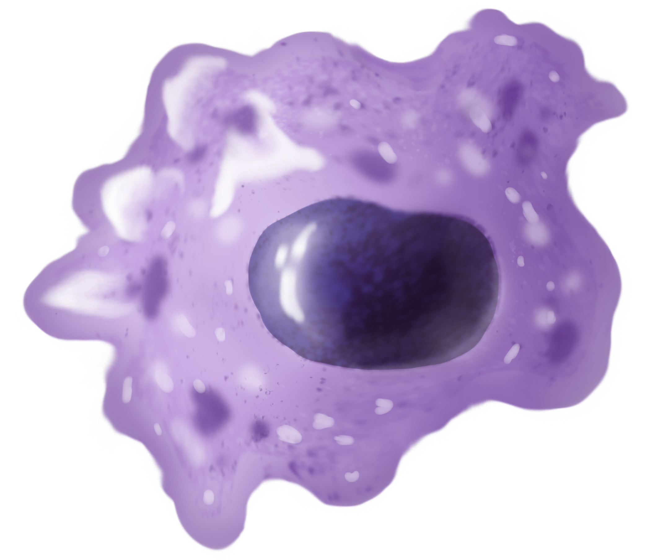
Macrophages (abbreviated as M φ, MΦ or MP) ( el, large eaters, from Greek ''μακρός'' (') = large, ''φαγεῖν'' (') = to eat) are a type of white blood cell of the immune system that engulfs and digests pathogens, such as cancer cells, microbes, cellular debris, and foreign substances, which do not have proteins that are specific to healthy body cells on their surface. The process is called phagocytosis, which acts to defend the host against infection and injury. These large phagocytes are found in essentially all tissues, where they patrol for potential pathogens by amoeboid movement. They take various forms (with various names) throughout the body (e.g., histiocytes, Kupffer cells, alveolar macrophages, microglia, and others), but all are part of the mononuclear phagocyte system. Besides phagocytosis, they play a critical role in nonspecific defense (innate immunity) and also help initiate specific defense mechanisms (adaptive immunity) by recruiting other immune ... [...More Info...] [...Related Items...] OR: [Wikipedia] [Google] [Baidu] |
Epiretinal Membrane
Epiretinal membrane or macular pucker is a disease of the eye in response to changes in the vitreous humor or more rarely, diabetes. Sometimes, as a result of immune system response to protect the retina, cells converge in the macular area as the vitreous ages and pulls away in posterior vitreous detachment (PVD). PVD can create minor damage to the retina, stimulating exudate, inflammation, and leucocyte response. These cells can form a transparent layer gradually and, like all scar tissue, tighten to create tension on the retina which may bulge and pucker, or even cause swelling or macular edema. Often this results in distortions of vision that are clearly visible as bowing and blurring when looking at lines on chart paper (or an Amsler grid) within the macular area, or central 1.0 degree of visual arc. Usually it occurs in one eye first, and may cause binocular diplopia or double vision if the image from one eye is too different from the image of the other eye. The distortions ... [...More Info...] [...Related Items...] OR: [Wikipedia] [Google] [Baidu] |
Retina Society Terminology Committee
The retina (from la, rete "net") is the innermost, light-sensitive layer of tissue of the eye of most vertebrates and some molluscs. The optics of the eye create a focused two-dimensional image of the visual world on the retina, which then processes that image within the retina and sends nerve impulses along the optic nerve to the visual cortex to create visual perception. The retina serves a function which is in many ways analogous to that of the film or image sensor in a camera. The neural retina consists of several layers of neurons interconnected by synapses and is supported by an outer layer of pigmented epithelial cells. The primary light-sensing cells in the retina are the photoreceptor cells, which are of two types: rods and cones. Rods function mainly in dim light and provide monochromatic vision. Cones function in well-lit conditions and are responsible for the perception of colour through the use of a range of opsins, as well as high-acuity vision used for tasks s ... [...More Info...] [...Related Items...] OR: [Wikipedia] [Google] [Baidu] |

