|
Mammary-type Myofibroblastoma
Mammary-type myofibroblastoma (MFB), also named mammary and extramammary myofibroblastoma, was first termed myofibrolastoma of the breast, or, more simply, either mammary myofibroblastoma (MMFB) or just myofibroblastoma. The change in this terminology occurred because the initial 1987 study and many subsequent studies found this tumor only in breast tissue. However, a 2001 study followed by numerous reports found tumors with the microscopic histopathology and other key features of mammary MFB in a wide range of organs and tissues. Further complicating the issue, early studies on MFB classified it as one of various types of spindle cell tumors that, except for MFB, were ill-defined. These other tumors, which have often been named interchangeably in different reports, are: myelofibroblastoma, benign spindle cell tumor, fibroma, spindle cell lipoma, myogenic stromal tumor, and solitary stromal tumor. Finally, studies suggest that spindle cell lipoma and cellular angiofibroma are variant ... [...More Info...] [...Related Items...] OR: [Wikipedia] [Google] [Baidu] |
Micrograph
A micrograph or photomicrograph is a photograph or digital image taken through a microscope or similar device to show a magnified image of an object. This is opposed to a macrograph or photomacrograph, an image which is also taken on a microscope but is only slightly magnified, usually less than 10 times. Micrography is the practice or art of using microscopes to make photographs. A micrograph contains extensive details of microstructure. A wealth of information can be obtained from a simple micrograph like behavior of the material under different conditions, the phases found in the system, failure analysis, grain size estimation, elemental analysis and so on. Micrographs are widely used in all fields of microscopy. Types Photomicrograph A light micrograph or photomicrograph is a micrograph prepared using an optical microscope, a process referred to as ''photomicroscopy''. At a basic level, photomicroscopy may be performed simply by connecting a camera to a microscope, th ... [...More Info...] [...Related Items...] OR: [Wikipedia] [Google] [Baidu] |
Breast Cancer Screening
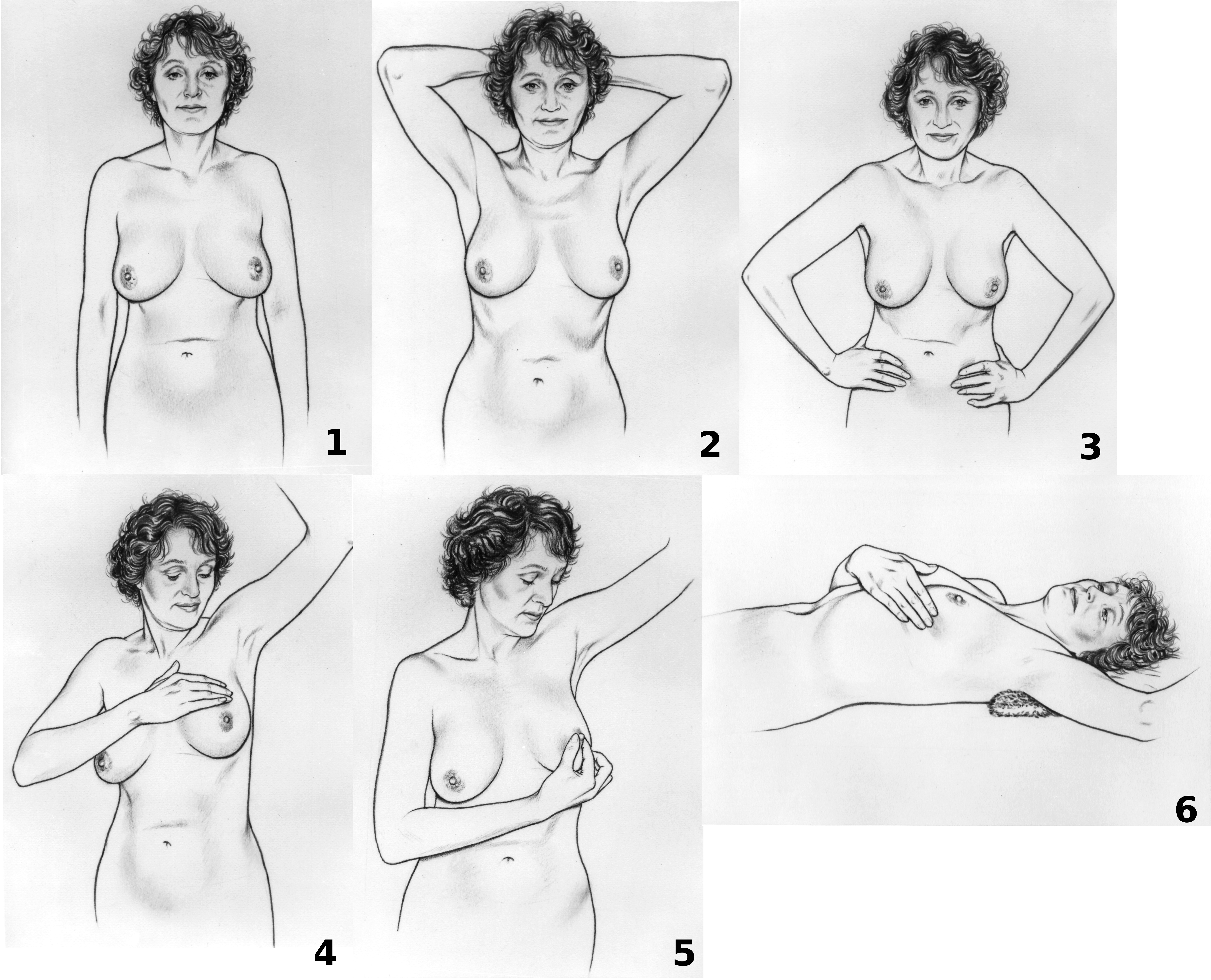
Breast cancer screening is the medical screening of asymptomatic, apparently healthy women for breast cancer in an attempt to achieve an earlier diagnosis. The assumption is that early detection will improve outcomes. A number of screening tests have been employed, including clinical and self breast exams, mammography, genetic screening, ultrasound, and magnetic resonance imaging. A clinical or self breast exam involves feeling the breast for breast lump, lumps or other abnormalities. Medical evidence, however, does not support its use in women with a typical risk for breast cancer. Universal screening with mammography is controversial as it may not reduce all-cause mortality and may cause harms through unnecessary treatments and medical procedures. Many national organizations recommend it for most older women. The United States Preventive Services Task Force recommends screening mammography in women at normal risk for breast cancer, every two years between the ages of 50 and 74 ... [...More Info...] [...Related Items...] OR: [Wikipedia] [Google] [Baidu] |
Medical Imaging
Medical imaging is the technique and process of imaging the interior of a body for clinical analysis and medical intervention, as well as visual representation of the function of some organs or tissues (physiology). Medical imaging seeks to reveal internal structures hidden by the skin and bones, as well as to diagnose and treat disease. Medical imaging also establishes a database of normal anatomy and physiology to make it possible to identify abnormalities. Although imaging of removed organs and tissues can be performed for medical reasons, such procedures are usually considered part of pathology instead of medical imaging. Measurement and recording techniques that are not primarily designed to produce images, such as electroencephalography (EEG), magnetoencephalography (MEG), electrocardiography (ECG), and others, represent other technologies that produce data susceptible to representation as a parameter graph versus time or maps that contain data about the measurement loca ... [...More Info...] [...Related Items...] OR: [Wikipedia] [Google] [Baidu] |
Vulva
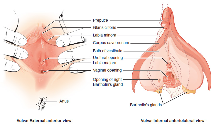
The vulva (plural: vulvas or vulvae; derived from Latin for wrapper or covering) consists of the external sex organ, female sex organs. The vulva includes the mons pubis (or mons veneris), labia majora, labia minora, clitoris, bulb of vestibule, vestibular bulbs, vulval vestibule, urinary meatus, the Vagina#Vaginal opening and hymen, vaginal opening, hymen, and Bartholin's gland, Bartholin's and Skene's gland, Skene's vestibular glands. The urinary meatus is also included as it opens into the vulval vestibule. Other features of the vulva include the pudendal cleft, sebaceous glands, the urogenital triangle (anterior part of the perineum), and pubic hair. The vulva includes the entrance to the vagina, which leads to the uterus, and provides a double layer of protection for this by the folds of the outer and inner labia. Pelvic floor muscles support the structures of the vulva. Other muscles of the urogenital triangle also give support. Blood supply to the vulva comes from the t ... [...More Info...] [...Related Items...] OR: [Wikipedia] [Google] [Baidu] |
Scrotum
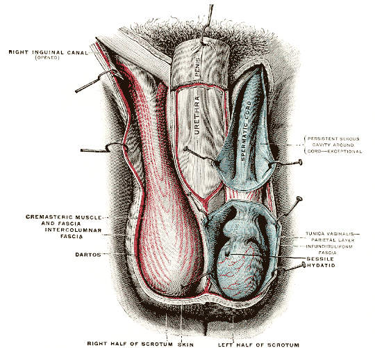
The scrotum or scrotal sac is an anatomical male reproductive structure located at the base of the penis that consists of a suspended dual-chambered sac of skin and smooth muscle. It is present in most terrestrial male mammals. The scrotum contains the external spermatic fascia, testes, epididymis, and ductus deferens. It is a distention of the perineum and carries some abdominal tissues into its cavity including the testicular artery, testicular vein, and pampiniform plexus. The perineal raphe is a small, vertical, slightly raised ridge of scrotal skin under which is found the scrotal septum. It appears as a thin longitudinal line that runs front to back over the entire scrotum. In humans and some other mammals the scrotum becomes covered with pubic hair at puberty. The scrotum will usually tighten during penile erection and when exposed to cold temperatures. One testis is typically lower than the other to avoid compression in the event of an impact. The scrotum is biologicall ... [...More Info...] [...Related Items...] OR: [Wikipedia] [Google] [Baidu] |
Retroperitoneal Space
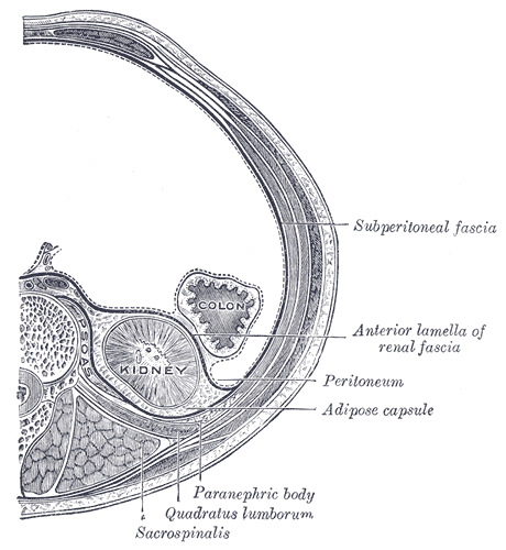
The retroperitoneal space (retroperitoneum) is the anatomical space (sometimes a potential space) behind (''retro'') the peritoneum. It has no specific delineating anatomical structures. Organs are retroperitoneal if they have peritoneum on their anterior side only. Structures that are not suspended by mesentery in the abdominal cavity and that lie between the parietal peritoneum and abdominal wall are classified as retroperitoneal. This is different from organs that are not retroperitoneal, which have peritoneum on their posterior side and are suspended by mesentery in the abdominal cavity. The retroperitoneum can be further subdivided into the following: *Perirenal (or perinephric) space *Anterior pararenal (or paranephric) space *Posterior pararenal (or paranephric) space Retroperitoneal structures Structures that lie behind the peritoneum are termed "retroperitoneal". Organs that were once suspended within the abdominal cavity by mesentery but migrated posterior to the p ... [...More Info...] [...Related Items...] OR: [Wikipedia] [Google] [Baidu] |
Abdominal Cavity
The abdominal cavity is a large body cavity in humans and many other animals that contains many organs. It is a part of the abdominopelvic cavity. It is located below the thoracic cavity, and above the pelvic cavity. Its dome-shaped roof is the thoracic diaphragm, a thin sheet of muscle under the lungs, and its floor is the pelvic inlet, opening into the pelvis. Structure Organs Organs of the abdominal cavity include the stomach, liver, gallbladder, spleen, pancreas, small intestine, kidneys, large intestine, and adrenal glands. Peritoneum The abdominal cavity is lined with a protective membrane termed the peritoneum. The inside wall is covered by the parietal peritoneum. The kidneys are located behind the peritoneum, in the retroperitoneum, outside the abdominal cavity. The viscera are also covered by visceral peritoneum. Between the visceral and parietal peritoneum is the peritoneal cavity, which is a potential space. It contains a serous fluid called peritoneal fluid tha ... [...More Info...] [...Related Items...] OR: [Wikipedia] [Google] [Baidu] |
Groin
In human anatomy, the groin (the adjective is ''inguinal'', as in inguinal canal) is the junctional area (also known as the inguinal region) between the abdomen and the thigh on either side of the pubic bone. This is also known as the medial compartment of the thigh that consists of the adductor muscles of the hip or the groin muscles. A pulled groin muscle usually refers to a painful injury sustained by straining the hip adductor muscles. These hip adductor muscles that make up the groin consist of the adductor brevis, adductor longus, adductor magnus, gracilis, and pectineus. These groin muscles adduct the thigh (bring the femur and knee closer to the midline). The groin is innervated by the obturator nerve, with two exceptions: the pectineus muscle is innervated by the femoral nerve, and the hamstring portion of adductor magnus is innervated by the tibial nerve. In the groin, underneath the skin, there are three to five deep inguinal lymph nodes that play a role in t ... [...More Info...] [...Related Items...] OR: [Wikipedia] [Google] [Baidu] |
Axillary Lymph Nodes
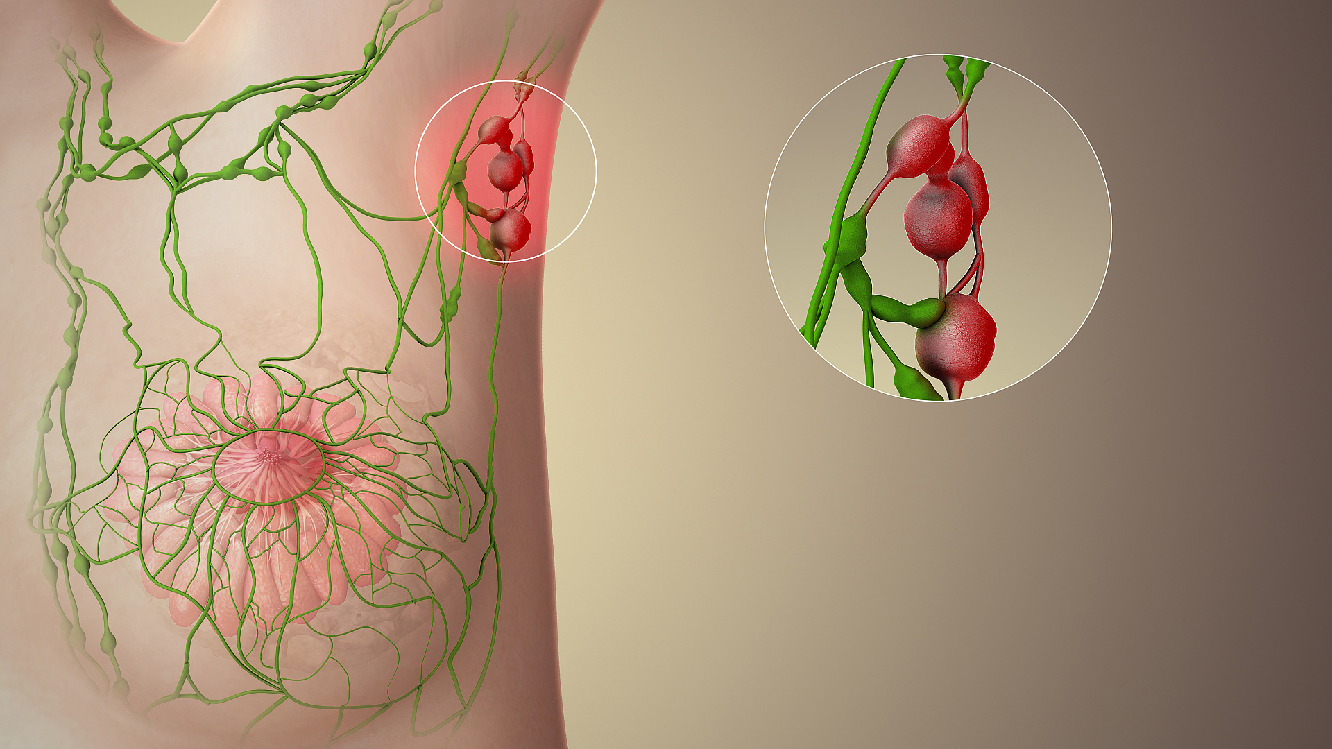
The axillary lymph nodes or armpit lymph nodes are lymph nodes in the human armpit. Between 20 and 49 in number, they drain lymph vessels from the lateral quadrants of the breast, the superficial lymph vessels from thin walls of the chest and the abdomen above the level of the navel, and the vessels from the upper limb. They are divided in several groups according to their location in the armpit. These lymph nodes are clinically significant in breast cancer, and metastases from the breast to the axillary lymph nodes are considered in the staging of the disease. Structure The axillary lymph nodes are arranged in six groups: # Anterior (pectoral) group: Lying along the lower border of the pectoralis minor behind the pectoralis major, these nodes receive lymph vessels from the lateral quadrants of the breast and superficial vessels from the anterolateral abdominal wall above the level of the umbilicus. # Posterior (subscapular) group: Lying in front of the subscapularis muscle, th ... [...More Info...] [...Related Items...] OR: [Wikipedia] [Google] [Baidu] |
Axilla
The axilla (also, armpit, underarm or oxter) is the area on the human body directly under the shoulder joint. It includes the axillary space, an anatomical space within the shoulder girdle between the arm and the thoracic cage, bounded superiorly by the imaginary plane between the superior borders of the first rib, clavicle and scapula (above which are considered part of the neck), medially by the serratus anterior muscle and thoracolumbar fascia, anteriorly by the pectoral muscles and posteriorly by the subscapularis, teres major and latissimus dorsi muscle. The soft skin covering the lateral axilla contains many hair and sweat glands. In humans, the formation of body odor happens mostly in the axilla. These odorant substances have been suggested by some to serve as pheromones, which play a role related to mate selection, although this is a controversial topic within the scientific community. The underarms seem more important than the pubic area for emitting body odor, whi ... [...More Info...] [...Related Items...] OR: [Wikipedia] [Google] [Baidu] |
Mammary Ridge
The mammary ridge or mammary crest is a primordium specific for the development of mammary glands. Development The mammary ridge is primordial for the mammary glands on the chest in humans, and is associated with mammary gland and breast development. In human embryogenesis the mammary ridge usually appears as a narrow, microscopic ectodermal thickening during the first seven weeks of pregnancy and grows caudally as a narrow, linear ridge. In many mammals, these glands first appear as elevated ridges along the milk lines, which then separate into individual buds located in regions lateral to the ventral midline. The location of these buds varies according to species: they are located in the thoracic region in primates, in the inguinal area in ungulates, and along the entire length of the trunk in rodents and pigs. A mammary ridge, or crest, usually stops growing at eight weeks and its length is regressed starting at the caudal end and extending cranially, so that what remains ... [...More Info...] [...Related Items...] OR: [Wikipedia] [Google] [Baidu] |
Gynecomastia
Gynecomastia (also spelled gynaecomastia) is the abnormal non-cancerous enlargement of one or both breasts in males due to the growth of breast tissue as a result of a hormone imbalance between estrogens and androgens. Updated by Brent Wisse (10 November 2018) Gynecomastia can cause significant psychological distress or unease. Gynecomastia can be normal in newborn babies due to exposure to estrogen from the mother, in adolescents going through puberty, in older men over age 50, and/or in obese men. Most occurrences of gynecomastia do not require diagnostic tests. Gynecomastia may be caused by abnormal hormone changes, any condition that leads to an increase in the ratio of estrogens/androgens such as liver disease, kidney failure, thyroid disease and some non-breast tumors. Alcohol and some drugs can also cause breast enlargement. Other causes may include Klinefelter syndrome, metabolic dysfunction, or a natural decline in testosterone production. This may occur even if the l ... [...More Info...] [...Related Items...] OR: [Wikipedia] [Google] [Baidu] |

