|
Muscular System Of The Horse
Types of muscle There are 3 types of muscle, all found within the equine: * Skeletal muscle: Contraction of these muscles leads to the muscle pulling a tendon, which in turn pulls a bone. Moving a bone results in either flexing or extending a joint. Skeletal muscles are usually arranged in pairs so that they oppose each other (they are "antagonists" irror image, with one flexing the joint (a flexor muscle) and the other extending it (extensor muscle). Therefore, one muscle of the pair must be relaxed in order for the other muscle in the pair to contract and bend the joint properly. A muscle or muscles and its/their tendon(s) that operate together to cause flexion or extension of a joint are referred to respectively as a flexor unit and an extensor unit. * Smooth: muscle which makes up automatic systems (digestive system, for example) * Cardiac: involuntary muscle the keeps the heart pumping and able to carry round oxygen to the muscles. Build of skeletal muscle Skeletal muscle is ... [...More Info...] [...Related Items...] OR: [Wikipedia] [Google] [Baidu] |
Skeletal Muscle
Skeletal muscles (commonly referred to as muscles) are organs of the vertebrate muscular system and typically are attached by tendons to bones of a skeleton. The muscle cells of skeletal muscles are much longer than in the other types of muscle tissue, and are often known as muscle fibers. The muscle tissue of a skeletal muscle is striated – having a striped appearance due to the arrangement of the sarcomeres. Skeletal muscles are voluntary muscles under the control of the somatic nervous system. The other types of muscle are cardiac muscle which is also striated and smooth muscle which is non-striated; both of these types of muscle tissue are classified as involuntary, or, under the control of the autonomic nervous system. A skeletal muscle contains multiple muscle fascicle, fascicles – bundles of muscle fibers. Each individual fiber, and each muscle is surrounded by a type of connective tissue layer of fascia. Muscle fibers are formed from the cell fusion, fusion of ... [...More Info...] [...Related Items...] OR: [Wikipedia] [Google] [Baidu] |
Hyoid
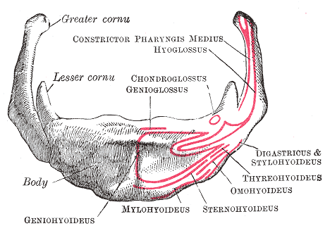
The hyoid bone (lingual bone or tongue-bone) () is a horseshoe-shaped bone situated in the anterior midline of the neck between the chin and the thyroid cartilage. At rest, it lies between the base of the mandible and the third cervical vertebra. Unlike other bones, the hyoid is only distantly articulated to other bones by muscles or ligaments. It is the only bone in the human body that is not connected to any other bones nearby. The hyoid is anchored by muscles from the anterior, posterior and inferior directions, and aids in tongue movement and swallowing. The hyoid bone provides attachment to the muscles of the floor of the mouth and the tongue above, the larynx below, and the epiglottis and pharynx behind. Its name is derived . Structure The hyoid bone is classed as an irregular bone and consists of a central part called the body, and two pairs of horns, the greater and lesser horns. Body The body of the hyoid bone is the central part of the hyoid bone. *At the front, ... [...More Info...] [...Related Items...] OR: [Wikipedia] [Google] [Baidu] |
Hyperkalemic Periodic Paralysis (equine)
Hyperkalemic periodic paralysis (HYPP, HyperKPP) is a genetic disorder that occurs in horses. It is also known as Impressive syndrome, after an index case in a horse named Impressive. It is an inherited autosomal dominant disorder that affects sodium channels in muscle cells and the ability to regulate potassium levels in the blood. It is characterized by muscle hyperexcitability or weakness which, exacerbated by potassium, heat or cold, can lead to uncontrolled shaking followed by paralysis. The mutation which causes this disorder is dominant on SCN4A with linkage to the sodium channel expressed in muscle. The mutation causes single amino acid changes in parts of the channel which are important for inactivation. In the presence of high potassium levels, including those induced by diet, sodium channels fail to inactivate properly. Equine hyperkalemic periodic paralysis occurs in 1 in 50 Quarter Horses and can be traced to a single ancestor, a stallion named Impressive. Sym ... [...More Info...] [...Related Items...] OR: [Wikipedia] [Google] [Baidu] |
Hypocalcemia
Hypocalcemia is a medical condition characterized by low calcium levels in the blood serum. The normal range of blood calcium is typically between 2.1–2.6 mmol/L (8.8–10.7 mg/dL, 4.3–5.2 mEq/L) while levels less than 2.1 mmol/L are defined as hypocalcemic. Mildly low levels that develop slowly often have no symptoms. Otherwise symptoms may include numbness, muscle spasms, seizures, confusion, or cardiac arrest. The most common cause for hypocalcemia is iatrogenic hypoparathyroidism. Other causes include other forms of hypoparathyroidism, vitamin D deficiency, kidney failure, pancreatitis, calcium channel blocker overdose, rhabdomyolysis, tumor lysis syndrome, and medications such as bisphosphonates or denosumab. Diagnosis should generally be confirmed with a corrected calcium or ionized calcium level. Specific changes may be seen on an electrocardiogram (ECG). Initial treatment for severe disease is with intravenous calcium chloride and possibly magnesium ... [...More Info...] [...Related Items...] OR: [Wikipedia] [Google] [Baidu] |
Glycogen Branching Enzyme Deficiency
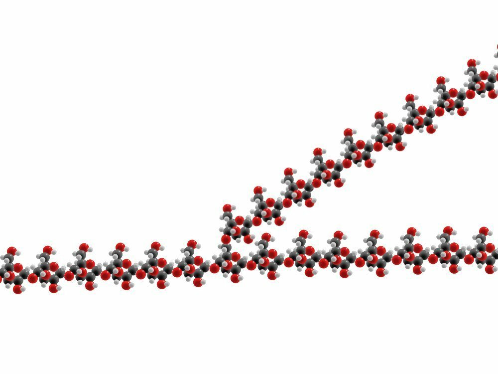
Glycogen-branching enzyme deficiency (GBED) is an inheritable glycogen storage disease affecting American Quarter Horses and American Paint Horses. It leads to abortion, stillbirths, or early death of affected animals. The human form of the disease is known as glycogen storage disease type IV. Pathophysiology Glycogen is a molecular polymer of glucose used to store energy. It is important for providing energy for skeletal and cardiac muscle contraction, and for maintaining glucose hemostasis in the blood. Molecules of glucose are linked into linear chains by α-1,4-glycosidic bonds. Additionally, branches of glucose are formed off of the chain via α-1,6-glycosidic bonds. 2 molecules of glucose are joined into an α-1,4-glycosidic bonds by an enzyme known as glycogen synthase. This bond may be broken by amylase when the body wishes to break down glycogen into glucose for energy. Glycogen branching enzyme is responsible for the required α-1,6-glycosidic bonds needed to start a bran ... [...More Info...] [...Related Items...] OR: [Wikipedia] [Google] [Baidu] |
Equine Polysaccharide Storage Myopathy
Equine polysaccharide storage myopathy (EPSM, PSSM, EPSSM) is a hereditary glycogen storage disease of horses that causes exertional rhabdomyolysis. It is currently known to affect the following breeds American Quarter Horses, American Paint Horses, Warmbloods, Cobs, Dales Ponies, Thoroughbreds, Arabians, New Forest ponies, and a large number of Heavy horse breeds. While incurable, PSSM can be managed with appropriate diet and exercise. There are currently 2 subtypes, known as Type 1 PSSM and Type 2 PSSM. Pathophysiology of glycogen storage disorders and sub-typing of PSSM Glycogen is a molecular polymer of glucose (a polysaccharide) used to store energy, and is important for maintaining glucose homeostasis in the blood, as well as for providing energy for skeletal muscle and cardiac muscle contraction. Molecules of glucose are linked into linear chains by α-1,4-glycosidic bonds. Additionally, branches of glucose are formed off of the chain by α-1,6-glycosidic bonds. 2 molecules ... [...More Info...] [...Related Items...] OR: [Wikipedia] [Google] [Baidu] |
Equine Exertional Rhabdomyolysis
Equine exertional rhabdomyolysis (ER) is a syndrome that affects the skeletal muscles within a horse. This syndrome causes the muscle to break down which is generally associated with exercise and diet regime. Depending on the severity, there are various types of ER, including sporadic (i.e., Tying-Up, Monday Morning Sickness/Disease, Azoturia) and chronic (i.e., Polysaccharide Storage Myopathy (PSSM) and Recurrent Exertional Rhabdomyolysis (RER)). Types of equine exertional rhabdomyolysis (ER) Equine Exertional Rhabdomyolysis (ER) is a general term used to define both sporadic - (infrequent) and chronic - (repeated) manifestations for the condition. The severity of the condition defines what type of ER a horse has. Sporadic equine exertional rhabdomyolysis (ER) The types of equine ER that are considered sporadic include tying-up, also commonly referred to as Monday morning sickness and/or Monday morning disease, and azoturia also known as black water disease, set fast, and/or p ... [...More Info...] [...Related Items...] OR: [Wikipedia] [Google] [Baidu] |
Compartment Syndrome
Compartment syndrome is a condition in which increased pressure within one of the body's anatomical compartments results in insufficient blood supply to tissue within that space. There are two main types: acute and chronic. Compartments of the leg or arm are most commonly involved. Symptoms of acute compartment syndrome (ACS) can include severe pain, poor pulses, decreased ability to move, numbness, or a pale color of the affected limb. It is most commonly due to physical trauma such as a bone fracture (up to 75% of cases) or crush injury, but it can also be caused by acute exertion during sport. It can also occur after blood flow returns following a period of poor blood flow. Diagnosis is generally based upon a person's symptoms and may be supported by measurement of intracompartmental pressure before, during, and after activity. Normal compartment pressure should be within 12-18 mmHg; anything greater than that is considered abnormal and would need treatment. Treatment i ... [...More Info...] [...Related Items...] OR: [Wikipedia] [Google] [Baidu] |
Bowed Tendon
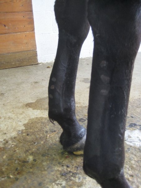
Tendinitis/tendonitis is inflammation of a tendon, often involving torn collagen fibers. A bowed tendon is a horseman's term for a tendon after a horse has sustained an injury that causes swelling in one or more tendons creating a "bowed" appearance. Description of tendinitis in horses Tendinitis usually involves disruption of the tendon fibers. It is most commonly seen in the superficial digital flexor tendon (SDFT) in a front leg—the tendon that runs down the back of the leg, closest to the surface. Tendinitis creating a "bow" is uncommon in the deep digital flexor tendon (DDFT) of a front leg, but is not uncommon in the pastern and foot regions. Tendinitis of the SDFT or DDFT in the hind leg is less common. When the SDFT is damaged, there is a thickening of the tendon, giving it a bowed appearance when the leg is viewed from the side. Bows usually occur in the middle of the tendon region, although they may also be seen in the upper third, right below the knee or hock (high ... [...More Info...] [...Related Items...] OR: [Wikipedia] [Google] [Baidu] |
Trochanter
A trochanter is a tubercle of the femur near its joint with the hip bone. In humans and most mammals, the trochanters serve as important muscle attachment sites. Humans are known to have three trochanters, though the anatomic "normal" includes only the greater and lesser trochanters. (The third trochanter is not present in all specimens.) Etymology "Trokhos" (Greek) = "wheel", with reference to the spherical femoral head which was first named "trokhanter". Later usage came to include the femoral neck. Structure In human anatomy, the trochanter is a part of the femur. It can refer to: * Greater trochanter * Lesser trochanter * Third trochanter, which is occasionally present Other animals * Fourth trochanter, of archosaur leg bones * Trochanter (arthropod leg), a segment of the arthropod leg See also * Intertrochanteric crest * Intertrochanteric line References External links * * {{Bones of lower extremity Trochanter A trochanter is a tubercle of the femur n ... [...More Info...] [...Related Items...] OR: [Wikipedia] [Google] [Baidu] |
Aponeurosis
An aponeurosis (; plural: ''aponeuroses'') is a type or a variant of the deep fascia, in the form of a sheet of pearly-white fibrous tissue that attaches sheet-like muscles needing a wide area of attachment. Their primary function is to join muscles and the body parts they act upon, whether bone or other muscles. They have a shiny, whitish-silvery color, are histologically similar to tendons, and are very sparingly supplied with blood vessels and nerves. When dissected, aponeuroses are papery and peel off by sections. The primary regions with thick aponeuroses are in the ventral abdominal region, the dorsal lumbar region, the ventriculus in birds, and the palmar (palms) and plantar (soles) regions. Anatomy Anterior abdominal aponeuroses The anterior abdominal aponeuroses are located just superficial to the rectus abdominis muscle. It has for its borders the external oblique, pectoralis muscles, and the latissimus dorsi. Posterior lumbar aponeuroses The posterior lumbar apo ... [...More Info...] [...Related Items...] OR: [Wikipedia] [Google] [Baidu] |
Abduction (kinesiology)
Motion, the process of movement, is described using specific anatomical terms. Motion includes movement of organs, joints, limbs, and specific sections of the body. The terminology used describes this motion according to its direction relative to the anatomical position of the body parts involved. Anatomists and others use a unified set of terms to describe most of the movements, although other, more specialized terms are necessary for describing unique movements such as those of the hands, feet, and eyes. In general, motion is classified according to the anatomical plane it occurs in. ''Flexion'' and ''extension'' are examples of ''angular'' motions, in which two axes of a joint are brought closer together or moved further apart. ''Rotational'' motion may occur at other joints, for example the shoulder, and are described as ''internal'' or ''external''. Other terms, such as ''elevation'' and ''depression'', describe movement above or below the horizontal plane. Many anatomic ... [...More Info...] [...Related Items...] OR: [Wikipedia] [Google] [Baidu] |


