|
Mucoepidermoid Carcinoma
Mucoepidermoid carcinoma (MEC) is the most common type of minor salivary gland malignancy in adults. Mucoepidermoid carcinoma can also be found in other organs, such as bronchi, lacrimal sac, and thyroid gland. Mucicarmine staining is one stain used by pathologist for detection. Signs and symptoms Presents as painless, slow-growing mass that is firm or hard. Most appear clinically as mixed tumors. Usually occurs at 30–50 years of age. More predilection towards female sex. Diagnosis Histology This tumor is not encapsulated and is characterized by squamous cells, mucus-secreting cells, and intermediate cells. Image:Mucoepidermoid carcinoma (2) HE stain.jpg, Histopathologic image of mucoepidermoid carcinoma of the major salivary gland. H & E stain Image:Mucoepidermoid carcinoma (3) HE stain.jpg, Histopathologic image of mucoepidermoid carcinoma of the major salivary gland. The same lesion as shown in a filename "Mucoepidermoid carcinoma (2) HE stain.jpg". H & E stain Image:Mu ... [...More Info...] [...Related Items...] OR: [Wikipedia] [Google] [Baidu] |
Micrograph
A micrograph or photomicrograph is a photograph or digital image taken through a microscope or similar device to show a magnify, magnified image of an object. This is opposed to a macrograph or photomacrograph, an image which is also taken on a microscope but is only slightly magnified, usually less than 10 times. Micrography is the practice or art of using microscopes to make photographs. A micrograph contains extensive details of microstructure. A wealth of information can be obtained from a simple micrograph like behavior of the material under different conditions, the phases found in the system, failure analysis, grain size estimation, elemental analysis and so on. Micrographs are widely used in all fields of microscopy. Types Photomicrograph A light micrograph or photomicrograph is a micrograph prepared using an optical microscope, a process referred to as ''photomicroscopy''. At a basic level, photomicroscopy may be performed simply by connecting a camera to a micros ... [...More Info...] [...Related Items...] OR: [Wikipedia] [Google] [Baidu] |
Chromosomal Translocation
In genetics, chromosome translocation is a phenomenon that results in unusual rearrangement of chromosomes. This includes balanced and unbalanced translocation, with two main types: reciprocal-, and Robertsonian translocation. Reciprocal translocation is a chromosome abnormality caused by exchange of parts between non-homologous chromosomes. Two detached fragments of two different chromosomes are switched. Robertsonian translocation occurs when two non-homologous chromosomes get attached, meaning that given two healthy pairs of chromosomes, one of each pair "sticks" and blends together homogeneously. A gene fusion may be created when the translocation joins two otherwise-separated genes. It is detected on cytogenetics or a karyotype of affected cells. Translocations can be balanced (in an even exchange of material with no genetic information extra or missing, and ideally full functionality) or unbalanced (where the exchange of chromosome material is unequal resulting in extra o ... [...More Info...] [...Related Items...] OR: [Wikipedia] [Google] [Baidu] |
Cytomegalovirus
''Cytomegalovirus'' (''CMV'') (from ''cyto-'' 'cell' via Greek - 'container' + 'big, megalo-' + -''virus'' via Latin 'poison') is a genus of viruses in the order '' Herpesvirales'', in the family '' Herpesviridae'', in the subfamily '' Betaherpesvirinae''. Humans and other primates serve as natural hosts. The 11 species in this genus include '' human betaherpesvirus 5'' (HCMV, human cytomegalovirus, HHV-5), which is the species that infects humans. Diseases associated with HHV-5 include mononucleosis and pneumonia. In the medical literature, most mentions of CMV without further specification refer implicitly to human CMV. Human CMV is the most studied of all cytomegaloviruses. MX2/MXB was identified as a restriction factor for herpesviruses, which acts at a very early stage of the replication cycle and MX2/MXB restriction of herpesvirus requires GTPase activity. Taxonomy Within the '' Herpesviridae'', CMV belongs to the '' Betaherpesvirinae'' subfamily, which also include ... [...More Info...] [...Related Items...] OR: [Wikipedia] [Google] [Baidu] |
Papillomavirus
''Papillomaviridae'' is a family of non- enveloped DNA viruses whose members are known as papillomaviruses. Several hundred species of papillomaviruses, traditionally referred to as "types", have been identified infecting all carefully inspected mammals, but also other vertebrates such as birds, snakes, turtles and fish. Infection by most papillomavirus types, depending on the type, is either asymptomatic (e.g. most Beta-PVs) or causes small benign tumors, known as papillomas or warts (e.g. human papillomavirus 1, HPV6 or HPV11). Papillomas caused by some types, however, such as human papillomaviruses 16 and 18, carry a risk of becoming cancerous. Papillomaviruses are usually considered as highly host- and tissue-tropic, and are thought to rarely be transmitted between species. Papillomaviruses replicate exclusively in the basal layer of the body surface tissues. All known papillomavirus types infect a particular body surface, typically the skin or mucosal epithelium of t ... [...More Info...] [...Related Items...] OR: [Wikipedia] [Google] [Baidu] |
MAML2
Mastermind-like protein 2 is a protein Proteins are large biomolecules and macromolecules that comprise one or more long chains of amino acid residues. Proteins perform a vast array of functions within organisms, including catalysing metabolic reactions, DNA replication, respon ... that in humans is encoded by the ''MAML2'' gene. See also Details on the activity of the N-terminal Protein domain, domain of Mastermind-like protein 2 may be found under MamL-1 domain, MamL-1. References Further reading * * * * * * * * * {{gene-11-stub ... [...More Info...] [...Related Items...] OR: [Wikipedia] [Google] [Baidu] |
CREB
CREB-TF (CREB, cAMP response element-binding protein) is a cellular transcription factor. It binds to certain DNA sequences called cAMP response elements (CRE), thereby increasing or decreasing the transcription of the genes. CREB was first described in 1987 as a cAMP-responsive transcription factor regulating the somatostatin gene. Genes whose transcription is regulated by CREB include: ''c-fos'', BDNF, tyrosine hydroxylase, numerous neuropeptides (such as somatostatin, enkephalin, VGF, corticotropin-releasing hormone), and genes involved in the mammalian circadian clock ( PER1, PER2). CREB is closely related in structure and function to CREM ( cAMP response element modulator) and ATF-1 ( activating transcription factor-1) proteins. CREB proteins are expressed in many animals, including humans. CREB has a well-documented role in neuronal plasticity and long-term memory formation in the brain and has been shown to be integral in the formation of spatial memory. CREB downr ... [...More Info...] [...Related Items...] OR: [Wikipedia] [Google] [Baidu] |
Fusion Gene
A fusion gene is a hybrid gene formed from two previously independent genes. It can occur as a result of translocation, interstitial deletion, or chromosomal inversion. Fusion genes have been found to be prevalent in all main types of human neoplasia. The identification of these fusion genes play a prominent role in being a diagnostic and prognostic marker. History The first fusion gene was described in cancer cells in the early 1980s. The finding was based on the discovery in 1960 by Peter Nowell and David Hungerford in Philadelphia of a small abnormal marker chromosome in patients with chronic myeloid leukemia—the first consistent chromosome abnormality detected in a human malignancy, later designated the Philadelphia chromosome. In 1973, Janet Rowley in Chicago showed that the Philadelphia chromosome had originated through a translocation between chromosomes 9 and 22, and not through a simple deletion of chromosome 22 as was previously thought. Several investigators i ... [...More Info...] [...Related Items...] OR: [Wikipedia] [Google] [Baidu] |
Thyroid Gland
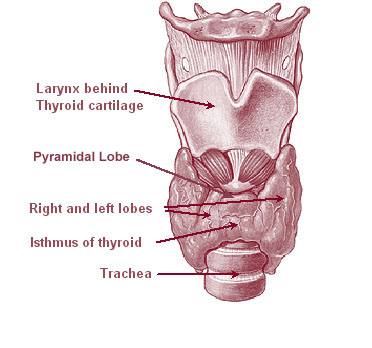
The thyroid, or thyroid gland, is an endocrine gland in vertebrates. In humans it is in the neck and consists of two connected lobes. The lower two thirds of the lobes are connected by a thin band of tissue called the thyroid isthmus. The thyroid is located at the front of the neck, below the Adam's apple. Microscopically, the functional unit of the thyroid gland is the spherical thyroid follicle, lined with follicular cells (thyrocytes), and occasional parafollicular cells that surround a lumen containing colloid. The thyroid gland secretes three hormones: the two thyroid hormones triiodothyronine (T3) and thyroxine (T4)and a peptide hormone, calcitonin. The thyroid hormones influence the metabolic rate and protein synthesis, and in children, growth and development. Calcitonin plays a role in calcium homeostasis. Secretion of the two thyroid hormones is regulated by thyroid-stimulating hormone (TSH), which is secreted from the anterior pituitary gland. TSH is regulated by ... [...More Info...] [...Related Items...] OR: [Wikipedia] [Google] [Baidu] |
FNA Specimen
Fine-needle aspiration (FNA) is a diagnostic procedure used to investigate lumps or masses. In this technique, a thin (23–25 gauge (0.52 to 0.64 mm outer diameter)), hollow needle is inserted into the mass for sampling of cells that, after being stained, are examined under a microscope (biopsy). The sampling and biopsy considered together are called fine-needle aspiration biopsy (FNAB) or fine-needle aspiration cytology (FNAC) (the latter to emphasize that any aspiration biopsy involves cytopathology, not histopathology). Fine-needle aspiration biopsies are very safe minor surgical procedures. Often, a major surgical (excisional or open) biopsy can be avoided by performing a needle aspiration biopsy instead, eliminating the need for hospitalization. In 1981, the first fine-needle aspiration biopsy in the United States was done at Maimonides Medical Center. Today, this procedure is widely used in the diagnosis of cancer and inflammatory conditions. Aspiration is safer an ... [...More Info...] [...Related Items...] OR: [Wikipedia] [Google] [Baidu] |
Lacrimal Sac
The lacrimal sac or lachrymal sac is the upper dilated end of the nasolacrimal duct, and is lodged in a deep groove formed by the lacrimal bone and frontal process of the maxilla. It connects the lacrimal canaliculi, which drain tears from the eye's surface, and the nasolacrimal duct, which conveys this fluid into the nasal cavity. Lacrimal sac occlusion leads to dacryocystitis. Structure It is oval in form and measures from 12 to 15 mm. in length; its upper end is closed and rounded; its lower is continued into the nasolacrimal duct. Its superficial surface is covered by a fibrous expansion derived from the medial palpebral ligament, and its deep surface is crossed by the lacrimal part of the orbicularis oculi, which is attached to the crest on the lacrimal bone. Histology Like the nasolacrimal duct, the sac is lined by stratified columnar epithelium with mucus-secreting goblet cells Goblet cells are simple columnar epithelial cells that secrete gel-forming muc ... [...More Info...] [...Related Items...] OR: [Wikipedia] [Google] [Baidu] |
Bronchi
A bronchus is a passage or airway in the lower respiratory tract that conducts air into the lungs. The first or primary bronchi pronounced (BRAN-KAI) to branch from the trachea at the carina are the right main bronchus and the left main bronchus. These are the widest bronchi, and enter the right lung, and the left lung at each hilum. The main bronchi branch into narrower secondary bronchi or lobar bronchi, and these branch into narrower tertiary bronchi or segmental bronchi. Further divisions of the segmental bronchi are known as 4th order, 5th order, and 6th order segmental bronchi, or grouped together as subsegmental bronchi. The bronchi, when too narrow to be supported by cartilage, are known as bronchioles. No gas exchange takes place in the bronchi. Structure The trachea (windpipe) divides at the carina into two main or primary bronchi, the left bronchus and the right bronchus. The carina of the trachea is located at the level of the sternal angle and the fifth thoraci ... [...More Info...] [...Related Items...] OR: [Wikipedia] [Google] [Baidu] |


