|
Motor Unit Plasticity
The motor unit consists of a voluntary alpha motoneuron and all of the collective muscle fibers that it controls, known as the effector muscle. The alpha motoneuron communicates with acetylcholine receptors on the motor end plate of the effector muscle. Reception of acetylcholine neurotransmitters on the motor end plate causes contraction of that effector muscle. Motor unit plasticity is defined as the ability of motoneurons and their respective effector muscles to physically and functionally change as a result of activity, age, and other factors. Motor unit plasticity has implications for improved athletic performance and resistance to immobility as a result of age. Recent advanced training techniques and physical therapy techniques that focus on improving neural function in addition to muscular function show promising results to improving athletic performances and extending mobility for the elderly. Plasticity due to Resistance Training Resistance training has been shown to dra ... [...More Info...] [...Related Items...] OR: [Wikipedia] [Google] [Baidu] |
Motor Unit
A motor unit is made up of a motor neuron and all of the skeletal muscle fibers innervated by the neuron's axon terminals, including the neuromuscular junctions between the neuron and the fibres. Groups of motor units often work together as a motor pool to coordinate the contractions of a single muscle. The concept was proposed by Charles Scott Sherrington. All muscle fibers in a motor unit are of the same fiber type. When a motor unit is activated, all of its fibers contract. In vertebrates, the force of a muscle contraction is controlled by the number of activated motor units. The number of muscle fibers within each unit can vary within a particular muscle and even more from muscle to muscle; the muscles that act on the largest body masses have motor units that contain more muscle fibers, whereas smaller muscles contain fewer muscle fibers in each motor unit. For instance, thigh muscles can have a thousand fibers in each unit, while extraocular muscles might have ten. Muscles ... [...More Info...] [...Related Items...] OR: [Wikipedia] [Google] [Baidu] |
Motoneuron
A motor neuron (or motoneuron or efferent neuron) is a neuron whose cell body is located in the motor cortex, brainstem or the spinal cord, and whose axon (fiber) projects to the spinal cord or outside of the spinal cord to directly or indirectly control effector organs, mainly muscles and glands. There are two types of motor neuron – upper motor neurons and lower motor neurons. Axons from upper motor neurons synapse onto interneurons in the spinal cord and occasionally directly onto lower motor neurons. The axons from the lower motor neurons are efferent nerve fibers that carry signals from the spinal cord to the effectors. Types of lower motor neurons are alpha motor neurons, beta motor neurons, and gamma motor neurons. A single motor neuron may innervate many muscle fibres and a muscle fibre can undergo many action potentials in the time taken for a single muscle twitch. Innervation takes place at a neuromuscular junction and twitches can become superimposed as a result o ... [...More Info...] [...Related Items...] OR: [Wikipedia] [Google] [Baidu] |
Acetylcholine Receptors
An acetylcholine receptor (abbreviated AChR) is an integral membrane protein that responds to the binding of acetylcholine, a neurotransmitter. Classification Like other transmembrane receptors, acetylcholine receptors are classified according to their "pharmacology," or according to their relative affinities and sensitivities to different molecules. Although all acetylcholine receptors, by definition, respond to acetylcholine, they respond to other molecules as well. *Nicotinic acetylcholine receptors (''nAChR'', also known as "ionotropic" acetylcholine receptors) are particularly responsive to nicotine. The nicotine ACh receptor is also a Na+, K+ and Ca2+ ion channel. *Muscarinic acetylcholine receptors (''mAChR'', also known as "metabotropic" acetylcholine receptors) are particularly responsive to muscarine. Nicotinic and muscarinic are two main kinds of "cholinergic" receptors. Receptor types Molecular biology has shown that the nicotinic and muscarinic receptors belong ... [...More Info...] [...Related Items...] OR: [Wikipedia] [Google] [Baidu] |
Motor End Plate
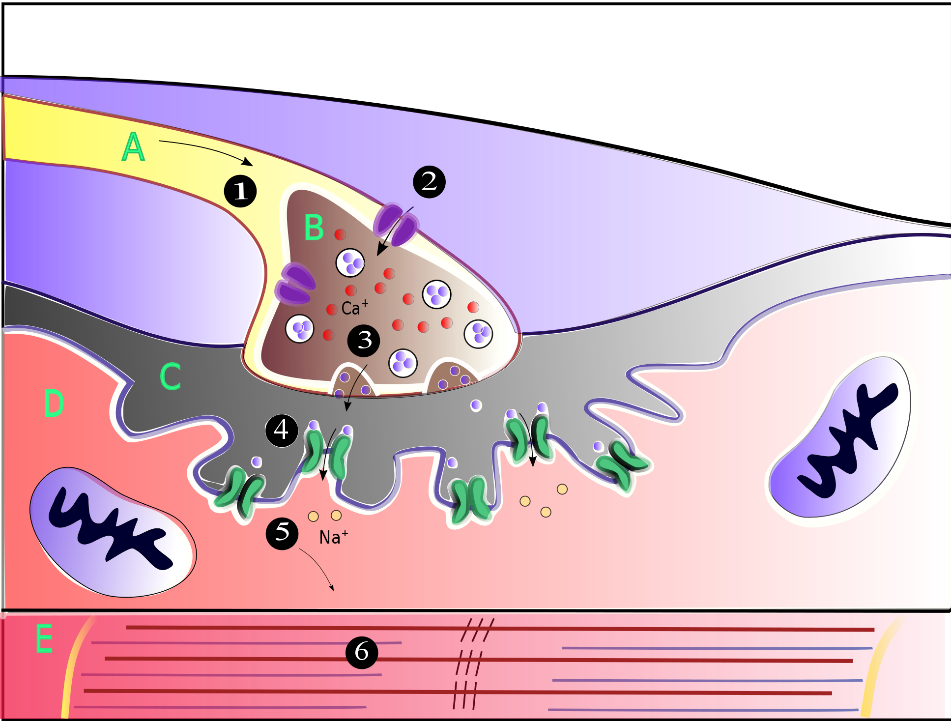
A neuromuscular junction (or myoneural junction) is a chemical synapse between a motor neuron and a muscle fiber. It allows the motor neuron to transmit a signal to the muscle fiber, causing muscle contraction. Muscles require innervation to function—and even just to maintain muscle tone, avoiding atrophy. In the neuromuscular system nerves from the central nervous system and the peripheral nervous system are linked and work together with muscles. Synaptic transmission at the neuromuscular junction begins when an action potential reaches the presynaptic terminal of a motor neuron, which activates voltage-gated calcium channels to allow calcium ions to enter the neuron. Calcium ions bind to sensor proteins (synaptotagmins) on synaptic vesicles, triggering vesicle fusion with the cell membrane and subsequent neurotransmitter release from the motor neuron into the synaptic cleft. In vertebrates, motor neurons release acetylcholine (ACh), a small molecule neurotransmitter, which ... [...More Info...] [...Related Items...] OR: [Wikipedia] [Google] [Baidu] |
Strength Training
Strength training or resistance training involves the performance of physical exercises that are designed to improve strength and endurance. It is often associated with the lifting of weights. It can also incorporate a variety of training techniques such as bodyweight exercises, isometrics, and plyometrics. Training works by progressively increasing the force output of the muscles and uses a variety of exercises and types of equipment. Strength training is primarily an anaerobic activity, although circuit training also is a form of aerobic exercise. Strength training can increase muscle, tendon, and ligament strength as well as bone density, metabolism, and the lactate threshold; improve joint and cardiac function; and reduce the risk of injury in athletes and the elderly. For many sports and physical activities, strength training is central or is used as part of their training regimen. Principles and training methods The basic principles of strength training involve ... [...More Info...] [...Related Items...] OR: [Wikipedia] [Google] [Baidu] |
Electromyography
Electromyography (EMG) is a technique for evaluating and recording the electrical activity produced by skeletal muscles. EMG is performed using an instrument called an electromyograph to produce a record called an electromyogram. An electromyograph detects the electric potential generated by muscle cells when these cells are electrically or neurologically activated. The signals can be analyzed to detect abnormalities, activation level, or recruitment order, or to analyze the biomechanics of human or animal movement. Needle EMG is an electrodiagnostic medicine technique commonly used by neurologists. Surface EMG is a non-medical procedure used to assess muscle activation by several professionals, including physiotherapists, kinesiologists and biomedical engineers. In Computer Science, EMG is also used as middleware in gesture recognition towards allowing the input of physical action to a computer as a form of human-computer interaction. Clinical uses EMG testing has a variety of ... [...More Info...] [...Related Items...] OR: [Wikipedia] [Google] [Baidu] |
Neural Synchronization
Neural oscillations, or brainwaves, are rhythmic or repetitive patterns of neural activity in the central nervous system. Neural tissue can generate oscillation, oscillatory activity in many ways, driven either by mechanisms within individual neurons or by interactions between neurons. In individual neurons, oscillations can appear either as oscillations in membrane potential or as rhythmic patterns of action potentials, which then produce oscillatory activation of post-synaptic potential, post-synaptic neurons. At the level of neural ensembles, synchronized activity of large numbers of neurons can give rise to macroscopic scale, macroscopic oscillations, which can be observed in an electroencephalography, electroencephalogram. Oscillatory activity in groups of neurons generally arises from feedback connections between the neurons that result in the synchronization of their firing patterns. The interaction between neurons can give rise to oscillations at a different frequency th ... [...More Info...] [...Related Items...] OR: [Wikipedia] [Google] [Baidu] |
Motor Unit Recruitment
Motor unit recruitment is the activation of additional motor units to accomplish an increase in contractile strength in a muscle. A motor unit consists of one motor neuron and all of the muscle fibers it stimulates. All muscles consist of a number of motor units and the fibers belonging to a motor unit are dispersed and intermingle amongst fibers of other units. The muscle fibers belonging to one motor unit can be spread throughout part, or most of the entire muscle, depending on the number of fibers and size of the muscle. When a motor neuron is activated, all of the muscle fibers innervated by the motor neuron are stimulated and contract. The activation of one motor neuron will result in a weak but distributed muscle contraction. The activation of more motor neurons will result in more muscle fibers being activated, and therefore a stronger muscle contraction. Motor unit recruitment is a measure of how many motor neurons are activated in a particular muscle, and therefore is a ... [...More Info...] [...Related Items...] OR: [Wikipedia] [Google] [Baidu] |
Fast Twitch Muscle
Skeletal muscles (commonly referred to as muscles) are organs of the vertebrate muscular system and typically are attached by tendons to bones of a skeleton. The muscle cells of skeletal muscles are much longer than in the other types of muscle tissue, and are often known as muscle fibers. The muscle tissue of a skeletal muscle is striated – having a striped appearance due to the arrangement of the sarcomeres. Skeletal muscles are voluntary muscles under the control of the somatic nervous system. The other types of muscle are cardiac muscle which is also striated and smooth muscle which is non-striated; both of these types of muscle tissue are classified as involuntary, or, under the control of the autonomic nervous system. A skeletal muscle contains multiple fascicles – bundles of muscle fibers. Each individual fiber, and each muscle is surrounded by a type of connective tissue layer of fascia. Muscle fibers are formed from the fusion of developmental myoblasts in a proc ... [...More Info...] [...Related Items...] OR: [Wikipedia] [Google] [Baidu] |
Slow Twitch Muscle
A muscle cell is also known as a myocyte when referring to either a cardiac muscle cell (cardiomyocyte), or a smooth muscle cell as these are both small cells. A skeletal muscle cell is long and threadlike with many nuclei and is called a muscle fiber. Muscle cells (including myocytes and muscle fibers) develop from embryonic precursor cells called myoblasts. Myoblasts fuse to form multinucleated skeletal muscle cells known as syncytia in a process known as myogenesis. Skeletal muscle cells and cardiac muscle cells both contain myofibrils and sarcomeres and form a striated muscle tissue. Cardiac muscle cells form the cardiac muscle in the walls of the heart chambers, and have a single central nucleus. Cardiac muscle cells are joined to neighboring cells by intercalated discs, and when joined in a visible unit they are described as a ''cardiac muscle fiber''. Smooth muscle cells control involuntary movements such as the peristalsis contractions in the esophagus and stomach. Smo ... [...More Info...] [...Related Items...] OR: [Wikipedia] [Google] [Baidu] |
Neuromuscular Junction
A neuromuscular junction (or myoneural junction) is a chemical synapse between a motor neuron and a muscle fiber. It allows the motor neuron to transmit a signal to the muscle fiber, causing muscle contraction. Muscles require innervation to function—and even just to maintain muscle tone, avoiding atrophy. In the neuromuscular system nerves from the central nervous system and the peripheral nervous system are linked and work together with muscles. Synaptic transmission at the neuromuscular junction begins when an action potential reaches the presynaptic terminal of a motor neuron, which activates voltage-gated calcium channels to allow calcium ions to enter the neuron. Calcium ions bind to sensor proteins (synaptotagmins) on synaptic vesicles, triggering vesicle fusion with the cell membrane and subsequent neurotransmitter release from the motor neuron into the synaptic cleft. In vertebrates, motor neurons release acetylcholine (ACh), a small molecule neurotransmitter, whic ... [...More Info...] [...Related Items...] OR: [Wikipedia] [Google] [Baidu] |


