|
Metanephrogenic Blastema
The metanephrogenic blastema or metanephric blastema (or metanephric mesenchyme, or metanephric mesoderm) is one of the two embryological structures that give rise to the kidney, the other being the ureteric bud. The metanephric blastema mostly develops into nephrons, but can also form parts of the collecting duct system. The system of tissue induction between the ureteric bud and the metanephric blastema is a reciprocal control system. GDNF, GDNF, glial cell-derived neurotrophic factor, is produced by the metanephric blastema and is essential in binding to the RET proto-oncogene, Ret receptor on the ureteric bud, which bifurcates and coalesces as a result to form the renal pelvis, major and minor Renal calyx, calyces and collecting ducts. Mutations in the ''EYA1'' gene, whose product regulates ''GDNF'' expression in the developing kidney, lead to the renal abnormalities of BOR syndrome (branchio-oto-renal syndrome). See also * Mesenchyme * Metanephros * Blastema * Kidney developm ... [...More Info...] [...Related Items...] OR: [Wikipedia] [Google] [Baidu] |
Wolffian Duct
The mesonephric duct (also known as the Wolffian duct, archinephric duct, Leydig's duct or nephric duct) is a paired organ that forms during the embryonic development of humans and other mammals and gives rise to male reproductive organs. Structure The mesonephric duct connects the primitive kidney, the '' mesonephros'', to the cloaca. It also serves as the primordium for male urogenital structures including the epididymis, vas deferens, and seminal vesicles. Development In both male and female the mesonephric duct develops into the trigone of urinary bladder, a part of the bladder wall, but the sexes differentiate in other ways during development of the urinary and reproductive organs. Male In a male, it develops into a system of connected organs between the efferent ducts of the testis and the prostate, namely the epididymis, the vas deferens, and the seminal vesicle. The prostate forms from the urogenital sinus and the efferent ducts form from the meso ... [...More Info...] [...Related Items...] OR: [Wikipedia] [Google] [Baidu] |
Collecting Duct System
The collecting duct system of the kidney consists of a series of tubules and ducts that physically connect nephrons to a minor calyx or directly to the renal pelvis. The collecting duct system is the last part of nephron and participates in electrolyte and fluid balance through reabsorption and excretion, processes regulated by the hormones aldosterone and vasopressin (antidiuretic hormone). There are several components of the collecting duct system, including the connecting tubules, cortical collecting ducts, and medullary collecting ducts. Structure Segments The segments of the system are as follows: Connecting tubule With respect to the renal corpuscle, the connecting tubule (CNT, or junctional tubule, or arcuate renal tubule) is the most proximal part of the collecting duct system. It is adjacent to the distal convoluted tubule, the most distal segment of the renal tubule. Connecting tubules from several adjacent nephrons merge to form cortical collecting tubules, and the ... [...More Info...] [...Related Items...] OR: [Wikipedia] [Google] [Baidu] |
Blastema
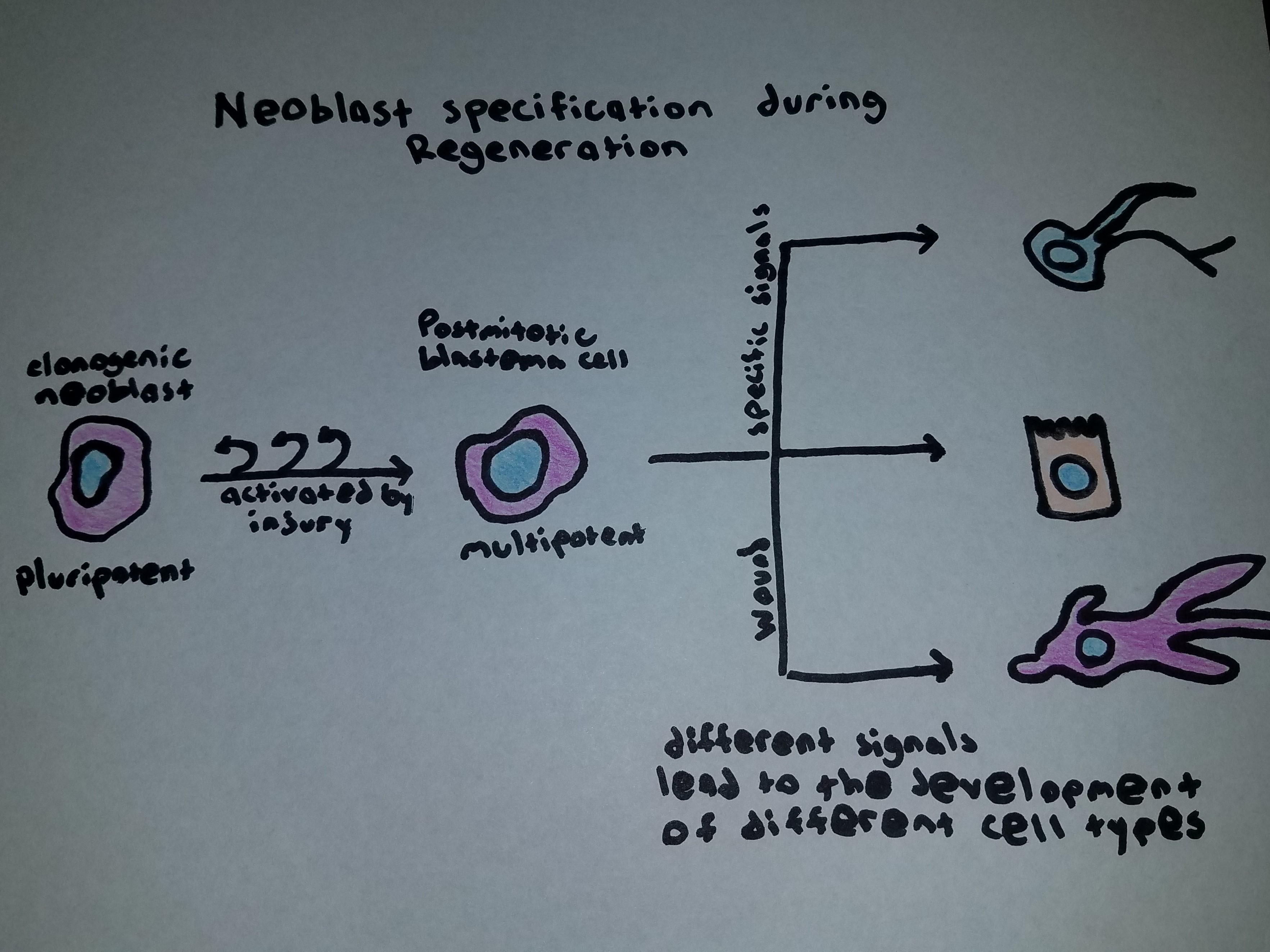
A blastema (Greek ''βλάστημα'', "offspring") is a mass of cells capable of growth and regeneration into organs or body parts. The changing definition of the word "blastema" has been reviewed by Holland (2021). A broad survey of how blastema has been used over time brings to light a somewhat involved history. The word entered the biomedical vocabulary in 1799 to designate a sinister acellular slime that was the starting point for the growth of cancers, themselves, at the time, thought to be acellular, as reviewed by Hajdu (2011, Cancer 118: 1155-1168). Then, during the early nineteenth century, the definition broadened to include growth zones (still considered acellular) in healthy, normally developing plant and animal embryos. Contemporaneously, cancer specialists dropped the term from their vocabulary, perhaps because they felt a term connoting a state of health and normalcy was not appropriate for describing a pathological condition. During the middle decades of the nine ... [...More Info...] [...Related Items...] OR: [Wikipedia] [Google] [Baidu] |
Metanephros
Kidney development, or nephrogenesis, describes the embryologic origins of the kidney, a major organ in the urinary system. This article covers a 3 part developmental process that is observed in most reptiles, birds and mammals, including humans. Nephrogenesis is often considered in the broader context of the development of the urinary and reproductive organs. Phases The development of the kidney proceeds through a series of successive phases, each marked by the development of a more advanced kidney: the archinephros, pronephros, mesonephros, and metanephros. The pronephros is the most immature form of kidney, while the metanephros is most developed. The metanephros persists as the definitive adult kidney. Archinephros The archinephros is considered as hypothetical or primitive kidney. Pronephros The pronephros develops in the cervical region of the embryo. During approximately day 22 of human gestation, the paired pronephri appears towards the cranial end of the intermediate m ... [...More Info...] [...Related Items...] OR: [Wikipedia] [Google] [Baidu] |
Mesenchyme
Mesenchyme () is a type of loosely organized animal embryonic connective tissue of undifferentiated cells that give rise to most tissues, such as skin, blood or bone. The interactions between mesenchyme and epithelium help to form nearly every organ in the developing embryo. Vertebrates Structure Mesenchyme is characterized morphologically by a prominent ground substance matrix containing a loose aggregate of reticular fibers and unspecialized mesenchymal stem cells. Mesenchymal cells can migrate easily (in contrast to epithelial cells, which lack mobility), are organized into closely adherent sheets, and are polarized in an apical-basal orientation. Development The mesenchyme originates from the mesoderm. From the mesoderm, the mesenchyme appears as an embryologically primitive "soup". This "soup" exists as a combination of the mesenchymal cells plus serous fluid plus the many different tissue proteins. Serous fluid is typically stocked with the many serous elements, such a ... [...More Info...] [...Related Items...] OR: [Wikipedia] [Google] [Baidu] |
Branchio-oto-renal Syndrome
Branchio-oto-renal syndrome (BOR) is an autosomal dominant genetic disorder involving the kidneys, ears, and neck. It often has also been described as Melnick-Fraser syndrome. Signs and symptoms The signs and symptoms of branchio-oto-renal syndrome are consistent with underdeveloped (hypoplastic) or absent kidneys with resultant chronic kidney disease or kidney failure. Ear anomalies include extra openings in front of the ears, extra pieces of skin in front of the ears (preauricular tags), or further malformation or absence of the outer ear ( pinna). Malformation or absence of the middle ear is also possible, individuals can have mild to profound hearing loss. People with BOR may also have cysts or fistulae along the sides of their neck. In some individuals and families, renal features are completely absent. The disease may then be termed "branchio-oto syndrome" (BO syndrome)., updated, 2015, Cause The cause of branchio-oto-renal syndrome are mutations in genes, EYA1, SIX1, and ... [...More Info...] [...Related Items...] OR: [Wikipedia] [Google] [Baidu] |
EYA1
Eyes absent homolog 1 is a protein that in humans is encoded by the ''EYA1'' gene. This gene encodes a member of the eyes absent (EYA) subfamily of proteins. The encoded protein may play a role in the developing kidney, branchial arches, eye, and ear. Mutations of this gene have been associated with branchiootorenal dysplasia syndrome, branchiootic syndrome, and sporadic cases of congenital cataracts and ocular anterior segment anomalies. A similar protein in mice can act as a transcriptional activator. Four transcript variants encoding three distinct isoforms have been identified for this gene. Interactions EYA1 has been shown to interact with SIX1 Homeobox protein SIX1 (Sine oculis homeobox homolog 1) is a protein that in humans is encoded by the ''SIX1'' gene. Function The vertebrate SIX genes are homologs of the Drosophila 'sine oculis' (so) gene, which is expressed primarily in the .... References Further reading * * * * * * * * * * * * * * * * * * * {{ge ... [...More Info...] [...Related Items...] OR: [Wikipedia] [Google] [Baidu] |
Renal Calyx
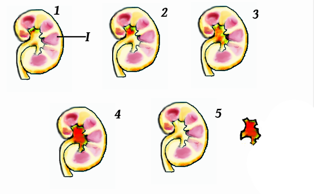
The renal calyces are chambers of the kidney through which urine passes. The minor calyces surround the apex of the renal pyramids. Urine formed in the kidney passes through a renal papilla at the apex into the minor calyx; two or three minor calyces converge to form a major calyx, through which urine passes before continuing through the renal pelvis into the ureter. Function Peristalsis of the smooth muscle originating in pace-maker cells originating in the walls of the calyces propels urine through the renal pelvis and ureters to the bladder. The initiation is caused by the increase in volume that stretches the walls of the calyces. This causes them to fire impulses which stimulate rhythmical contraction and relaxation, called peristalsis. Parasympathetic innervation enhances the peristalsis while sympathetic innervation inhibits it. Clinical significance A "staghorn calculus" is a kidney stone that may extend into the renal calyces. A renal diverticulum is diverticulum of ... [...More Info...] [...Related Items...] OR: [Wikipedia] [Google] [Baidu] |
Renal Pelvis
The renal pelvis or pelvis of the kidney is the funnel-like dilated part of the ureter in the kidney. It is formed by the covnvergence of the major calyces, acting as a funnel for urine flowing from the major calyces to the ureter. It has a mucous membrane and is covered with transitional epithelium and an underlying lamina propria of loose-to-dense connective tissue. The renal pelvis is situated within the renal sinus alongside the other structures of the renal sinus. The renal pelvis is the location of several kinds of kidney cancer and is affected by infection in pyelonephritis. Clinical significance The renal pelvis is the location of several kinds of kidney cancer and is affected by infection in pyelonephritis. A large "staghorn" kidney stone may block all or part of the renal pelvis. The size of the renal pelvis plays a major role in the grading of hydronephrosis. Normally, the anteroposterior diameter of the renal pelvis is less than 4 mm in fetuses up to 32 weeks ... [...More Info...] [...Related Items...] OR: [Wikipedia] [Google] [Baidu] |
RET Proto-oncogene
The ''RET'' proto-oncogene encodes a receptor tyrosine kinase for members of the glial cell line-derived neurotrophic factor (GDNF) family of extracellular signalling molecules. ''RET'' loss of function mutations are associated with the development of Hirschsprung's disease, while gain of function mutations are associated with the development of various types of human cancer, including medullary thyroid carcinoma, multiple endocrine neoplasias type 2A and 2B, pheochromocytoma and parathyroid hyperplasia. Structure ''RET'' is an abbreviation for "rearranged during transfection", as the DNA sequence of this gene was originally found to be rearranged within a 3T3 fibroblast cell line following its transfection with DNA taken from human lymphoma cells. The human gene ''RET'' is localized to chromosome 10 (10q11.2) and contains 21 exons. The natural alternative splicing of the ''RET'' gene results in the production of 3 different isoforms of the protein RET. RET51, RET43 and RET9 ... [...More Info...] [...Related Items...] OR: [Wikipedia] [Google] [Baidu] |
GDNF
Glial cell line-derived neurotrophic factor (GDNF) is a protein that, in humans, is encoded by the ''GDNF'' gene. GDNF is a small protein that potently promotes the survival of many types of neurons. It signals through GFRα receptors, particularly GFRα1. It is also responsible for the determination of spermatogonia into primary spermatocytes, i.e. it is received by RET proto-oncogene (RET) and by forming gradient with SCF it divides the spermatogonia into two cells. As the result there is retention of spermatogonia and formation of spermatocyte. GDNF family of ligands (GFL) GDNF was discovered in 1991, and is the first member of the GDNF family of ligands (GFL) found. Function GDNF is highly distributed throughout both the peripheral and central nervous system. It can be secreted by astrocytes, oligodendrocytes, Schwann cells, motor neurons, and skeletal muscle during the development and growth of neurons and other peripheral cells. The GDNF gene encodes a highly conserved ... [...More Info...] [...Related Items...] OR: [Wikipedia] [Google] [Baidu] |
Nephrons
The nephron is the minute or microscopic structural and functional unit of the kidney. It is composed of a renal corpuscle and a renal tubule. The renal corpuscle consists of a tuft of capillaries called a glomerulus and a cup-shaped structure called Bowman's capsule. The renal tubule extends from the capsule. The capsule and tubule are connected and are composed of epithelial cells with a lumen. A healthy adult has 1 to 1.5 million nephrons in each kidney. Blood is filtered as it passes through three layers: the endothelial cells of the capillary wall, its basement membrane, and between the foot processes of the podocytes of the lining of the capsule. The tubule has adjacent peritubular capillaries that run between the descending and ascending portions of the tubule. As the fluid from the capsule flows down into the tubule, it is processed by the epithelial cells lining the tubule: water is reabsorbed and substances are exchanged (some are added, others are removed); first with t ... [...More Info...] [...Related Items...] OR: [Wikipedia] [Google] [Baidu] |


