|
Metanephric Adenoma
Metanephric adenoma (MA) is a rare, benign tumour of the kidney, that can have a microscopic appearance similar to a nephroblastoma (Wilms tumours), or a papillary renal cell carcinoma. It should not be confused with the pathologically unrelated, yet similar sounding, '' mesonephric adenoma''. Symptoms The symptoms may be similar to those classically associated with renal cell carcinoma, and may include polycythemia, abdominal pain, hematuria Hematuria or haematuria is defined as the presence of blood or red blood cells in the urine. “Gross hematuria” occurs when urine appears red, brown, or tea-colored due to the presence of blood. Hematuria may also be subtle and only detectable w ... and a palpable mass. Mean age at onset is around 40 years with a range of 5 to 83 years and the mean size of the tumour is 5.5 cm with a range 0.3 to 15 cm (1). Polycythemia is more frequent in MA than in any other type of renal tumour. Of further relevance is that this tumour is mo ... [...More Info...] [...Related Items...] OR: [Wikipedia] [Google] [Baidu] |
Micrograph
A micrograph or photomicrograph is a photograph or digital image taken through a microscope or similar device to show a magnified image of an object. This is opposed to a macrograph or photomacrograph, an image which is also taken on a microscope but is only slightly magnified, usually less than 10 times. Micrography is the practice or art of using microscopes to make photographs. A micrograph contains extensive details of microstructure. A wealth of information can be obtained from a simple micrograph like behavior of the material under different conditions, the phases found in the system, failure analysis, grain size estimation, elemental analysis and so on. Micrographs are widely used in all fields of microscopy. Types Photomicrograph A light micrograph or photomicrograph is a micrograph prepared using an optical microscope, a process referred to as ''photomicroscopy''. At a basic level, photomicroscopy may be performed simply by connecting a camera to a microscope, th ... [...More Info...] [...Related Items...] OR: [Wikipedia] [Google] [Baidu] |
Renal Cell Carcinoma
Renal cell carcinoma (RCC) is a kidney cancer that originates in the lining of the proximal convoluted tubule, a part of the very small tubes in the kidney that transport primary urine. RCC is the most common type of kidney cancer in adults, responsible for approximately 90–95% of cases. RCC occurrence shows a male predominance over women with a ratio of 1.5:1. RCC most commonly occurs between 6th and 7th decade of life. Initial treatment is most commonly either partial or complete removal of the affected kidney(s). Where the cancer has not metastasised (spread to other organs) or burrowed deeper into the tissues of the kidney, the five-year survival rate is 65–90%, but this is lowered considerably when the cancer has spread. The body is remarkably good at hiding the symptoms and as a result people with RCC often have advanced disease by the time it is discovered. The initial symptoms of RCC often include blood in the urine (occurring in 40% of affected persons at the time th ... [...More Info...] [...Related Items...] OR: [Wikipedia] [Google] [Baidu] |
H&E Stain
Hematoxylin and eosin stain ( or haematoxylin and eosin stain or hematoxylin-eosin stain; often abbreviated as H&E stain or HE stain) is one of the principal tissue stains used in histology. It is the most widely used stain in medical diagnosis and is often the gold standard. For example, when a pathologist looks at a biopsy of a suspected cancer, the histological section is likely to be stained with H&E. H&E is the combination of two histological stains: hematoxylin and eosin. The hematoxylin stains cell nuclei a purplish blue, and eosin stains the extracellular matrix and cytoplasm pink, with other structures taking on different shades, hues, and combinations of these colors. Hence a pathologist can easily differentiate between the nuclear and cytoplasmic parts of a cell, and additionally, the overall patterns of coloration from the stain show the general layout and distribution of cells and provides a general overview of a tissue sample's structure. Thus, pattern recogniti ... [...More Info...] [...Related Items...] OR: [Wikipedia] [Google] [Baidu] |
Benign
Malignancy () is the tendency of a medical condition to become progressively worse. Malignancy is most familiar as a characterization of cancer. A ''malignant'' tumor contrasts with a non-cancerous benign tumor, ''benign'' tumor in that a malignancy is not self-limited in its growth, is capable of invading into adjacent tissues, and may be capable of spreading to distant tissues. A benign tumor has none of those properties. Malignancy in cancers is characterized by anaplasia, invasiveness, and metastasis. Malignant tumors are also characterized by genome instability, so that cancers, as assessed by whole genome sequencing, frequently have between 10,000 and 100,000 mutations in their entire genomes. Cancers usually show tumour heterogeneity, containing multiple subclones. They also frequently have reduced expression of DNA repair enzymes due to Epigenetics#DNA repair epigenetics in cancer, epigenetic methylation of DNA repair genes or altered MicroRNA#DNA repair and cancer, micr ... [...More Info...] [...Related Items...] OR: [Wikipedia] [Google] [Baidu] |
Tumour
A neoplasm () is a type of abnormal and excessive growth of tissue. The process that occurs to form or produce a neoplasm is called neoplasia. The growth of a neoplasm is uncoordinated with that of the normal surrounding tissue, and persists in growing abnormally, even if the original trigger is removed. This abnormal growth usually forms a mass, when it may be called a tumor. ICD-10 classifies neoplasms into four main groups: benign neoplasms, in situ neoplasms, malignant neoplasms, and neoplasms of uncertain or unknown behavior. Malignant neoplasms are also simply known as cancers and are the focus of oncology. Prior to the abnormal growth of tissue, as neoplasia, cells often undergo an abnormal pattern of growth, such as metaplasia or dysplasia. However, metaplasia or dysplasia does not always progress to neoplasia and can occur in other conditions as well. The word is from Ancient Greek 'new' and 'formation, creation'. Types A neoplasm can be benign, potentially ma ... [...More Info...] [...Related Items...] OR: [Wikipedia] [Google] [Baidu] |
Kidney
The kidneys are two reddish-brown bean-shaped organs found in vertebrates. They are located on the left and right in the retroperitoneal space, and in adult humans are about in length. They receive blood from the paired renal arteries; blood exits into the paired renal veins. Each kidney is attached to a ureter, a tube that carries excreted urine to the bladder. The kidney participates in the control of the volume of various body fluids, fluid osmolality, acid–base balance, various electrolyte concentrations, and removal of toxins. Filtration occurs in the glomerulus: one-fifth of the blood volume that enters the kidneys is filtered. Examples of substances reabsorbed are solute-free water, sodium, bicarbonate, glucose, and amino acids. Examples of substances secreted are hydrogen, ammonium, potassium and uric acid. The nephron is the structural and functional unit of the kidney. Each adult human kidney contains around 1 million nephrons, while a mouse kidney contains on ... [...More Info...] [...Related Items...] OR: [Wikipedia] [Google] [Baidu] |
Nephroblastoma
Wilms' tumor or Wilms tumor, also known as nephroblastoma, is a cancer of the kidneys that typically occurs in children, rarely in adults.; and occurs most commonly as a renal tumor in child patients. It is named after Max Wilms, the German surgeon (1867–1918) who first described it. Approximately 650 cases are diagnosed in the U.S. annually. The majority of cases occur in children with no associated genetic syndromes; however, a minority of children with Wilms' tumor have a congenital abnormality. It is highly responsive to treatment, with about 90 percent of children being cured. Signs and symptoms Typical signs and symptoms of Wilms' tumor include the following: * a painless, palpable abdominal mass * loss of appetite * abdominal pain * fever * nausea and vomiting * blood in the urine (in about 20% of cases) * high blood pressure in some cases (especially if synchronous or metachronous bilateral kidney involvement) * Rarely as varicoceleErginel B, Vural S, Akın M, K ... [...More Info...] [...Related Items...] OR: [Wikipedia] [Google] [Baidu] |
Mesonephric Adenoma
Nephrogenic adenoma is a benign growth typically found in the urinary bladder. It is thought to result from displacement and implantation of renal tubular cells, as this entity in kidney transplant recipients has been shown to be kidney donor derived. This entity should not be confused with the similar-sounding ''metanephric adenoma''. Diagnosis Nephrogenic adenomas are diagnosed under the microscope by pathologists. Microscopically the tumor shows closely packed small tubular structures in edematous stroma. The tubules show considerable variation in size and shape resembling convoluted tubules of the kidney. The single layer of cells lining the tubules are cuboidal with a scant to moderate amount of cytoplasm. In some areas they may have a hobnail appearance. Image: Nephrogenic adenoma - low mag.jpg , Low mag. Image: Nephrogenic adenoma - high mag.jpg , High mag. Treatment Even though there is no evidence of malignant potential, transurethral resection is recommended tog ... [...More Info...] [...Related Items...] OR: [Wikipedia] [Google] [Baidu] |
Polycythemia
Polycythemia (also known as polycythaemia) is a laboratory finding in which the hematocrit (the volume percentage of red blood cells in the blood) and/or hemoglobin concentration are increased in the blood. Polycythemia is sometimes called erythrocytosis, and there is significant overlap in the two findings, but the terms are not the same: polycythemia describes any increase in hematocrit and/or hemoglobin, while erythrocytosis describes an increase specifically in the number of red blood cells in the blood. Polycythemia has many causes. It can describe an increase in the number of red blood cells ("absolute polycythemia") or to a decrease in the volume of plasma ("relative polycythemia"). Absolute polycythemia can be due to genetic mutations in the bone marrow ("primary polycythemia"), physiologic adaptations to one's environment, medications, and/or other health conditions. Laboratory studies such as serum Erythropoietin, erythropoeitin levels and genetic testing might be helpfu ... [...More Info...] [...Related Items...] OR: [Wikipedia] [Google] [Baidu] |
Abdominal Pain
Abdominal pain, also known as a stomach ache, is a symptom Signs and symptoms are the observed or detectable signs, and experienced symptoms of an illness, injury, or condition. A sign for example may be a higher or lower temperature than normal, raised or lowered blood pressure or an abnormality showin ... associated with both non-serious and serious medical issues. Common causes of pain in the abdomen include gastroenteritis and irritable bowel syndrome. About 15% of people have a more serious underlying condition such as appendicitis, leaking or ruptured abdominal aortic aneurysm, diverticulitis, or ectopic pregnancy. In a third of cases the exact cause is unclear. Given that a variety of diseases can cause some form of abdominal pain, a systematic approach to the examination of a person and the formulation of a differential diagnosis remains important. Differential diagnosis The most frequent reasons for abdominal pain are gastroenteritis (13%), irritable bowel syndr ... [...More Info...] [...Related Items...] OR: [Wikipedia] [Google] [Baidu] |
Hematuria
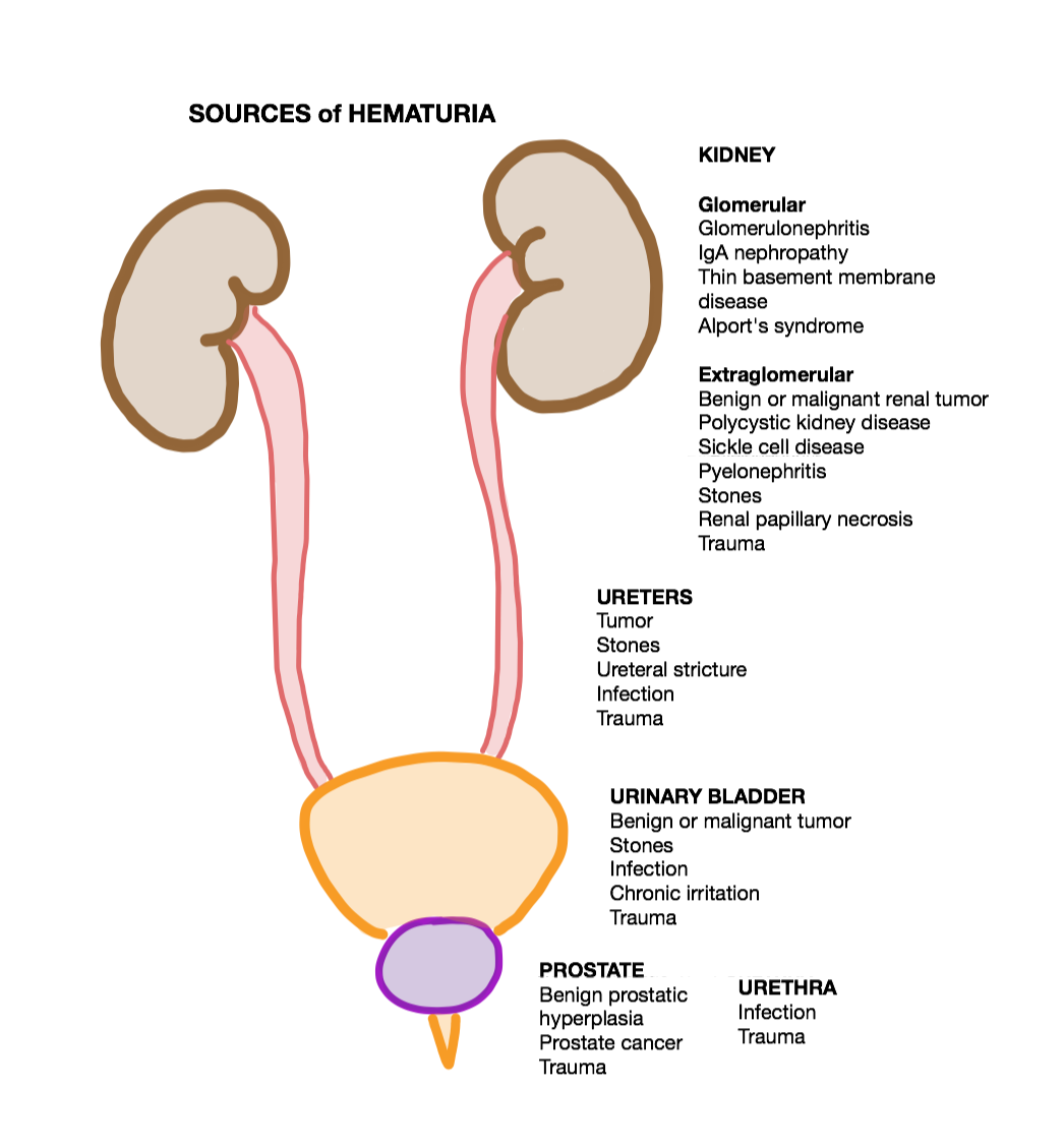
Hematuria or haematuria is defined as the presence of blood or red blood cells in the urine. “Gross hematuria” occurs when urine appears red, brown, or tea-colored due to the presence of blood. Hematuria may also be subtle and only detectable with a microscope or laboratory test. Blood that enters and mixes with the urine can come from any location within the urinary system, including the kidney, ureter, urinary bladder, urethra, and in men, the prostate. Common causes of hematuria include urinary tract infection (UTI), kidney stones, viral illness, trauma, bladder cancer, and exercise. These causes are grouped into glomerular and non-glomerular causes, depending on the involvement of the glomerulus of the kidney. But not all red urine is hematuria. Other substances such as certain medications and foods (e.g. blackberries, beets, food dyes) can cause urine to appear red. Menstruation in women may also cause the appearance of hematuria and may result in a positive urine dipstick ... [...More Info...] [...Related Items...] OR: [Wikipedia] [Google] [Baidu] |

_Nephrectomy.jpg)


