|
Melanization
Melanin (; from el, μέλας, melas, black, dark) is a broad term for a group of natural pigments found in most organisms. Eumelanin is produced through a multistage chemical process known as melanogenesis, where the oxidation of the amino acid tyrosine is followed by polymerization. The melanin pigments are produced in a specialized group of cells known as melanocytes. Functionally, eumelanin serves as protection against UV radiation. There are five basic types of melanin: eumelanin, pheomelanin, neuromelanin, allomelanin and pyomelanin. The most common type is eumelanin, of which there are two types— brown eumelanin and black eumelanin. Pheomelanin, which is produced when melanocytes are malfunctioning due to derivation of the gene to its recessive format is a cysteine-derivative that contains poly benzothiazine portions that are largely responsible for the of red yellow tint given to some skin or hair colors. Neuromelanin is found in the brain. Research has been u ... [...More Info...] [...Related Items...] OR: [Wikipedia] [Google] [Baidu] |
Pheomelanin
Melanin (; from el, μέλας, melas, black, dark) is a broad term for a group of natural pigments found in most organisms. Eumelanin is produced through a multistage chemical process known as melanogenesis, where the oxidation of the amino acid tyrosine is followed by polymerization. The melanin pigments are produced in a specialized group of cells known as melanocytes. Functionally, eumelanin serves as protection against UV radiation. There are five basic types of melanin: eumelanin, pheomelanin, neuromelanin, allomelanin and pyomelanin. The most common type is eumelanin, of which there are two types— brown eumelanin and black eumelanin. Pheomelanin, which is produced when melanocytes are malfunctioning due to derivation of the gene to its recessive format is a cysteine-derivative that contains poly benzothiazine portions that are largely responsible for the of red yellow tint given to some skin or hair colors. Neuromelanin is found in the brain. Research has been under ... [...More Info...] [...Related Items...] OR: [Wikipedia] [Google] [Baidu] |
Eumelanin
Melanin (; from el, μέλας, melas, black, dark) is a broad term for a group of natural pigments found in most organisms. Eumelanin is produced through a multistage chemical process known as melanogenesis, where the oxidation of the amino acid tyrosine is followed by polymerization. The melanin pigments are produced in a specialized group of cells known as melanocytes. Functionally, eumelanin serves as protection against UV radiation. There are five basic types of melanin: eumelanin, pheomelanin, neuromelanin, allomelanin and pyomelanin. The most common type is eumelanin, of which there are two types— brown eumelanin and black eumelanin. Pheomelanin, which is produced when melanocytes are malfunctioning due to derivation of the gene to its recessive format is a cysteine-derivative that contains poly benzothiazine portions that are largely responsible for the of red yellow tint given to some skin or hair colors. Neuromelanin is found in the brain. Research has been u ... [...More Info...] [...Related Items...] OR: [Wikipedia] [Google] [Baidu] |
UV Radiation
Ultraviolet (UV) is a form of electromagnetic radiation with wavelength from 10 nm (with a corresponding frequency around 30 PHz) to 400 nm (750 THz), shorter than that of visible light, but longer than X-rays. UV radiation is present in sunlight, and constitutes about 10% of the total electromagnetic radiation output from the Sun. It is also produced by electric arcs and specialized lights, such as mercury-vapor lamps, tanning lamps, and black lights. Although long-wavelength ultraviolet is not considered an ionizing radiation because its photons lack the energy to ionize atoms, it can cause chemical reactions and causes many substances to glow or fluoresce. Consequently, the chemical and biological effects of UV are greater than simple heating effects, and many practical applications of UV radiation derive from its interactions with organic molecules. Short-wave ultraviolet light damages DNA and sterilizes surfaces with which it comes into contact. For h ... [...More Info...] [...Related Items...] OR: [Wikipedia] [Google] [Baidu] |
Adrenal Gland
The adrenal glands (also known as suprarenal glands) are endocrine glands that produce a variety of hormones including adrenaline and the steroids aldosterone and cortisol. They are found above the kidneys. Each gland has an outer cortex which produces steroid hormones and an inner medulla. The adrenal cortex itself is divided into three main zones: the zona glomerulosa, the zona fasciculata and the zona reticularis. The adrenal cortex produces three main types of steroid hormones: mineralocorticoids, glucocorticoids, and androgens. Mineralocorticoids (such as aldosterone) produced in the zona glomerulosa help in the regulation of blood pressure and electrolyte balance. The glucocorticoids cortisol and cortisone are synthesized in the zona fasciculata; their functions include the regulation of metabolism and immune system suppression. The innermost layer of the cortex, the zona reticularis, produces androgens that are converted to fully functional sex hormones in the gonads ... [...More Info...] [...Related Items...] OR: [Wikipedia] [Google] [Baidu] |
Zona Reticularis
The zona reticularis (sometimes, reticulate zone) is the innermost layer of the adrenal cortex, lying deep to the zona fasciculata and superficial to the adrenal medulla. The cells are arranged cords that project in different directions giving a net-like appearance (L. reticulum - net). Cells in the zona reticularis produce precursor androgens including dehydroepiandrosterone (DHEA) and androstenedione from cholesterol. DHEA is further converted to DHEA-sulfate via a sulfotransferase, SULT2A1. These precursors are not further converted in the adrenal cortex if the cells lack 17β-Hydroxysteroid dehydrogenase. Instead, they are released into the blood stream and taken up in the testis and ovaries to produce testosterone and the estrogens respectively. ACTH partially regulates adrenal androgen secretion, also CRH. In humans the reticularis layer does contain 17α-hydroxylase; this hydroxylates pregnenolone, which is then converted to cortisol by a mixed function oxidase. In rode ... [...More Info...] [...Related Items...] OR: [Wikipedia] [Google] [Baidu] |
Locus Coeruleus
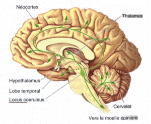
The locus coeruleus () (LC), also spelled locus caeruleus or locus ceruleus, is a nucleus in the pons of the brainstem involved with physiological responses to stress and panic. It is a part of the reticular activating system. The locus coeruleus, which in Latin means "blue spot", is the principal site for brain synthesis of norepinephrine (noradrenaline). The locus coeruleus and the areas of the body affected by the norepinephrine it produces are described collectively as the locus coeruleus-noradrenergic system or LC-NA system. Norepinephrine may also be released directly into the blood from the adrenal medulla. Anatomy The locus coeruleus (LC) is located in the posterior area of the rostral pons in the lateral floor of the fourth ventricle. It is composed of mostly medium-size neurons. Melanin granules inside the neurons of the LC contribute to its blue colour. Thus, it is also known as the nucleus pigmentosus pontis, meaning "heavily pigmented nucleus of the pons." The n ... [...More Info...] [...Related Items...] OR: [Wikipedia] [Google] [Baidu] |
Brainstem
The brainstem (or brain stem) is the posterior stalk-like part of the brain that connects the cerebrum with the spinal cord. In the human brain the brainstem is composed of the midbrain, the pons, and the medulla oblongata. The midbrain is continuous with the thalamus of the diencephalon through the tentorial notch, and sometimes the diencephalon is included in the brainstem. The brainstem is very small, making up around only 2.6 percent of the brain's total weight. It has the critical roles of regulating cardiac, and respiratory function, helping to control heart rate and breathing rate. It also provides the main motor and sensory nerve supply to the face and neck via the cranial nerves. Ten pairs of cranial nerves come from the brainstem. Other roles include the regulation of the central nervous system and the body's sleep cycle. It is also of prime importance in the conveyance of motor and sensory pathways from the rest of the brain to the body, and from the body back to t ... [...More Info...] [...Related Items...] OR: [Wikipedia] [Google] [Baidu] |
Adrenal Medulla
The adrenal medulla ( la, medulla glandulae suprarenalis) is part of the adrenal gland. It is located at the center of the gland, being surrounded by the adrenal cortex. It is the innermost part of the adrenal gland, consisting of chromaffin cells that secrete catecholamines, including epinephrine (adrenaline), norepinephrine (noradrenaline), and a small amount of dopamine, in response to stimulation by sympathetic preganglionic neurons. Structure The adrenal medulla consists of irregularly shaped cells grouped around blood vessels. These cells are intimately connected with the sympathetic division of the autonomic nervous system (ANS). These adrenal medullary cells are modified postganglionic neurons, and preganglionic autonomic nerve fibers lead to them directly from the central nervous system. The adrenal medulla affects energy availability, heart rate, and basal metabolic rate. Recent research indicates that the adrenal medulla may receive input from higher-order cognitive ... [...More Info...] [...Related Items...] OR: [Wikipedia] [Google] [Baidu] |
Inner Ear
The inner ear (internal ear, auris interna) is the innermost part of the vertebrate ear. In vertebrates, the inner ear is mainly responsible for sound detection and balance. In mammals, it consists of the bony labyrinth, a hollow cavity in the temporal bone of the skull with a system of passages comprising two main functional parts: * The cochlea, dedicated to hearing; converting sound pressure patterns from the outer ear into electrochemical impulses which are passed on to the brain via the auditory nerve. * The vestibular system, dedicated to balance The inner ear is found in all vertebrates, with substantial variations in form and function. The inner ear is innervated by the eighth cranial nerve in all vertebrates. Structure The labyrinth can be divided by layer or by region. Bony and membranous labyrinths The bony labyrinth, or osseous labyrinth, is the network of passages with bony walls lined with periosteum. The three major parts of the bony labyrinth are the vestib ... [...More Info...] [...Related Items...] OR: [Wikipedia] [Google] [Baidu] |
Stria Vascularis Of Cochlear Duct
The stria vascularis of the cochlear duct is a capillary loop in the upper portion of the spiral ligament (the outer wall of the cochlear duct). It produces endolymph for the scala media in the cochlea. Structure The stria vascularis is part of the lateral wall of the cochlear duct. It is a somewhat stratified epithelium containing primarily three cell types: * marginal cells, which are involved in K+ transport, and line the endolymphatic space of the scala media. * intermediate cells, which are pigment-containing cells scattered among capillaries. * basal cells, which separate the stria vascularis from the underlying spiral ligament. They are connected to basal cells with Gap junction, gap junctions. The stria vascularis also contains pericytes, melanocytes, and endothelial cells. It also contains intraepithelial capillaries - it is the only epithelial tissue that is not avascular (completely lacking Blood vessel, blood vessels and Lymphatic vessel, lymphatic vessels). Func ... [...More Info...] [...Related Items...] OR: [Wikipedia] [Google] [Baidu] |
Iris (anatomy)
In humans and most mammals and birds, the iris (plural: ''irides'' or ''irises'') is a thin, annular structure in the eye, responsible for controlling the diameter and size of the pupil, and thus the amount of light reaching the retina. Eye color is defined by the iris. In optical terms, the pupil is the eye's aperture, while the iris is the diaphragm. Structure The iris consists of two layers: the front pigmented fibrovascular layer known as a stroma and, beneath the stroma, pigmented epithelial cells. The stroma is connected to a sphincter muscle (sphincter pupillae), which contracts the pupil in a circular motion, and a set of dilator muscles ( dilator pupillae), which pull the iris radially to enlarge the pupil, pulling it in folds. The sphincter pupillae is the opposing muscle of the dilator pupillae. The pupil's diameter, and thus the inner border of the iris, changes size when constricting or dilating. The outer border of the iris does not change size. The constricti ... [...More Info...] [...Related Items...] OR: [Wikipedia] [Google] [Baidu] |


