|
Meesmann Juvenile Epithelial Corneal Dystrophy
Meesmann corneal dystrophy (MECD) is a rare hereditary autosomal dominant disease that is characterized as a type of corneal dystrophy and a keratin disease. MECD is characterized by the formation of microcysts in the outermost layer of the cornea, known as the anterior corneal epithelium. The anterior corneal epithelium also becomes fragile. This usually affects both eyes rather than a single eye and worsens over time. There are two phenotypes, Meesmann corneal dystrophy 1 (MECD1) and Meesmann corneal dystrophy 2 (MECD2), which affect the genes KRT3 and KRT12, respectively. A heterozygous mutation in either of these genes will lead to a single phenotype. Many with Meesmann corneal dystrophy are asymptomatic or experience mild symptoms. It is named after the German ophthalmologist Alois Meesmann (1888-1969). It is often considered as the "Meesmann-Wilke syndrome", after the joint contribution of Meesmann and Wilke in 1939.A. Meesmann, F. Wilke. Klinische und anatomische Untersuchun ... [...More Info...] [...Related Items...] OR: [Wikipedia] [Google] [Baidu] |
Ophthalmology
Ophthalmology ( ) is a surgical subspecialty within medicine that deals with the diagnosis and treatment of eye disorders. An ophthalmologist is a physician who undergoes subspecialty training in medical and surgical eye care. Following a medical degree, a doctor specialising in ophthalmology must pursue additional postgraduate residency training specific to that field. This may include a one-year integrated internship that involves more general medical training in other fields such as internal medicine or general surgery. Following residency, additional specialty training (or fellowship) may be sought in a particular aspect of eye pathology. Ophthalmologists prescribe medications to treat eye diseases, implement laser therapy, and perform surgery when needed. Ophthalmologists provide both primary and specialty eye care - medical and surgical. Most ophthalmologists participate in academic research on eye diseases at some point in their training and many include research as part ... [...More Info...] [...Related Items...] OR: [Wikipedia] [Google] [Baidu] |
Massive Parallel Sequencing
Massive parallel sequencing or massively parallel sequencing is any of several high-throughput approaches to DNA sequencing using the concept of massively parallel processing; it is also called next-generation sequencing (NGS) or second-generation sequencing. Some of these technologies emerged between 1994 and 1998 and have been commercially available since 2005. These technologies use miniaturized and parallelized platforms for sequencing of 1 million to 43 billion short reads (50 to 400 bases each) per instrument run. Many NGS platforms differ in engineering configurations and sequencing chemistry. They share the technical paradigm of massive parallel sequencing via spatially separated, clonally amplified DNA templates or single DNA molecules in a flow cell. This design is very different from that of Sanger sequencing—also known as capillary sequencing or first-generation sequencing—which is based on electrophoretic separation of chain-termination products produced in individ ... [...More Info...] [...Related Items...] OR: [Wikipedia] [Google] [Baidu] |
Thiel–Behnke Dystrophy
Thiel–Behnke dystrophy is a rare form of corneal dystrophy affecting the layer that supports corneal epithelium. The dystrophy was first described in 1967 and initially suspected to denote the same entity as the earlier-described Reis-Bucklers dystrophy, but following a study in 1995 by Kuchle et al. the two look-alike dystrophies were deemed separate disorders. Presentation To clarify whether Thiel–Behnke corneal dystrophy is a separate entity from Reis-Bucklers corneal dystrophy, Kuchle et al. (1995) examined 28 corneal specimens with a clinically suspected diagnosis of corneal dystrophy of the Bowman layer by light and electron microscopy and reviewed the literature and concluded that two distinct autosomal dominant corneal dystrophy of Bowman layer (CBD) exist and proposed the designation CDB type I (geographic or 'true' Reis-Bucklers dystrophy) and CDB type II (honeycomb-shaped or Thiel–Behnke dystrophy). Visual loss is significantly greater in CDB I, and recurrences ... [...More Info...] [...Related Items...] OR: [Wikipedia] [Google] [Baidu] |
Reis–Bucklers Corneal Dystrophy
Reis-Bücklers corneal dystrophy is a disease of the eye, a rare corneal dystrophy of unknown cause, in which the Bowman's layer of the cornea undergoes disintegration. The disorder is inherited in an autosomal dominant fashion, and is associated with mutations in the gene TGFB1. Reis-Bücklers dystrophy causes a cloudiness in the corneas of both eyes, which may occur as early as 1 year of age, but usually develops by 4 to 5 years of age. It is usually evident within the first decade of life. This cloudiness, or opacity, causes the corneal epithelium to become elevated, which leads to corneal opacities. The corneal erosions may prompt attacks of redness and swelling in the eye (ocular hyperemia), eye pain, and photophobia. Significant vision loss may occur. Reis-Bücklers dystrophy is diagnosed by clinical history physical examination of the eye. Laboratory and imaging studies are not necessary. Treatment may include a complete or partial corneal transplant, or photorefracti ... [...More Info...] [...Related Items...] OR: [Wikipedia] [Google] [Baidu] |
Epithelial Basement Membrane Dystrophy
Epithelial basement membrane dystrophy (EBMD) is a disorder of the eye that can cause pain and dryness. It is sometimes included in the group of corneal dystrophies. It diverges from the formal definition of corneal dystrophy since it is non-familial in most cases. It also has a fluctuating course, while for a typical corneal dystrophy the course is progressive. When it is considered part of this group, it is the most common type of corneal dystrophy. Signs and symptoms Patients may complain of severe problems with dry eyes, or with visual obscurations. It can also be asymptomatic, and only discovered because of subtle lines and marks seen during an eye exam. EBMD is a bilateral anterior corneal dystrophy characterized by grayish epithelial fingerprint lines, geographic map-like lines, and dots (or microcysts) on slit-lamp examination. Findings are variable and can change with time. While the disorder is usually asymptomatic, up to 10% of patients may have recurrent corneal ... [...More Info...] [...Related Items...] OR: [Wikipedia] [Google] [Baidu] |
List Of Cutaneous Conditions Caused By Mutations In Keratins
There are many different keratin proteins normally expressed in the human integumentary system. Mutations in keratin proteins in the skin can cause disease. Of note, other structural proteins in the epidermis of the skin that are closely related to keratins may also cause disease if mutated. Examples include: Footnotes See also * List of keratins expressed in the human integumentary system * List of cutaneous conditions caused by problems with junctional proteins * List of target antigens in pemphigoid * List of target antigens in pemphigus * Cutaneous conditions with immunofluorescence findings * List of cutaneous conditions * List of genes mutated in cutaneous conditions * List of histologic stains that aid in diagnosis of cutaneous conditions * Keratoderma Keratoderma is a hornlike skin condition. Classification The keratodermas are classified into the following subgroups:Freedberg, et al. (2003). ''Fitzpatrick's Dermatology in General Medicine''. (6th ed.). ... [...More Info...] [...Related Items...] OR: [Wikipedia] [Google] [Baidu] |
Corneal Dystrophy
Corneal dystrophy is a group of rare hereditary disorders characterised by bilateral abnormal deposition of substances in the transparent front part of the eye called the cornea. Signs and symptoms Corneal dystrophy may not significantly affect vision in the early stages. However, it does require proper evaluation and treatment for restoration of optimal vision. Corneal dystrophies usually manifest themselves during the first or second decade but sometimes later. It appears as grayish white lines, circles, or clouding of the cornea. Corneal dystrophy can also have a crystalline appearance. There are over 20 corneal dystrophies that affect all parts of the cornea. These diseases share many traits: * They are usually inherited. * They affect the right and left eyes equally. * They are not caused by outside factors, such as injury or diet. * Most progress gradually. * Most usually begin in one of the five corneal layers and may later spread to nearby layers. * Most do not affect oth ... [...More Info...] [...Related Items...] OR: [Wikipedia] [Google] [Baidu] |
Penetrating Keratoplasty
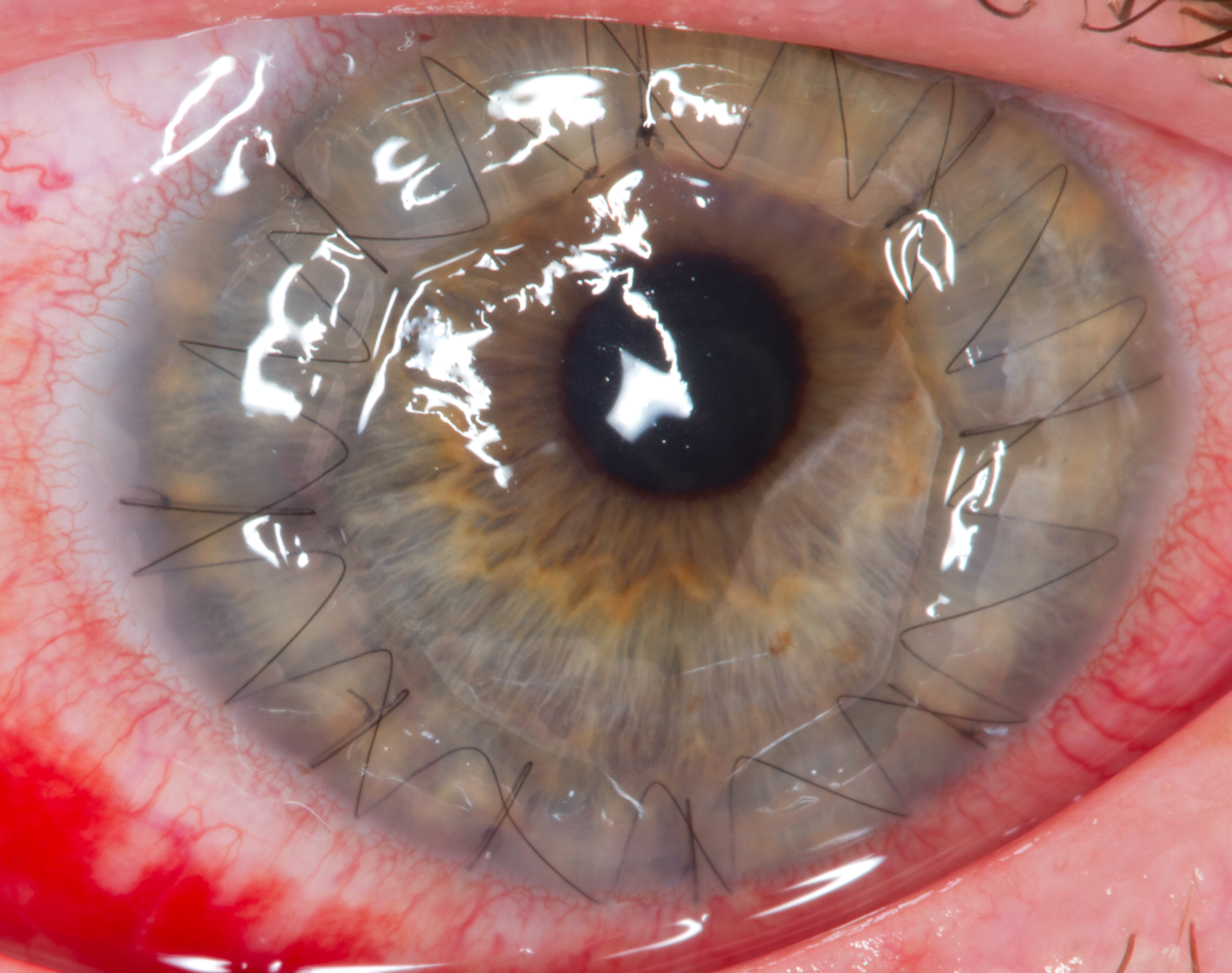
Corneal transplantation, also known as corneal grafting, is a surgical procedure where a damaged or diseased cornea is replaced by donated corneal tissue (the graft). When the entire cornea is replaced it is known as penetrating keratoplasty and when only part of the cornea is replaced it is known as lamellar keratoplasty. Keratoplasty simply means surgery to the cornea. The graft is taken from a recently deceased individual with no known diseases or other factors that may affect the chance of survival of the donated tissue or the health of the recipient. The cornea is the transparent front part of the eye that covers the iris, pupil and anterior chamber. The surgical procedure is performed by ophthalmologists, physicians who specialize in eyes, and is often done on an outpatient basis. Donors can be of any age, as is shown in the case of Janis Babson, who donated her eyes after dying at the age of 10. Corneal transplantation is performed when medicines, keratoconus conservati ... [...More Info...] [...Related Items...] OR: [Wikipedia] [Google] [Baidu] |
Phototherapeutic Keratectomy
Phototherapeutic keratectomy (PTK) is a type of eye surgery that uses a laser to treat various ocular disorders by removing tissue from the cornea. PTK allows the removal of superficial corneal opacities and surface irregularities. It is similar to photorefractive keratectomy Photorefractive keratectomy (PRK) and laser-assisted sub-epithelial keratectomy (or laser epithelial keratomileusis) (LASEK) are laser eye surgery procedures intended to correct a person's vision, reducing dependency on glasses or contact lenses. ..., which is used for the treatment of refractive conditions. The common indications for PTK are corneal dystrophies, scars, opacities, and bullous keratopathy. TK in the developing worldttp://www.ophthalmologyweb.com/JournalUpdates.aspx?spid=23&jid=15009 References External links "Facts About The Cornea and Corneal Disease" - National Eye Institute {{eye-stub Eye surgery ... [...More Info...] [...Related Items...] OR: [Wikipedia] [Google] [Baidu] |
Electron Microscope
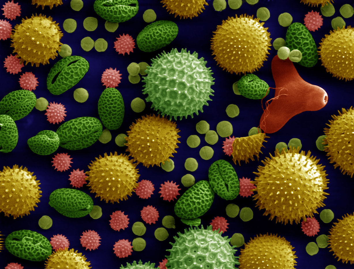
An electron microscope is a microscope that uses a beam of accelerated electrons as a source of illumination. As the wavelength of an electron can be up to 100,000 times shorter than that of visible light photons, electron microscopes have a higher resolving power than light microscopes and can reveal the structure of smaller objects. A scanning transmission electron microscope has achieved better than 50 pm resolution in annular dark-field imaging mode and magnifications of up to about 10,000,000× whereas most light microscopes are limited by diffraction to about 200 nm resolution and useful magnifications below 2000×. Electron microscopes use shaped magnetic fields to form electron optical lens systems that are analogous to the glass lenses of an optical light microscope. Electron microscopes are used to investigate the ultrastructure of a wide range of biological and inorganic specimens including microorganisms, cells, large molecules, biopsy samples, ... [...More Info...] [...Related Items...] OR: [Wikipedia] [Google] [Baidu] |
Light Microscopy
Microscopy is the technical field of using microscopes to view objects and areas of objects that cannot be seen with the naked eye (objects that are not within the resolution range of the normal eye). There are three well-known branches of microscopy: optical microscope, optical, electron microscope, electron, and scanning probe microscopy, along with the emerging field of X-ray microscopy. Optical microscopy and electron microscopy involve the diffraction, reflection (physics), reflection, or refraction of electromagnetic radiation/electron beams interacting with the Laboratory specimen, specimen, and the collection of the scattered radiation or another signal in order to create an image. This process may be carried out by wide-field irradiation of the sample (for example standard light microscopy and transmission electron microscope, transmission electron microscopy) or by scanning a fine beam over the sample (for example confocal laser scanning microscopy and scanning electro ... [...More Info...] [...Related Items...] OR: [Wikipedia] [Google] [Baidu] |
Slit Lamp
A slit lamp is an instrument consisting of a high-intensity light source that can be focused to shine a thin sheet of light into the eye. It is used in conjunction with a biomicroscope. The lamp facilitates an examination of the anterior segment and posterior segment of the human eye, which includes the eyelid, sclera, conjunctiva, iris, natural crystalline lens, and cornea. The binocular slit-lamp examination provides a stereoscopic magnified view of the eye structures in detail, enabling anatomical diagnoses to be made for a variety of eye conditions. A second, hand-held lens is used to examine the retina. History Two conflicting trends emerged in the development of the slit lamp. One trend originated from clinical research and aimed to apply the increasingly complex and advanced technology of the time. [...More Info...] [...Related Items...] OR: [Wikipedia] [Google] [Baidu] |


