|
Medial Vestibulospinal Tract
The medial vestibulospinal tract is one of the descending spinal tracts of the ventromedial funiculus of the spinal cord. It is found only in the cervical spine and above. The medial part of the vestibulospinal tract is the smaller part, and is primarily made of fibers from the medial vestibular nucleus. It projects bilaterally down the spinal cord and triggers the ventral horn of the cervical spinal circuits, particularly controlling lower motor neurons associated with the spinal accessory nerve (CN XI). Additionally, the pathway projects superiorly to the paramedian pontine reticular formation The paramedian pontine reticular formation, also known as PPRF or paraabducens nucleus, is part of the pontine reticular formation, a brain region without clearly defined borders in the center of the pons. It is involved in the coordination of eye ..., indirectly innervating the nuclei of CN VI and III. Through this superior projection, the medial vestibulospinal tract is involved in " ... [...More Info...] [...Related Items...] OR: [Wikipedia] [Google] [Baidu] |
Anatomical Position
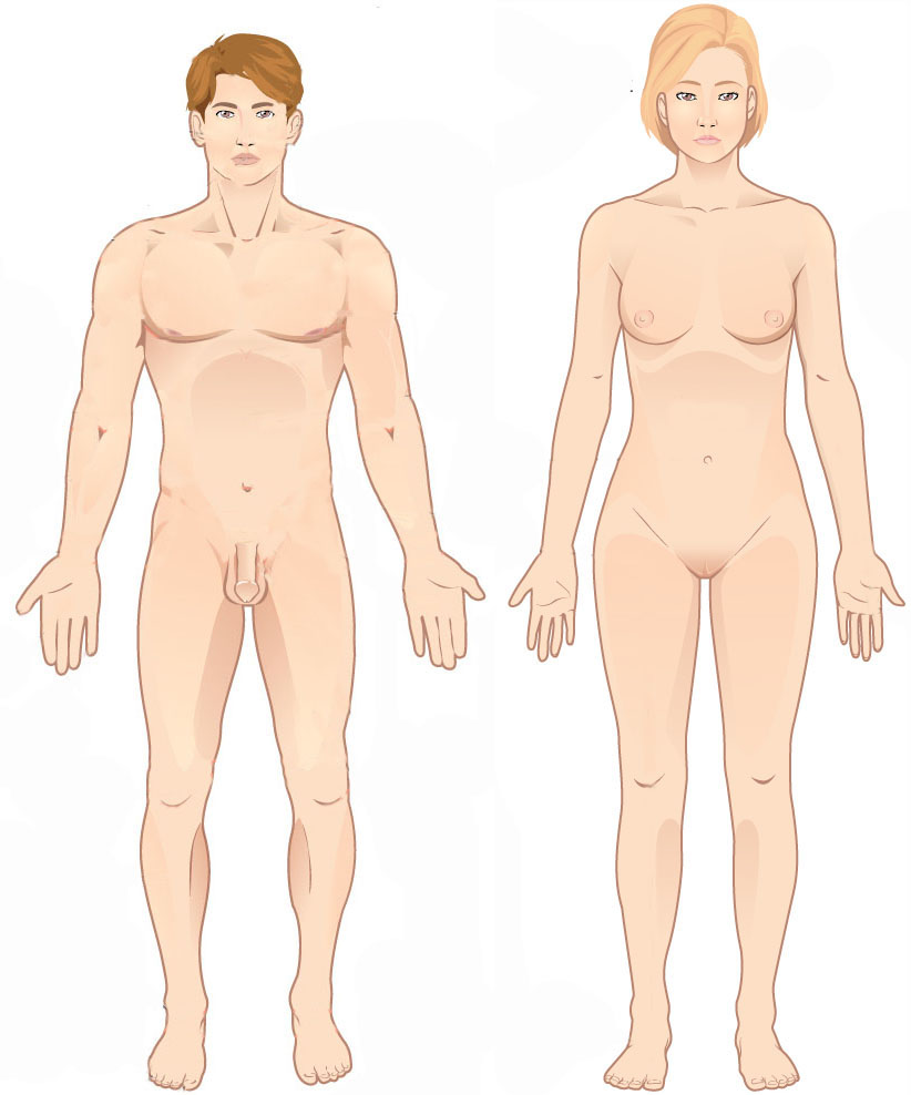
The standard anatomical position, or standard anatomical model, is the scientifically agreed upon reference position for anatomical location terms. Standard anatomical positions are used to standardise the position of appendages of animals with respect to the main body of the organism. In medical disciplines, all references to a location on or in the body are made based upon the standard anatomical position. A straight position is assumed when describing a proximo-distal axis (towards or away from a point of attachment). This helps avoid confusion in terminology when referring to the same organism in different postures. For example, if the elbow is flexed, the hand remains distal to the shoulder even if it approaches the shoulder. Human anatomy In standard anatomical position, the human body is standing erect and at rest. Unlike the situation in other vertebrates, the limbs are placed in positions reminiscent of the supine position imposed on cadavers during autopsy. Therefo ... [...More Info...] [...Related Items...] OR: [Wikipedia] [Google] [Baidu] |
Spinal Cord
The spinal cord is a long, thin, tubular structure made up of nervous tissue, which extends from the medulla oblongata in the brainstem to the lumbar region of the vertebral column (backbone). The backbone encloses the central canal of the spinal cord, which contains cerebrospinal fluid. The brain and spinal cord together make up the central nervous system (CNS). In humans, the spinal cord begins at the occipital bone, passing through the foramen magnum and then enters the spinal canal at the beginning of the cervical vertebrae. The spinal cord extends down to between the first and second lumbar vertebrae, where it ends. The enclosing bony vertebral column protects the relatively shorter spinal cord. It is around long in adult men and around long in adult women. The diameter of the spinal cord ranges from in the cervical and lumbar regions to in the thoracic area. The spinal cord functions primarily in the transmission of nerve signals from the motor cortex to the body, ... [...More Info...] [...Related Items...] OR: [Wikipedia] [Google] [Baidu] |
Paramedian Pontine Reticular Formation
The paramedian pontine reticular formation, also known as PPRF or paraabducens nucleus, is part of the pontine reticular formation, a brain region without clearly defined borders in the center of the pons. It is involved in the coordination of eye movements, particularly horizontal gaze and saccades. Input, output, and function The PPRF is located anterior and lateral to the medial longitudinal fasciculus (MLF). It receives input from the superior colliculus via the predorsal bundle and from the frontal eye fields via frontopontine fibers. The rostral PPRF probably coordinates vertical saccades; the caudal PPRF may be the generator of horizontal saccades. In particular, activity of the excitatory burst neurons (EBNs) in the PPRF generates the "pulse" movement that initiates a saccade. In the case of horizontal saccades the "pulse" information is conveyed via axonal fibres to the abducens nucleus, initiating lateral eye movements. The angular velocity of the eye during horizontal ... [...More Info...] [...Related Items...] OR: [Wikipedia] [Google] [Baidu] |
