|
Medial Tibial Stress Syndrome
A shin splint, also known as medial tibial stress syndrome, is pain along the inside edge of the shinbone (tibia) due to inflammation of tissue in the area. Generally this is between the middle of the lower leg and the ankle. The pain may be dull or sharp, and is generally brought on by high-impact exercise that overloads the tibia. It generally resolves during periods of rest. Complications may include stress fractures. Shin splints typically occur due to excessive physical activity. Groups that are commonly affected include runners, dancers, gymnasts, and military personnel. The underlying mechanism is not entirely clear. Diagnosis is generally based on the symptoms, with medical imaging done to rule out other possible causes. Shin splints are generally treated by rest followed by a gradual return to exercise over a period of weeks. Other measures such as nonsteroidal anti-inflammatory drugs (NSAIDs), cold packs, physical therapy, and compression may be used. Shoe insoles may ... [...More Info...] [...Related Items...] OR: [Wikipedia] [Google] [Baidu] |
Tibia
The tibia (; ), also known as the shinbone or shankbone, is the larger, stronger, and anterior (frontal) of the two bones in the leg below the knee in vertebrates (the other being the fibula, behind and to the outside of the tibia); it connects the knee with the ankle. The tibia is found on the medial side of the leg next to the fibula and closer to the median plane. The tibia is connected to the fibula by the interosseous membrane of leg, forming a type of fibrous joint called a syndesmosis with very little movement. The tibia is named for the flute ''tibia''. It is the second largest bone in the human body, after the femur. The leg bones are the strongest long bones as they support the rest of the body. Structure In human anatomy, the tibia is the second largest bone next to the femur. As in other vertebrates the tibia is one of two bones in the lower leg, the other being the fibula, and is a component of the knee and ankle joints. The ossification or formation of the bone ... [...More Info...] [...Related Items...] OR: [Wikipedia] [Google] [Baidu] |
Compliant Mechanism
In mechanical engineering, a compliant mechanism is a flexible mechanism that achieves force and motion transmission through elastic body deformation. It gains some or all of its motion from the relative flexibility of its members rather than from rigid-body joints alone. These may be monolithic (single-piece) or jointless structures. Some common devices that use compliant mechanisms are backpack latches and paper clips. One of the oldest examples of using compliant structures is the bow and arrow. Design methods Compliant mechanisms are usually designed using two techniques: Kinematics approach Kinematic analysis can be used to design a compliant mechanism by creating a pseudo- rigid-body model of the mechanism. In this model, flexible segments are modeled as rigid links connected to revolute joints with torsional springs. Other structures can be modeled as a combination of rigid links, springs, and dampers. Structural optimization approach In this method, computationa ... [...More Info...] [...Related Items...] OR: [Wikipedia] [Google] [Baidu] |
Popliteal Artery Entrapment Syndrome
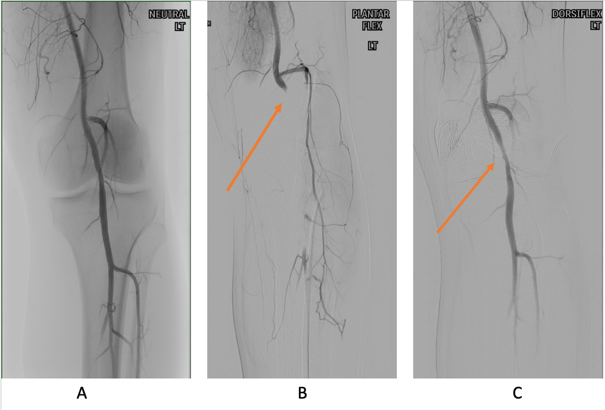
The popliteal artery entrapment syndrome (PAES) is an uncommon pathology that occurs when the popliteal artery is compressed by the surrounding popliteal fossa myofascial structures. This results in claudication and chronic leg ischemia. This condition mainly occurs more in young athletes than in the elderlies. Elderlies, who present with similar symptoms, are more likely to be diagnosed with peripheral artery disease with associated atherosclerosis. Patients with PAES mainly present with intermittent feet and calf pain associated with exercises and relieved with rest. PAES can be diagnosed with a combination of medical history, physical examination, and advanced imaging modalities such as duplex ultrasound, computer tomography, or magnetic resonance angiography. Management can range from non-intervention to open surgical decompression with a generally good prognosis. Complications of untreated PAES can include stenotic artery degeneration, complete popliteal artery occlusion, dis ... [...More Info...] [...Related Items...] OR: [Wikipedia] [Google] [Baidu] |
Nerve Entrapment
Nerve compression syndrome, or compression neuropathy, or nerve entrapment syndrome, is a medical condition caused by direct pressure on a nerve. It is known colloquially as a ''trapped nerve'', though this may also refer to nerve root compression (by a herniated disc, for example). Its symptoms include pain, tingling, numbness and muscle weakness. The symptoms affect just one particular part of the body, depending on which nerve is affected. Nerve conduction studies help to confirm the diagnosis. In some cases, surgery may help to relieve the pressure on the nerve but this does not always relieve all the symptoms. Nerve injury by a single episode of physical trauma is in one sense a compression neuropathy but is not usually included under this heading. Syndromes * Upper limb * Lower limb, abdomen and pelvis Signs and symptoms Tingling, numbness, and/or a burning sensation in the area of the body affected by the corresponding nerve. These experiences may occur directly fol ... [...More Info...] [...Related Items...] OR: [Wikipedia] [Google] [Baidu] |
Compartment Syndrome
Compartment syndrome is a condition in which increased pressure within one of the body's anatomical compartments results in insufficient blood supply to tissue within that space. There are two main types: acute and chronic. Compartments of the leg or arm are most commonly involved. Symptoms of acute compartment syndrome (ACS) can include severe pain, poor pulses, decreased ability to move, numbness, or a pale color of the affected limb. It is most commonly due to physical trauma such as a bone fracture (up to 75% of cases) or crush injury, but it can also be caused by acute exertion during sport. It can also occur after blood flow returns following a period of poor blood flow. Diagnosis is generally based upon a person's symptoms and may be supported by measurement of intracompartmental pressure before, during, and after activity. Normal compartment pressure should be within 12-18 mmHg; anything greater than that is considered abnormal and would need treatment. Treatment i ... [...More Info...] [...Related Items...] OR: [Wikipedia] [Google] [Baidu] |
Stress Fractures
A stress fracture is a fatigue-induced bone fracture caused by repeated stress over time. Instead of resulting from a single severe impact, stress fractures are the result of accumulated injury from repeated submaximal loading, such as running or jumping. Because of this mechanism, stress fractures are common overuse injuries in athletes. Stress fractures can be described as small cracks in the bone, or hairline fractures. Stress fractures of the foot are sometimes called "march fractures" because of the injury's prevalence among heavily marching soldiers. Stress fractures most frequently occur in weight-bearing bones of the lower extremities, such as the tibia and fibula (bones of the lower leg), metatarsal and navicular bones (bones of the foot). Less common are stress fractures to the femur, pelvis, and sacrum. Treatment usually consists of rest followed by a gradual return to exercise over a period of months. Signs and symptoms Stress fractures are typically discovered after ... [...More Info...] [...Related Items...] OR: [Wikipedia] [Google] [Baidu] |
Physical Examination
In a physical examination, medical examination, or clinical examination, a medical practitioner examines a patient for any possible medical signs or symptoms of a medical condition. It generally consists of a series of questions about the patient's medical history followed by an examination based on the reported symptoms. Together, the medical history and the physical examination help to determine a diagnosis and devise the treatment plan. These data then become part of the medical record. Types Routine The ''routine physical'', also known as ''general medical examination'', ''periodic health evaluation'', ''annual physical'', ''comprehensive medical exam'', ''general health check'', ''preventive health examination'', ''medical check-up'', or simply ''medical'', is a physical examination performed on an asymptomatic patient for medical screening purposes. These are normally performed by a pediatrician, family practice physician, physician assistant, a certified nurse pr ... [...More Info...] [...Related Items...] OR: [Wikipedia] [Google] [Baidu] |
Periosteum
The periosteum is a membrane that covers the outer surface of all bones, except at the articular surfaces (i.e. the parts within a joint space) of long bones. Endosteum lines the inner surface of the medullary cavity of all long bones. Structure The periosteum consists of an outer fibrous layer, and an inner cambium layer (or osteogenic layer). The fibrous layer is of dense irregular connective tissue, containing fibroblasts, while the cambium layer is highly cellular containing progenitor cells that develop into osteoblasts. These osteoblasts are responsible for increasing the width of a long bone and the overall size of the other bone types. After a bone fracture, the progenitor cells develop into osteoblasts and chondroblasts, which are essential to the healing process. The outer fibrous layer and the inner cambium layer is differentiated under electron micrography. As opposed to osseous tissue, the periosteum has nociceptors, sensory neurons that make it very sensitive to ... [...More Info...] [...Related Items...] OR: [Wikipedia] [Google] [Baidu] |
Fascia
A fascia (; plural fasciae or fascias; adjective fascial; from Latin: "band") is a band or sheet of connective tissue, primarily collagen, beneath the skin that attaches to, stabilizes, encloses, and separates muscles and other internal organs. Fascia is classified by layer, as superficial fascia, deep fascia, and ''visceral'' or ''parietal'' fascia, or by its function and anatomical location. Like ligaments, aponeuroses, and tendons, fascia is made up of fibrous connective tissue containing closely packed bundles of collagen fibers oriented in a wavy pattern parallel to the direction of pull. Fascia is consequently flexible and able to resist great unidirectional tension forces until the wavy pattern of fibers has been straightened out by the pulling force. These collagen fibers are produced by fibroblasts located within the fascia. Fasciae are similar to ligaments and tendons as they have collagen as their major component. They differ in their location and function: ligament ... [...More Info...] [...Related Items...] OR: [Wikipedia] [Google] [Baidu] |
Sharpey's Fibres
Sharpey's fibres (bone fibres, or perforating fibres) are a Matrix (biology), matrix of connective tissue consisting of bundles of strong predominantly type I Collagen, collagen fibres connecting periosteum to bone. They are part of the outer fibrous layer of periosteum, entering into the outer circumferential and interstitial Lamellae (zoology), lamellae of bone tissue. Sharpey's fibres are also used to attach muscle to the periosteum of bone by merging with the fibrous periosteum and underlying bone as well. A good example is the attachment of the rotator cuff muscles to the blade of the scapula. In the Tooth, teeth, Sharpey's fibres are the terminal ends of principal fibres (of the periodontal ligament) that insert into the cementum and into the periosteum of the alveolar bone. A study on rats suggests that the three-dimensional structure of Sharpey's fibres intensifies the continuity between the periodontal ligament fibre and the Dental alveolus, alveolar bone (tooth socket), ... [...More Info...] [...Related Items...] OR: [Wikipedia] [Google] [Baidu] |
Flexor Digitorum Longus
The flexor digitorum longus muscle is situated on the tibial side of the Human leg, leg. At its origin it is thin and pointed, but it gradually increases in size as it descends. It serves to flex the second, third, fourth, and fifth toes. Structure The flexor digitorum longus muscle arises from the posterior surface of the body of the tibia, from immediately below the soleal line to within 7 or 8 cm of its lower extremity, medial to the tibial origin of the tibialis posterior muscle. It also arises from the fascia covering the tibialis posterior muscle. The fibers end in a tendon, which runs nearly the whole length of the posterior surface of the muscle. This tendon passes behind the medial malleolus, in a groove, common to it and the tibialis posterior, but separated from the latter by a fibrous septum, each tendon being contained in a special compartment lined by a separate mucous sheath. The tendon of the tibialis posterior and the tendon of the flexor digitorum longus cr ... [...More Info...] [...Related Items...] OR: [Wikipedia] [Google] [Baidu] |


