|
Marginal-zone B Cell
Marginal zone B cells (MZ B cells) are noncirculating mature B cells that in humans segregate anatomically into the marginal zone (MZ) of the spleen and certain other types of lymphoid tissue. The MZ B cells within this region typically express low-affinity polyreactive B-cell receptors (BCR), high levels of IgM, Toll-like receptors (TLRs), CD21, CD1, CD9, CD27 with low to negligible levels of secreted-IgD, CD23, CD5, and CD11b that help to distinguish them phenotypically from follicular (FO) B cells and B1 B cells. MZ B cells are innate-like B cells specialized to mount rapid T-independent, but also T-dependent responses against blood-borne pathogens. They are also known to be the main producers of IgM antibodies in humans. Development and differentiation The spleen's marginal zone contains multiple subtypes of macrophages and dendritic cells interlaced with the MZ B cells; it is not fully formed until 2 to 3 weeks after birth in rodents and 1 to 2 years in humans. In h ... [...More Info...] [...Related Items...] OR: [Wikipedia] [Google] [Baidu] |
Peyer's Patch
Peyer's patches (or aggregated lymphoid nodules) are organized lymphoid follicles, named after the 17th-century Swiss anatomist Johann Conrad Peyer. * Reprinted as: * Peyer referred to Peyer's patches as ''plexus'' or ''agmina glandularum'' (clusters of glands). From (Peyer, 1681), p. 7: ''"Tenui a perfectiorum animalium Intestina accuratius perlustranti, crebra hinc inde, variis intervallis, corpusculorum glandulosorum Agmina sive Plexus se produnt, diversae Magnitudinis atque Figurae."'' (I knew from careful study of more advanced animals, the intestines bear — often here and there, at various intervals — clusters of glandular small bodies or "plexuses" of diverse size and shape.) From p. 15: ''"(has Plexus seu agmina Glandularum voco)"'' (I call them "plexuses" or clusters of glands) He described their appearance. From p. 8: ''"Horum vero Plexuum facies modo in orbem concinnata; modo in Ovi aut Olivae oblongam, aliamve angulosam ac magis anomalam disposita figuram cer ... [...More Info...] [...Related Items...] OR: [Wikipedia] [Google] [Baidu] |
VCAM-1
Vascular cell adhesion protein 1 also known as vascular cell adhesion molecule 1 (VCAM-1) or cluster of differentiation 106 (CD106) is a protein that in humans is encoded by the ''VCAM1'' gene. VCAM-1 functions as a cell adhesion molecule. Structure VCAM-1 is a member of the immunoglobulin superfamily, the superfamily of proteins including antibodies and T-cell receptors. The VCAM-1 gene contains six or seven immunoglobulin domains, and is expressed on both large and small blood vessels only after the endothelial cells are stimulated by cytokines. It is alternatively spliced into two known RNA transcripts that encode different isoforms in humans. The gene product is a cell surface sialoglycoprotein, a type I membrane protein that is a member of the Ig superfamily. Function The VCAM-1 protein mediates the adhesion of lymphocytes, monocytes, eosinophils, and basophils to vascular endothelium. It also functions in leukocyte-endothelial cell signal transduction, and it may ... [...More Info...] [...Related Items...] OR: [Wikipedia] [Google] [Baidu] |
VLA-4
Integrin α4β1 (very late antigen-4) is an integrin dimer. It is composed of CD49d (alpha 4) and CD29 (beta 1). The alpha 4 subunit is 155 kDa, and the beta 1 subunit is 150 kDa. Function The integrin VLA-4 is expressed on the cell surfaces of stem cells, progenitor cells, T and B cells, monocytes, natural killer cells, eosinophils, but not neutrophils. It functions to promote an inflammatory response by the immune system by assisting in the movement of leukocytes to tissue that requires inflammation. It is a key player in cell adhesion. However, VLA-4 does not adhere to its appropriate ligands until the leukocytes are activated by chemotactic agents or other stimuli (often produced by the endothelium or other cells at the site of injury). VLA-4's primary ligands include VCAM-1 and fibronectin. One activating chemokine is SDF-1. Following SDF-1 binding, the integrin undergoes a conformational change of the alpha and beta domains that is necessary to confer high binding a ... [...More Info...] [...Related Items...] OR: [Wikipedia] [Google] [Baidu] |
ICAM-1
ICAM-1 (Intercellular Adhesion Molecule 1) also known as CD54 (Cluster of Differentiation 54) is a protein that in humans is encoded by the ''ICAM1'' gene. This gene encodes a cell surface glycoprotein which is typically expressed on endothelial cells and cells of the immune system. It binds to integrins of type CD11a / CD18, or CD11b / CD18 and is also exploited by rhinovirus as a receptor for entry into respiratory epithelium. Structure ICAM-1 is a member of the immunoglobulin superfamily, the superfamily of proteins including antibodies and T-cell receptors. ICAM-1 is a transmembrane protein possessing an amino-terminus extracellular domain, a single transmembrane domain, and a carboxy-terminus cytoplasmic domain. The structure of ICAM-1 is characterized by heavy glycosylation, and the protein’s extracellular domain is composed of multiple loops created by disulfide bridges within the protein. The dominant secondary structure of the protein is the beta sheet, leading resea ... [...More Info...] [...Related Items...] OR: [Wikipedia] [Google] [Baidu] |
LFA-1
Lymphocyte function-associated antigen 1 (LFA-1) is an integrin found on lymphocytes and other leukocytes. LFA-1 plays a key role in emigration, which is the process by which leukocytes leave the bloodstream to enter the tissues. LFA-1 also mediates firm arrest of leukocytes. Additionally, LFA-1 is involved in the process of cytotoxic T cell mediated killing as well as antibody mediated killing by granulocytes and monocytes. As of 2007, LFA-1 has 6 known ligands: ICAM-1, ICAM-2, ICAM-3, ICAM-4, ICAM-5, and JAM-A. LFA-1/ICAM-1 interactions have recently been shown to stimulate signaling pathways that influence T cell differentiation. LFA-1 belongs to the integrin superfamily of adhesion molecules. Structure LFA-1 is a heterodimeric glycoprotein with non-covalently linked subunits. LFA-1 has two subunits designated as the alpha subunit and beta subunit. The alpha subunit was named aL in 1983. The alpha subunit is designated CD11a; and the beta subunit, unique to leukocytes, is bet ... [...More Info...] [...Related Items...] OR: [Wikipedia] [Google] [Baidu] |
Follicular Dendritic Cells
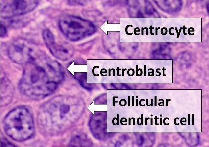
Follicular dendritic cells (FDC) are cells of the immune system found in primary and secondary lymph follicles (lymph nodes) of the B cell areas of the lymphoid tissue. Unlike dendritic cells (DC), FDCs are not derived from the bone-marrow hematopoietic stem cell, but are of mesenchymal origin. Possible functions of FDC include: organizing lymphoid tissue's cells and microarchitecture, capturing antigen to support B cell, promoting debris removal from germinal centers, and protecting against autoimmunity. Disease processes that FDC may contribute include primary FDC-tumor, chronic inflammatory conditions, HIV-1 infection development, and neuroinvasive scrapie. Location and molecular markers Follicular DCs are a non-migratory population found in primary and secondary follicles of the B cell areas of lymph nodes, spleen, and mucosa-associated lymphoid tissue (MALT). They form a stable network due to intercellular connections between FDCs processes and intimate interaction ... [...More Info...] [...Related Items...] OR: [Wikipedia] [Google] [Baidu] |
Granulocyte
Granulocytes are cells in the innate immune system characterized by the presence of specific granules in their cytoplasm. Such granules distinguish them from the various agranulocytes. All myeloblastic granulocytes are polymorphonuclear. They have varying shapes (morphology) of the nucleus (segmented, irregular; often lobed into three segments); and are referred to as polymorphonuclear leukocytes (PMN, PML, or PMNL). In common terms, ''polymorphonuclear granulocyte'' refers specifically to " neutrophil granulocytes", the most abundant of the granulocytes; the other types ( eosinophils, basophils, and mast cells) have varying morphology. Granulocytes are produced via granulopoiesis in the bone marrow. Types There are four types of granulocytes (full name polymorphonuclear granulocytes): * Basophils * Eosinophils * Neutrophils * Mast cells Except for the mast cells, their names are derived from their staining characteristics; for example, the most abundant granulocyte ... [...More Info...] [...Related Items...] OR: [Wikipedia] [Google] [Baidu] |
T Cell
A T cell is a type of lymphocyte. T cells are one of the important white blood cells of the immune system and play a central role in the adaptive immune response. T cells can be distinguished from other lymphocytes by the presence of a T-cell receptor (TCR) on their cell surface. T cells are born from hematopoietic stem cells, found in the bone marrow. Developing T cells then migrate to the thymus gland to develop (or mature). T cells derive their name from the thymus. After migration to the thymus, the precursor cells mature into several distinct types of T cells. T cell differentiation also continues after they have left the thymus. Groups of specific, differentiated T cell subtypes have a variety of important functions in controlling and shaping the immune response. One of these functions is immune-mediated cell death, and it is carried out by two major subtypes: CD8+ "killer" and CD4+ "helper" T cells. (These are named for the presence of the cell surface proteins ... [...More Info...] [...Related Items...] OR: [Wikipedia] [Google] [Baidu] |
NOTCH2
Neurogenic locus notch homolog protein 2 (Notch 2) is a protein that in humans is encoded by the ''NOTCH2'' gene. NOTCH2 is associated with Alagille syndrome and Hajdu–Cheney syndrome. Function Notch 2 is a member of the notch family. Members of this Type 1 transmembrane protein family share structural characteristics including an extracellular domain consisting of multiple epidermal growth factor-like (EGF) repeats, and an intracellular domain consisting of multiple, different domain types. Notch family members play a role in a variety of developmental processes by controlling cell fate decisions. The Notch signaling network is an evolutionarily conserved intercellular signaling pathway that regulates interactions between physically adjacent cells. In ''Drosophila'', notch interaction with its cell-bound ligands (delta, serrate) establishes an intercellular signaling pathway that plays a key role in development. Homologues of the notch-ligands have also been identified in hum ... [...More Info...] [...Related Items...] OR: [Wikipedia] [Google] [Baidu] |
PTK2B
Protein tyrosine kinase 2 beta is an enzyme that in humans is encoded by the ''PTK2B'' gene. Function This gene encodes a cytoplasmic protein tyrosine kinase that is involved in calcium-induced regulation of ion channels and activation of the map kinase signaling pathway. The encoded protein may represent an important signaling intermediate between neuropeptide-activated receptors or neurotransmitters that increase calcium flux and the downstream signals that regulate neuronal activity. The encoded protein undergoes rapid tyrosine phosphorylation and activation in response to increases in the intracellular calcium concentration , nicotinic acetylcholine receptor activation, membrane depolarization, or protein kinase C activation. In addition, SOCE-induced Pyk2 activation mediates disassembly of endothelial adherens junctions, via tyrosine (Y1981-residue) phosphorylation of VE-PTP. This protein has been shown to bind a CRK-associated substrate, a nephrocystin, a GTPase regu ... [...More Info...] [...Related Items...] OR: [Wikipedia] [Google] [Baidu] |
Pathogen-associated Molecular Pattern
Pathogen-associated molecular patterns (PAMPs) are small molecular motifs conserved within a class of microbes. They are recognized by toll-like receptors (TLRs) and other pattern recognition receptors (PRRs) in both plants and animals. A vast array of different types of molecules can serve as PAMPs, including glycans and glycoconjugates. PAMPs activate innate immune responses, protecting the host from infection, by identifying some conserved nonself molecules. Bacterial lipopolysaccharides (LPSs), endotoxins found on the cell membranes of gram-negative bacteria, are considered to be the prototypical class of PAMPs. LPSs are specifically recognised by TLR4, a recognition receptor of the innate immune system. Other PAMPs include bacterial flagellin (recognized by TLR5), lipoteichoic acid from gram-positive bacteria (recognized by TLR2), peptidoglycan (recognized by TLR2), and nucleic acid variants normally associated with viruses, such as double-stranded RNA ( dsRNA), recogni ... [...More Info...] [...Related Items...] OR: [Wikipedia] [Google] [Baidu] |


