|
Major Aortopulmonary Collateral Artery
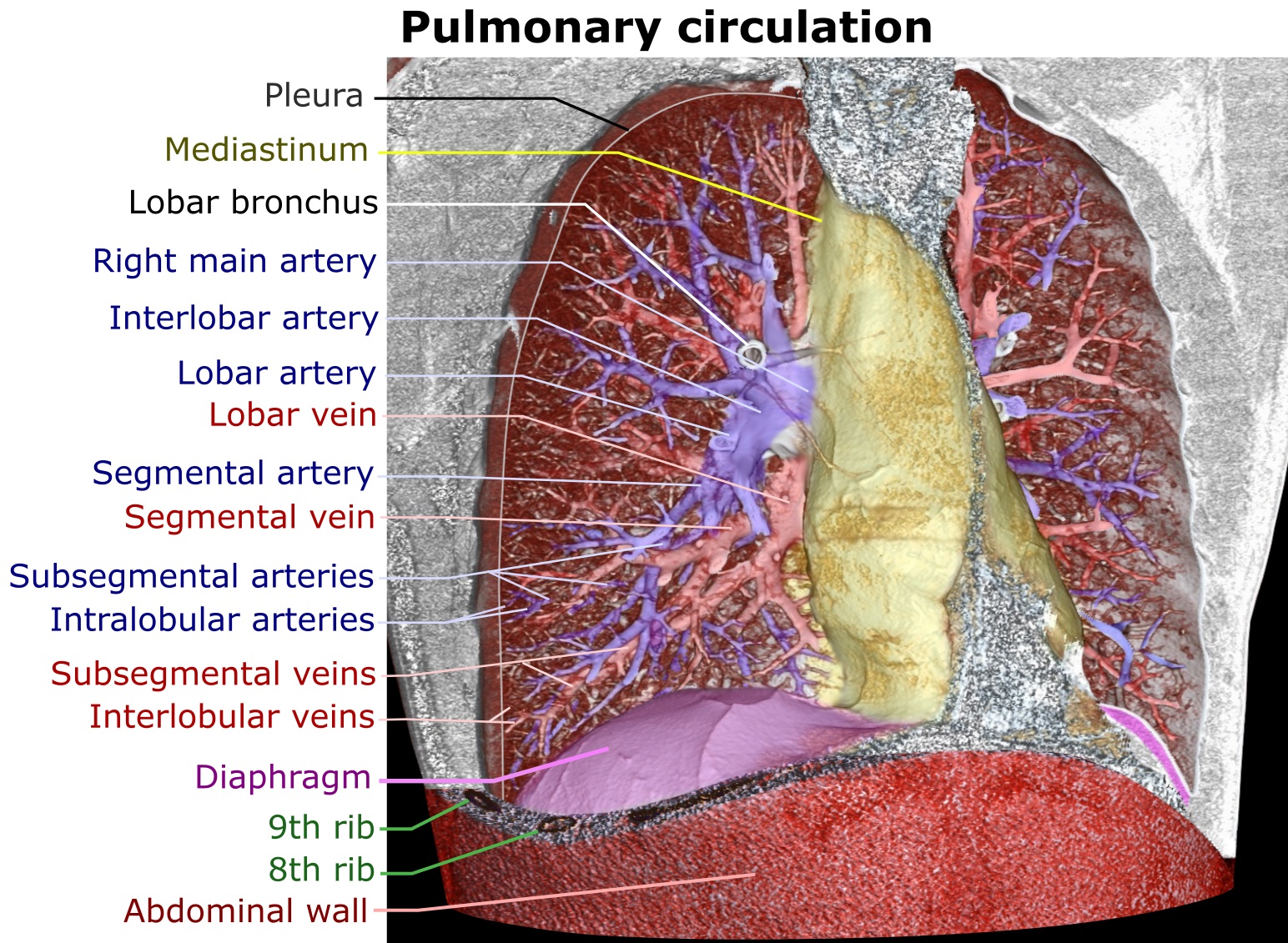
Major aortopulmonary collateral arteries (or MAPCAs) are arteries that develop to supply blood to the lungs when native pulmonary circulation is underdeveloped. Instead of coming from the pulmonary trunk, supply develops from the aorta and other systemic arteries. Pathogenesis and anatomy Major aortopulmonary collateral arteries (MAPCAs) develop early in embryonic life but regress as the normal pulmonary arteries (vessels that will supply deoxygenated blood to the lungs) develop. In certain heart conditions the pulmonary arteries do not develop. The collaterals continue to grow, and can become the main supply of blood to the lungs. Though it is usually associated with congenital heart diseases with decreased pulmonary blood flow like tetralogy of Fallot or pulmonary atresia it may be seen sometimes in isolation i.e. not associated with any congenital heart disease in that case it is termed as isolated aortopulmonary collateral artery. In these cases it may be one of the cause of con ... [...More Info...] [...Related Items...] OR: [Wikipedia] [Google] [Baidu] |
Pulmonary Trunk
A pulmonary artery is an artery in the pulmonary circulation that carries deoxygenated blood from the right side of the heart to the lungs. The largest pulmonary artery is the ''main pulmonary artery'' or ''pulmonary trunk'' from the heart, and the smallest ones are the arterioles, which lead to the capillaries that surround the pulmonary alveoli. Structure The pulmonary arteries are blood vessels that carry systemic venous blood from the right ventricle of the heart to the microcirculation of the lungs. Unlike in other organs where arteries supply oxygenated blood, the blood carried by the pulmonary arteries is deoxygenated, as it is venous blood returning to the heart. The main pulmonary arteries emerge from the right side of the heart, and then split into smaller arteries that progressively divide and become arterioles, eventually narrowing into the capillary microcirculation of the lungs where gas exchange occurs. Pulmonary trunk In order of blood flow, the pulmonary art ... [...More Info...] [...Related Items...] OR: [Wikipedia] [Google] [Baidu] |
Aorta
The aorta ( ) is the main and largest artery in the human body, originating from the left ventricle of the heart and extending down to the abdomen, where it splits into two smaller arteries (the common iliac arteries). The aorta distributes oxygenated blood to all parts of the body through the systemic circulation. Structure Sections In anatomical sources, the aorta is usually divided into sections. One way of classifying a part of the aorta is by anatomical compartment, where the thoracic aorta (or thoracic portion of the aorta) runs from the heart to the diaphragm. The aorta then continues downward as the abdominal aorta (or abdominal portion of the aorta) from the diaphragm to the aortic bifurcation. Another system divides the aorta with respect to its course and the direction of blood flow. In this system, the aorta starts as the ascending aorta, travels superiorly from the heart, and then makes a hairpin turn known as the aortic arch. Following the aortic arch ... [...More Info...] [...Related Items...] OR: [Wikipedia] [Google] [Baidu] |
Embryo
An embryo is an initial stage of development of a multicellular organism. In organisms that reproduce sexually, embryonic development is the part of the life cycle that begins just after fertilization of the female egg cell by the male sperm cell. The resulting fusion of these two cells produces a single-celled zygote that undergoes many cell divisions that produce cells known as blastomeres. The blastomeres are arranged as a solid ball that when reaching a certain size, called a morula, takes in fluid to create a cavity called a blastocoel. The structure is then termed a blastula, or a blastocyst in mammals. The mammalian blastocyst hatches before implantating into the endometrial lining of the womb. Once implanted the embryo will continue its development through the next stages of gastrulation, neurulation, and organogenesis. Gastrulation is the formation of the three germ layers that will form all of the different parts of the body. Neurulation forms the nervous ... [...More Info...] [...Related Items...] OR: [Wikipedia] [Google] [Baidu] |
Pulmonary Atresia With Ventricular Septal Defect
Pulmonary atresia with ventricular septal defect is a rare birth defect characterized by pulmonary valve atresia occurring alongside a defect on the right ventricular outflow tract. It is a type of congenital heart disease/defect, and one of the two recognized subtypes of pulmonary atresia, the other being pulmonary atresia with intact ventricular septum. Signs and symptoms The condition consists of atresia affecting the pulmonary valve and a hypoplastic right ventricular outflow tract. The ventricular septal defect doesn't impede the in and outflowing of blood in the ventricular septum, which helps it form during fetal life. The spectrum of symptoms exhibited by children with this condition depends on the severity of the condition, while some barely show symptoms, others might develop complications such as congestive heart failure. In symptomatic children, symptoms become apparent soon after birth, these usually consist of the following: * Cyanosis *Breathing difficul ... [...More Info...] [...Related Items...] OR: [Wikipedia] [Google] [Baidu] |
Tetralogy Of Fallot
Tetralogy of Fallot (TOF), formerly known as Steno-Fallot tetralogy, is a congenital heart defect characterized by four specific cardiac defects. Classically, the four defects are: *pulmonary stenosis, which is narrowing of the exit from the right ventricle; * a ventricular septal defect, which is a hole allowing blood to flow between the two ventricles; * right ventricular hypertrophy, which is thickening of the right ventricular muscle; and * an overriding aorta, which is where the aorta expands to allow blood from both ventricles to enter. At birth, children may be asymptomatic or present with many severe symptoms. Later in infancy, there are typically episodes of bluish colour to the skin due to a lack of sufficient oxygenation, known as cyanosis. When affected babies cry or have a bowel movement, they may undergo a "tet spell" where they turn cyanotic, have difficulty breathing, become limp, and occasionally lose consciousness. Other symptoms may include a heart murmur, ... [...More Info...] [...Related Items...] OR: [Wikipedia] [Google] [Baidu] |


