|
Macular Corneal Dystrophy
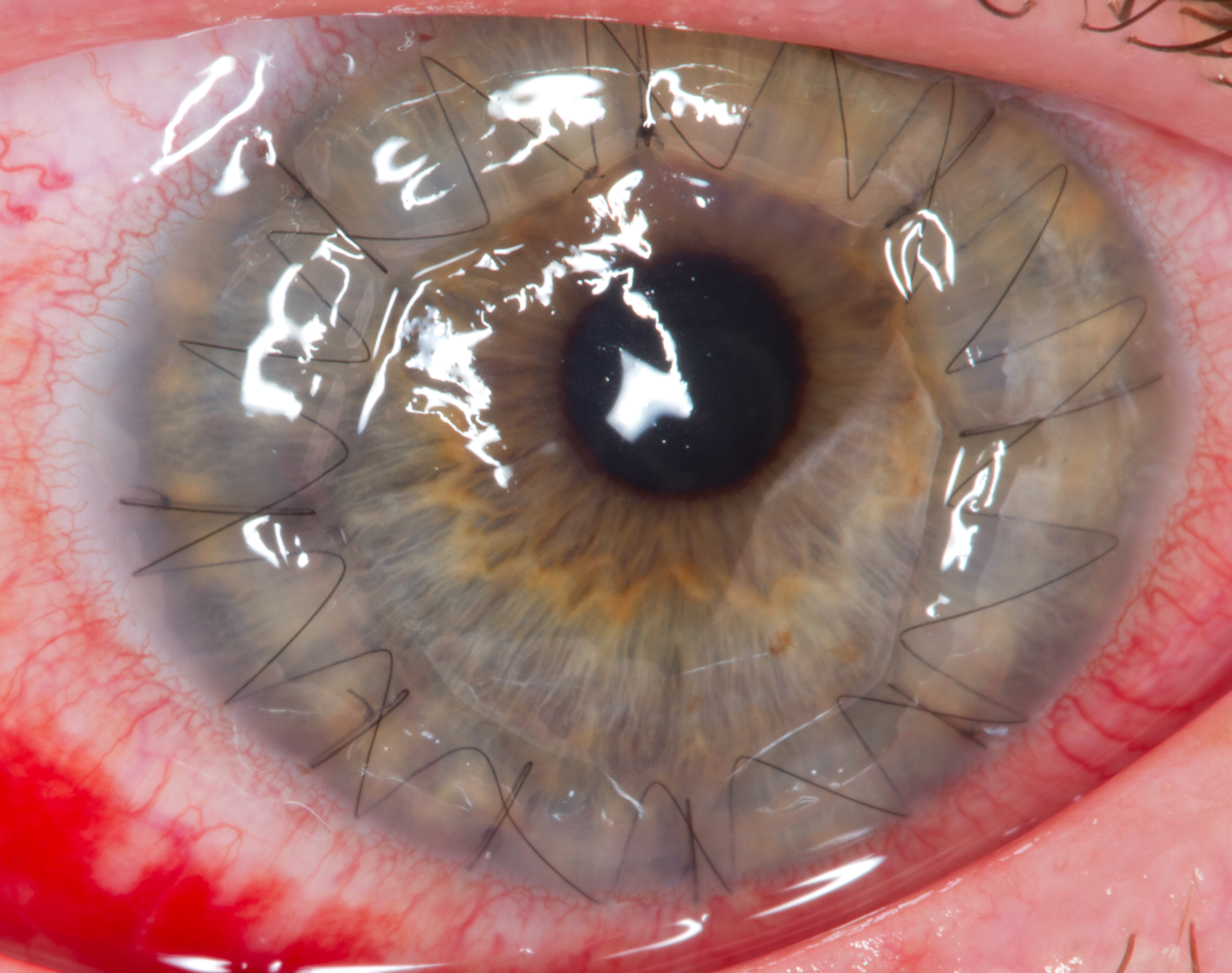
Macular corneal dystrophy, also known as Fehr corneal dystrophy named for German ophthalmologist Oskar Fehr (1871-1959), is a rare pathological condition affecting the stroma of cornea. The first signs are usually noticed in the first decade of life, and progress afterwards, with opacities developing in the cornea and attacks of pain. The condition was first described by Arthur Groenouw in 1890.Groenouw A. Knötchenförmige Hornhauttrübungen (noduli corneae). Arch Augenheilkunde. 1890;21:281–289. Signs and symptoms Onset occurs in the first decade, usually between ages 5 and 9. The disorder is progressive, vision changes with ageing from 2nd decade to 3rd visual impairment may seen in 4th and 5th decade severe visual impairment can be seen Minute, gray, punctate opacities develop. Corneal sensitivity is usually reduced. Painful attacks with photophobia, foreign body sensations, and recurrent erosions occur in most patients. Macular corneal dystrophy is very common in Iceland an ... [...More Info...] [...Related Items...] OR: [Wikipedia] [Google] [Baidu] |
Ophthalmologist
Ophthalmology ( ) is a surgery, surgical subspecialty within medicine that deals with the diagnosis and treatment of eye disorders. An ophthalmologist is a physician who undergoes subspecialty training in medical and surgical eye care. Following a medical degree, a doctor specialising in ophthalmology must pursue additional postgraduate residency (medicine), residency training specific to that field. This may include a one-year integrated internship that involves more general medical training in other fields such as internal medicine or general surgery. Following residency, additional specialty training (or fellowship) may be sought in a particular aspect of eye pathology. Ophthalmologists prescribe medications to treat eye diseases, implement laser therapy, and perform surgery when needed. Ophthalmologists provide both primary and specialty eye care - medical and surgical. Most ophthalmologists participate in academic research on eye diseases at some point in their training an ... [...More Info...] [...Related Items...] OR: [Wikipedia] [Google] [Baidu] |
Stroma Of Cornea
The stroma of the cornea (or substantia propria) is a fibrous, tough, unyielding, perfectly transparent and the thickest layer of the cornea of the eye. It is between Bowman's membrane anteriorly, and Descemet's membrane posteriorly. At its centre, human corneal stroma is composed of about 200 flattened ''lamellæ'' (layers of collagen fibrils), superimposed one on another. They are each about 1.5-2.5 μm in thickness. The anterior lamellæ interweave more than posterior lamellæ. The fibrils of each lamella are parallel with one another, but at different angles to those of adjacent lamellæ. The lamellæ are produced by keratocytes (corneal connective tissue cells), which occupy about 10% of the substantia propria. Apart from the cells, the major non-aqueous constituents of the stroma are collagen fibrils and proteoglycans. The collagen fibrils are made of a mixture of type I and type V collagens. These molecules are tilted by about 15 degrees to the fibril axis, and because of ... [...More Info...] [...Related Items...] OR: [Wikipedia] [Google] [Baidu] |
Arthur Groenouw
Arthur Groenouw (27 March 1862 – 1945) was a German ophthalmologist born in Bosatz, a village near Racibórz, Ratibor. He studied medicine in University of Breslau, Breslau, and was an assistant to physiologist Rudolf Heidenhain (1834–1897) and ophthalmologist Wilhelm Uhthoff (1853–1927). In 1892 he was habilitated for ophthalmology in Breslau, and in 1899 attained the title of professor. In 1890 Groenouw described two different types of corneal dystrophy, of which he wrote about in an article titled "''Knötchenförmige Hornhauttrübungen''" (noduli corneae). At the time, he believed that the two types were variations of the same disease. Later on, his findings on corneal dystrophy were classified as two separate syndromes: * "Granular corneal dystrophy type I, Groenouw Type I": Granular type of corneal dystrophy. Characterized by discrete grey opacities scattered over the surface of the cornea. * "Macular corneal dystrophy, Groenouw Type II": Macular type of corneal d ... [...More Info...] [...Related Items...] OR: [Wikipedia] [Google] [Baidu] |
Keratan Sulfate
Keratan sulfate (KS), also called keratosulfate, is any of several sulfated glycosaminoglycans (structural carbohydrates) that have been found especially in the cornea, cartilage, and bone. It is also synthesized in the central nervous system where it participates both in development and in the glial scar formation following an injury. Keratan sulfates are large, highly hydrated molecules which in joints can act as a cushion to absorb mechanical shock. Structure Like other glycosaminoglycans keratan sulfate is a linear polymer that consists of a repeating disaccharide unit. Keratan sulfate occurs as a proteoglycan (PG) in which KS chains are attached to cell-surface or extracellular matrix proteins, termed core proteins. KS core proteins include lumican, keratocan, mimecan, fibromodulin, PRELP, osteoadherin, and aggrecan. The basic repeating disaccharide unit within keratan sulfate is -3 Galβ1-4 GlcNAc6Sβ1-. This can be sulfated at carbon position 6 (C6) of either or both t ... [...More Info...] [...Related Items...] OR: [Wikipedia] [Google] [Baidu] |
CHST6
Carbohydrate sulfotransferase 6 is an enzyme that in humans is encoded by the ''CHST6'' gene. It codes for an enzyme necessary for the production of keratan sulfate. Mutations in the gene lead to macular corneal dystrophy Macular corneal dystrophy, also known as Fehr corneal dystrophy named for German ophthalmologist Oskar Fehr (1871-1959), is a rare pathological condition affecting the stroma of cornea. The first signs are usually noticed in the first decade of li .... References External links * Further reading * * * * * * * * * * * * * * * * * * {{gene-16-stub ... [...More Info...] [...Related Items...] OR: [Wikipedia] [Google] [Baidu] |
Orphanet J Rare Dis
The ''Orphanet Journal of Rare Diseases'' is a peer-reviewed open access medical journal covering research on rare diseases. It was established in 2006 and the editor-in-chief is Francesc Palau (Hospital Sant Joan de Déu Barcelona and CIBERER, Spain). It is an official journal of Orphanet and is published by BioMed Central, which is part of Springer Nature. Aims, scope and content By its own definition, Orphanet Journal of Rare Diseases is an open access, peer-reviewed journal that encompasses all aspects of rare diseases and orphan drugs. The journal publishes reviews on specific rare diseases, as well as articles on clinical trial outcome reports (positive or negative), and articles on public health issues in the field of rare diseases and orphan drugs. Readers can find contributions which are directly from patients affected by a rare disease or about events, such as the Rare Disease Day. Readers also have the possibility to search articles according to their subject or have t ... [...More Info...] [...Related Items...] OR: [Wikipedia] [Google] [Baidu] |
Corneal Transplantation
Corneal transplantation, also known as corneal grafting, is a surgical procedure where a damaged or diseased cornea is replaced by donated corneal tissue (the graft). When the entire cornea is replaced it is known as penetrating keratoplasty and when only part of the cornea is replaced it is known as lamellar keratoplasty. Keratoplasty simply means surgery to the cornea. The graft is taken from a recently deceased individual with no known diseases or other factors that may affect the chance of survival of the donated tissue or the health of the recipient. The cornea is the transparent front part of the eye that covers the iris, pupil and anterior chamber. The surgical procedure is performed by ophthalmologists, physicians who specialize in eyes, and is often done on an outpatient basis. Donors can be of any age, as is shown in the case of Janis Babson, who donated her eyes after dying at the age of 10. Corneal transplantation is performed when medicines, keratoconus conservat ... [...More Info...] [...Related Items...] OR: [Wikipedia] [Google] [Baidu] |
Corneal Dystrophy
Corneal dystrophy is a group of rare hereditary disorders characterised by bilateral abnormal deposition of substances in the transparent front part of the eye called the cornea. Signs and symptoms Corneal dystrophy may not significantly affect vision in the early stages. However, it does require proper evaluation and treatment for restoration of optimal vision. Corneal dystrophies usually manifest themselves during the first or second decade but sometimes later. It appears as grayish white lines, circles, or clouding of the cornea. Corneal dystrophy can also have a crystalline appearance. There are over 20 corneal dystrophies that affect all parts of the cornea. These diseases share many traits: * They are usually inherited. * They affect the right and left eyes equally. * They are not caused by outside factors, such as injury or diet. * Most progress gradually. * Most usually begin in one of the five corneal layers and may later spread to nearby layers. * Most do not affect oth ... [...More Info...] [...Related Items...] OR: [Wikipedia] [Google] [Baidu] |

