|
Lymph Sacs
Lymph sacs are a part of the development of the lymphatic system, known as lymphangiogenesis. The lymph sacs are precursors of the lymph vessels. These sacs develop through the processes of vasculogenesis and angiogenesis. However, there is evidence of both of these processes in different organisms. In mice, it is thought that the lymphatic components form through an angiogenic process. But, there is evidence from bird embryos that gives rise to the idea that lymphatic vessels arise in the embryos through a vasculogenesis-like process from the lymphangioblastic endothelial precursor cells. The development of the lymphatic system has been a highly debated topic in developmental biology for a long time. Previously, it was debated whether the lymph sacs developed from the venous system, or if they came from spaces in the mesenchyme, which come together in a centripetal direction and secondarily opened into the veins. However, more recent research has shown that the formati ... [...More Info...] [...Related Items...] OR: [Wikipedia] [Google] [Baidu] |
Lymphatic System
The lymphatic system, or lymphoid system, is an organ system in vertebrates that is part of the immune system, and complementary to the circulatory system. It consists of a large network of lymphatic vessels, lymph nodes, lymphatic or lymphoid organs, and lymphoid tissues. The vessels carry a clear fluid called lymph (the Latin word ''lympha'' refers to the deity of fresh water, "Lympha") back towards the heart, for re-circulation. Unlike the circulatory system that is a closed system, the lymphatic system is open. The human circulatory system processes an average of 20 litres of blood per day through capillary filtration, which removes plasma from the blood. Roughly 17 litres of the filtered blood is reabsorbed directly into the blood vessels, while the remaining three litres are left in the interstitial fluid. One of the main functions of the lymphatic system is to provide an accessory return route to the blood for the surplus three litres. The other main function is that of ... [...More Info...] [...Related Items...] OR: [Wikipedia] [Google] [Baidu] |
Lymphangiogenesis
Lymphangiogenesis is the formation of lymphatic vessels from pre-existing lymphatic vessels in a method believed to be similar to angiogenesis (blood vessel development). Lymphangiogenesis plays an important physiological role in homeostasis, metabolism and immunity. Impaired or excessive lymphatic vessel formation has been implicated in a number of pathological conditions including neoplasm metastasis, oedema, rheumatoid arthritis, psoriasis, lymphangiomatosis and impaired wound healing. The role of the lymphatic system in these diseases has received renewed interest largely due to the discovery of lymphatic endothelial cell (LEC)-specific markers such as podoplanin, LYVE-1, PROX1, desmoplakin and VEGF-C receptor VEGFR-3. These specific markers have enabled insights into functional and molecular lymphatic biology. There are several known pro-lymphangiogenesis inducers such as VEGF-C, hyaluronic acid and ephrin-B2 Ephrin-B2 is a protein that in humans is encoded by the ''EFNB2'' ... [...More Info...] [...Related Items...] OR: [Wikipedia] [Google] [Baidu] |
Lymph Vessel
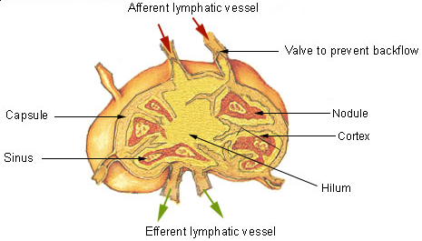
The lymphatic vessels (or lymph vessels or lymphatics) are thin-walled vessels (tubes), structured like blood vessels, that carry lymph. As part of the lymphatic system, lymph vessels are complementary to the cardiovascular system. Lymph vessels are lined by endothelial cells, and have a thin layer of smooth muscle, and adventitia that binds the lymph vessels to the surrounding tissue. Lymph vessels are devoted to the propulsion of the lymph from the lymph capillaries, which are mainly concerned with the absorption of interstitial fluid from the tissues. Lymph capillaries are slightly bigger than their counterpart capillaries of the vascular system. Lymph vessels that carry lymph to a lymph node are called afferent lymph vessels, and those that carry it from a lymph node are called efferent lymph vessels, from where the lymph may travel to another lymph node, may be returned to a vein, or may travel to a larger lymph duct. Lymph ducts drain the lymph into one of the subclavian ve ... [...More Info...] [...Related Items...] OR: [Wikipedia] [Google] [Baidu] |
Vasculogenesis
Vasculogenesis is the process of blood vessel formation, occurring by a '' de novo'' production of endothelial cells. It is sometimes paired with angiogenesis, as the first stage of the formation of the vascular network, closely followed by angiogenesis. Process In the sense distinguished from angiogenesis, vasculogenesis is different in one aspect: whereas angiogenesis is the formation of new blood vessels from pre-existing ones, vasculogenesis is the formation of new blood vessels, in blood islands, when there are no pre-existing ones. For example, if a monolayer of endothelial cells begins sprouting to form capillaries, angiogenesis is occurring. Vasculogenesis, in contrast, is when endothelial precursor cells (angioblasts) migrate and differentiate in response to local cues (such as growth factors and extracellular matrices) to form new blood vessels. These vascular trees are then pruned and extended through angiogenesis. Occurrences Vasculogenesis occurs during embryologic ... [...More Info...] [...Related Items...] OR: [Wikipedia] [Google] [Baidu] |
Angiogenesis
Angiogenesis is the physiological process through which new blood vessels form from pre-existing vessels, formed in the earlier stage of vasculogenesis. Angiogenesis continues the growth of the vasculature by processes of sprouting and splitting. Vasculogenesis is the embryonic formation of endothelial cells from mesoderm cell precursors, and from neovascularization, although discussions are not always precise (especially in older texts). The first vessels in the developing embryo form through vasculogenesis, after which angiogenesis is responsible for most, if not all, blood vessel growth during development and in disease. Angiogenesis is a normal and vital process in growth and development, as well as in wound healing and in the formation of granulation tissue. However, it is also a fundamental step in the transition of tumors from a benign state to a malignant one, leading to the use of angiogenesis inhibitors in the treatment of cancer. The essential role of angiogenesis in ... [...More Info...] [...Related Items...] OR: [Wikipedia] [Google] [Baidu] |
Mesenchyme
Mesenchyme () is a type of loosely organized animal embryonic connective tissue of undifferentiated cells that give rise to most tissues, such as skin, blood or bone. The interactions between mesenchyme and epithelium help to form nearly every organ in the developing embryo. Vertebrates Structure Mesenchyme is characterized morphologically by a prominent ground substance matrix containing a loose aggregate of reticular fibers and unspecialized mesenchymal stem cells. Mesenchymal cells can migrate easily (in contrast to epithelial cells, which lack mobility), are organized into closely adherent sheets, and are polarized in an apical-basal orientation. Development The mesenchyme originates from the mesoderm. From the mesoderm, the mesenchyme appears as an embryologically primitive "soup". This "soup" exists as a combination of the mesenchymal cells plus serous fluid plus the many different tissue proteins. Serous fluid is typically stocked with the many serous elements, such a ... [...More Info...] [...Related Items...] OR: [Wikipedia] [Google] [Baidu] |
Jugular Vein
The jugular veins are veins that take deoxygenated blood from the head back to the heart via the superior vena cava. The internal jugular vein descends next to the internal carotid artery and continues posteriorly to the sternocleidomastoid muscle. Structure and Function There are two sets of jugular veins: external and internal. The left and right external jugular veins drain into the subclavian veins. The internal jugular veins join with the subclavian veins more medially to form the brachiocephalic veins. Finally, the left and right brachiocephalic veins join to form the superior vena cava, which delivers deoxygenated blood to the right atrium of the heart. The Jugular veins help carry blood from the heart to and from the brain. An average human brain weighs about 3 pounds, and gets about 15%-20% of the blood that the heart pumps out. It is important for the brain to get enough blood for many reasons. The jugu ... [...More Info...] [...Related Items...] OR: [Wikipedia] [Google] [Baidu] |
Anterior Cardinal Vein
The anterior cardinal veins (precardinal veins) contribute to the formation of the internal jugular veins and together with the common cardinal vein form the superior vena cava. The anastomosis between the two anterior cardinal veins develops into the left brachiocephalic vein. Additional images Image:Gray459.png, Human embryo of about fourteen days, with yolk sac The yolk sac is a membranous sac attached to an embryo, formed by cells of the hypoblast layer of the bilaminar embryonic disc. This is alternatively called the umbilical vesicle by the Terminologia Embryologica (TE), though ''yolk sac'' is .... References External links * Diagram at nature.com Embryology of cardiovascular system {{circulatory-stub ... [...More Info...] [...Related Items...] OR: [Wikipedia] [Google] [Baidu] |
Lymph Node
A lymph node, or lymph gland, is a kidney-shaped organ of the lymphatic system and the adaptive immune system. A large number of lymph nodes are linked throughout the body by the lymphatic vessels. They are major sites of lymphocytes that include B and T cells. Lymph nodes are important for the proper functioning of the immune system, acting as filters for foreign particles including cancer cells, but have no detoxification function. In the lymphatic system a lymph node is a secondary lymphoid organ. A lymph node is enclosed in a fibrous capsule and is made up of an outer cortex and an inner medulla. Lymph nodes become inflamed or enlarged in various diseases, which may range from trivial throat infections to life-threatening cancers. The condition of lymph nodes is very important in cancer staging, which decides the treatment to be used and determines the prognosis. Lymphadenopathy refers to glands that are enlarged or swollen. When inflamed or enlarged, lymph nodes can be ... [...More Info...] [...Related Items...] OR: [Wikipedia] [Google] [Baidu] |
Cysterna Chyli
The cisterna chyli (or cysterna chyli, and etymologically more correct, receptaculum chyli) is a dilated sac at the lower end of the thoracic duct in most mammals into which lymph from the intestinal trunk and two lumbar lymphatic trunks flow. It receives fatty chyle from the intestines and thus acts as a conduit for the lipid products of digestion. It is the most common drainage trunk of most of the body's lymphatics. The cisterna chyli is a retro-peritoneal structure. Structure In humans, the cisterna chyli is located posterior to the abdominal aorta on the anterior aspect of the bodies of the first and second lumbar vertebrae (L1 and L2). There it forms the beginning of the primary lymph vessel, the thoracic duct, which transports lymph and chyle from the abdomen via the aortic opening of the diaphragm up to the junction of left subclavian vein and internal jugular veins. Other animals In dogs, the cisterna chyli is located to the left and often ventral to the aorta; in cat ... [...More Info...] [...Related Items...] OR: [Wikipedia] [Google] [Baidu] |
Transcription Factor
In molecular biology, a transcription factor (TF) (or sequence-specific DNA-binding factor) is a protein that controls the rate of transcription of genetic information from DNA to messenger RNA, by binding to a specific DNA sequence. The function of TFs is to regulate—turn on and off—genes in order to make sure that they are expressed in the desired cells at the right time and in the right amount throughout the life of the cell and the organism. Groups of TFs function in a coordinated fashion to direct cell division, cell growth, and cell death throughout life; cell migration and organization (body plan) during embryonic development; and intermittently in response to signals from outside the cell, such as a hormone. There are up to 1600 TFs in the human genome. Transcription factors are members of the proteome as well as regulome. TFs work alone or with other proteins in a complex, by promoting (as an activator), or blocking (as a repressor) the recruitment of RNA ... [...More Info...] [...Related Items...] OR: [Wikipedia] [Google] [Baidu] |


.jpg)