|
Lumbar Lordosis
Lordosis is historically defined as an ''abnormal'' inward curvature of the lumbar spine. However, the terms ''lordosis'' and ''lordotic'' are also used to refer to the normal inward curvature of the lumbar and cervical regions of the human spine. Similarly, kyphosis historically refers to ''abnormal'' convex curvature of the spine. The normal outward (convex) curvature in the thoracic and sacral regions is also termed ''kyphosis'' or ''kyphotic''. The term comes from the Greek lordōsis, from ''lordos'' ("bent backward"). Lordosis in the human spine makes it easier for humans to bring the bulk of their mass over the pelvis. This allows for a much more efficient walking gait than that of other primates, whose inflexible spines cause them to resort to an inefficient forward leaning "bent-knee, bent-waist" gait. As such, lordosis in the human spine is considered one of the primary physiological adaptations of the human skeleton that allows for human gait to be as energetically ... [...More Info...] [...Related Items...] OR: [Wikipedia] [Google] [Baidu] |
X-ray
An X-ray, or, much less commonly, X-radiation, is a penetrating form of high-energy electromagnetic radiation. Most X-rays have a wavelength ranging from 10 picometers to 10 nanometers, corresponding to frequencies in the range 30 petahertz to 30 exahertz ( to ) and energies in the range 145 eV to 124 keV. X-ray wavelengths are shorter than those of UV rays and typically longer than those of gamma rays. In many languages, X-radiation is referred to as Röntgen radiation, after the German scientist Wilhelm Conrad Röntgen, who discovered it on November 8, 1895. He named it ''X-radiation'' to signify an unknown type of radiation.Novelline, Robert (1997). ''Squire's Fundamentals of Radiology''. Harvard University Press. 5th edition. . Spellings of ''X-ray(s)'' in English include the variants ''x-ray(s)'', ''xray(s)'', and ''X ray(s)''. The most familiar use of X-rays is checking for fractures (broken bones), but X-rays are also used in other ways. ... [...More Info...] [...Related Items...] OR: [Wikipedia] [Google] [Baidu] |
Hamstring
In human anatomy, a hamstring () is any one of the three posterior thigh muscles in between the hip and the knee (from medial to lateral: semimembranosus, semitendinosus and biceps femoris). The hamstrings are susceptible to injury. In quadrupeds, the hamstring is the single large tendon found behind the knee or comparable area. Criteria The common criteria of any hamstring muscles are: # Muscles should originate from ischial tuberosity. # Muscles should be inserted over the knee joint, in the tibia or in the fibula. # Muscles will be innervated by the tibial branch of the sciatic nerve. # Muscle will participate in flexion of the knee joint and extension of the hip joint. Those muscles which fulfill all of the four criteria are called true hamstrings. The adductor magnus reaches only up to the adductor tubercle of the femur, but it is included amongst the hamstrings because the tibial collateral ligament of the knee joint morphologically is the degenerated tendon of this m ... [...More Info...] [...Related Items...] OR: [Wikipedia] [Google] [Baidu] |
Pelvic Tilt
Pelvic tilt is the orientation of the pelvis in respect to the thighbones and the rest of the body. The pelvis can tilt towards the front, back, or either side of the body. Anterior pelvic tilt and posterior pelvic tilt are very common abnormalities in regard to the orientation of the pelvis. Forms *Anterior pelvic tilt is when the front of the pelvis drops in relationship to the back of the pelvis. For example, this happens when the hip flexors shorten and the hip extensors lengthen. It is also called lumbar hyperlordosis. *Posterior pelvic tilt is the opposite, when the front of the pelvis rises and the back of the pelvis drops. For example, this happens when the hip flexors lengthen and the hip extensors shorten, particularly the gluteus maximus which is the primary extensor of the hip. *Lateral pelvic tilt describes tilting toward either right or left and is associated with scoliosis or people who have legs of different length. It can also happen when one leg is bent whil ... [...More Info...] [...Related Items...] OR: [Wikipedia] [Google] [Baidu] |
Vitamin D
Vitamin D is a group of fat-soluble secosteroids responsible for increasing intestinal absorption of calcium, magnesium, and phosphate, and many other biological effects. In humans, the most important compounds in this group are vitamin D3 (cholecalciferol) and vitamin D2 (ergocalciferol). The major natural source of the vitamin is synthesis of cholecalciferol in the lower layers of epidermis of the skin through a chemical reaction that is dependent on sun exposure (specifically UVB radiation). Cholecalciferol and ergocalciferol can be ingested from the diet and supplements. Only a few foods, such as the flesh of fatty fish, naturally contain significant amounts of vitamin D. In the U.S. and other countries, cow's milk and plant-derived milk substitutes are fortified with vitamin D, as are many breakfast cereals. Mushrooms exposed to ultraviolet light contribute useful amounts of vitamin D2. Dietary recommendations typically assume that all of a person's vitamin D is taken ... [...More Info...] [...Related Items...] OR: [Wikipedia] [Google] [Baidu] |
Rickets
Rickets is a condition that results in weak or soft bones in children, and is caused by either dietary deficiency or genetic causes. Symptoms include bowed legs, stunted growth, bone pain, large forehead, and trouble sleeping. Complications may include bone deformities, bone pseudofractures and fractures, muscle spasms, or an abnormally curved spine. The most common cause of rickets is a vitamin D deficiency, although hereditary genetic forms also exist. This can result from eating a diet without enough vitamin D, dark skin, too little sun exposure, exclusive breastfeeding without vitamin D supplementation, celiac disease, and certain genetic conditions. Other factors may include not enough calcium or phosphorus. The underlying mechanism involves insufficient calcification of the growth plate. Diagnosis is generally based on blood tests finding a low calcium, low phosphorus, and a high alkaline phosphatase together with X-rays. Prevention for exclusively breastfed bab ... [...More Info...] [...Related Items...] OR: [Wikipedia] [Google] [Baidu] |
Visceral Fat
Adipose tissue, body fat, or simply fat is a loose connective tissue composed mostly of adipocytes. In addition to adipocytes, adipose tissue contains the stromal vascular fraction (SVF) of cells including preadipocytes, fibroblasts, vascular endothelial cells and a variety of immune cells such as adipose tissue macrophages. Adipose tissue is derived from preadipocytes. Its main role is to store energy in the form of lipids, although it also cushions and insulates the body. Far from being hormonally inert, adipose tissue has, in recent years, been recognized as a major endocrine organ, as it produces hormones such as leptin, estrogen, resistin, and cytokines (especially TNFα). In obesity, adipose tissue is also implicated in the chronic release of pro-inflammatory markers known as adipokines, which are responsible for the development of metabolic syndrome, a constellation of diseases including, but not limited to, type 2 diabetes, cardiovascular disease and atherosclerosis. T ... [...More Info...] [...Related Items...] OR: [Wikipedia] [Google] [Baidu] |
Human Back
The human back, also called the dorsum, is the large posterior area of the human body, rising from the top of the buttocks to the back of the neck. It is the surface of the body opposite from the chest and the abdomen. The vertebral column runs the length of the back and creates a central area of recession. The breadth of the back is created by the shoulders at the top and the pelvis at the bottom. Back pain is a common medical condition, generally benign in origin. Structure The central feature of the human back is the vertebral column, specifically the length from the top of the thoracic vertebrae to the bottom of the lumbar vertebrae, which houses the spinal cord in its spinal canal, and which generally has some curvature that gives shape to the back. The ribcage extends from the spine at the top of the back (with the top of the ribcage corresponding to the T1 vertebra), more than halfway down the length of the back, leaving an area with less protection between the bot ... [...More Info...] [...Related Items...] OR: [Wikipedia] [Google] [Baidu] |
Swayback
Swayback, also known clinically as lordosis, refers to abnormal bent-back postures in humans and in quadrupeds, especially horses. Extreme lordosis can cause physical damage to the spinal cord and associated ligaments and tendons which can lead to severe pain. Moderate lordosis does not generally impact a horse’s usefulness and does not necessarily cause lameness. Humans Swayback posture in humans is characterised by the posterior displacement of the rib cage in comparison to the pelvis. It looks like the person has a hyperextension of the lower back, however this is not necessarily the case. Most sway-back exhibits a posteriorly tilted pelvis; the lumbar region is usually flat (too flexed) and not hyperlordotic (too extended). Horses Usually called "swayback", soft back, or low back, an excessive downward bend in the back is an undesirable conformation trait. Swayback is caused in part from a loss of muscle tone in both the back and abdominal muscles, plus a weakening and str ... [...More Info...] [...Related Items...] OR: [Wikipedia] [Google] [Baidu] |
Osteoporosis
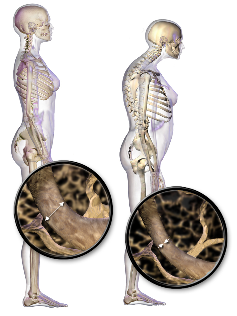
Osteoporosis is a systemic skeletal disorder characterized by low bone mass, micro-architectural deterioration of bone tissue leading to bone fragility, and consequent increase in fracture risk. It is the most common reason for a broken bone among the elderly. Bones that commonly break include the vertebrae in the spine, the bones of the forearm, and the hip. Until a broken bone occurs there are typically no symptoms. Bones may weaken to such a degree that a break may occur with minor stress or spontaneously. After the broken bone heals, the person may have chronic pain and a decreased ability to carry out normal activities. Osteoporosis may be due to lower-than-normal maximum bone mass and greater-than-normal bone loss. Bone loss increases after the menopause due to lower levels of estrogen, and after ' andropause' due to lower levels of testosterone. Osteoporosis may also occur due to a number of diseases or treatments, including alcoholism, anorexia, hyperthyroidism, k ... [...More Info...] [...Related Items...] OR: [Wikipedia] [Google] [Baidu] |
Spondylolisthesis
Spondylolisthesis is the displacement of one spinal vertebra compared to another. While some medical dictionaries define spondylolisthesis specifically as the forward or anterior displacement of a vertebra over the vertebra inferior to it (or the sacrum), it is often defined in medical textbooks as displacement in any direction.Introduction to chapter 17 in: Page 250 in: Spondylolisthesis is graded based upon the degree of slippage of one vertebral body relative to the subsequent adjacent vertebral body. Spondylolisthesis is classified as one of the six major etiologies: degenerative, traumatic, dysplastic, [...More Info...] [...Related Items...] OR: [Wikipedia] [Google] [Baidu] |
Achondroplasia
Achondroplasia is a genetic disorder with an autosomal dominant pattern of inheritance whose primary feature is dwarfism. In those with the condition, the arms and legs are short, while the torso is typically of normal length. Those affected have an average adult height of for males and for females. Other features can include an enlarged head and prominent forehead. Complications can include sleep apnea or recurrent ear infections. Achondroplasia includes short-limb skeletal dysplasia with severe combined immunodeficiency. Achondroplasia is caused by a mutation in the fibroblast growth factor receptor 3 (''FGFR3'') gene that results in its protein being overactive. Achondroplasia results in impaired endochondral bone growth (bone growth within cartilage). The disorder has an autosomal dominant mode of inheritance, meaning only one mutated copy of the gene is required for the condition to occur. About 80% of cases occur in children of parents of average stature and resu ... [...More Info...] [...Related Items...] OR: [Wikipedia] [Google] [Baidu] |


