|
List Of Keratins Expressed In The Human Integumentary System
There are many different keratin proteins normally expressed in the human integumentary system. See also * List of cutaneous conditions caused by mutations in keratins * List of target antigens in pemphigoid * List of target antigens in pemphigus * Cutaneous conditions with immunofluorescence findings * List of cutaneous conditions * List of genes mutated in cutaneous conditions * List of histologic stains that aid in diagnosis of cutaneous conditions A number of histologic stains are used in the field of dermatology that aid in the diagnosis of conditions of or affecting the human integumentary system. Footnotes See also * List of conditions associated with café au lait macules * ... References * * {{DEFAULTSORT:Keratins expressed in the human integumentary system Dermatology-related lists ... [...More Info...] [...Related Items...] OR: [Wikipedia] [Google] [Baidu] |
Keratin
Keratin () is one of a family of structural fibrous proteins also known as ''scleroproteins''. Alpha-keratin (α-keratin) is a type of keratin found in vertebrates. It is the key structural material making up scales, hair, nails, feathers, horns, claws, hooves, and the outer layer of skin among vertebrates. Keratin also protects epithelial cells from damage or stress. Keratin is extremely insoluble in water and organic solvents. Keratin monomers assemble into bundles to form intermediate filaments, which are tough and form strong unmineralized epidermal appendages found in reptiles, birds, amphibians, and mammals. Excessive keratinization participate in fortification of certain tissues such as in horns of cattle and rhinos, and armadillos' osteoderm. The only other biological matter known to approximate the toughness of keratinized tissue is chitin. Keratin comes in two types, the primitive, softer forms found in all vertebrates and harder, derived forms found only amon ... [...More Info...] [...Related Items...] OR: [Wikipedia] [Google] [Baidu] |
Integumentary System
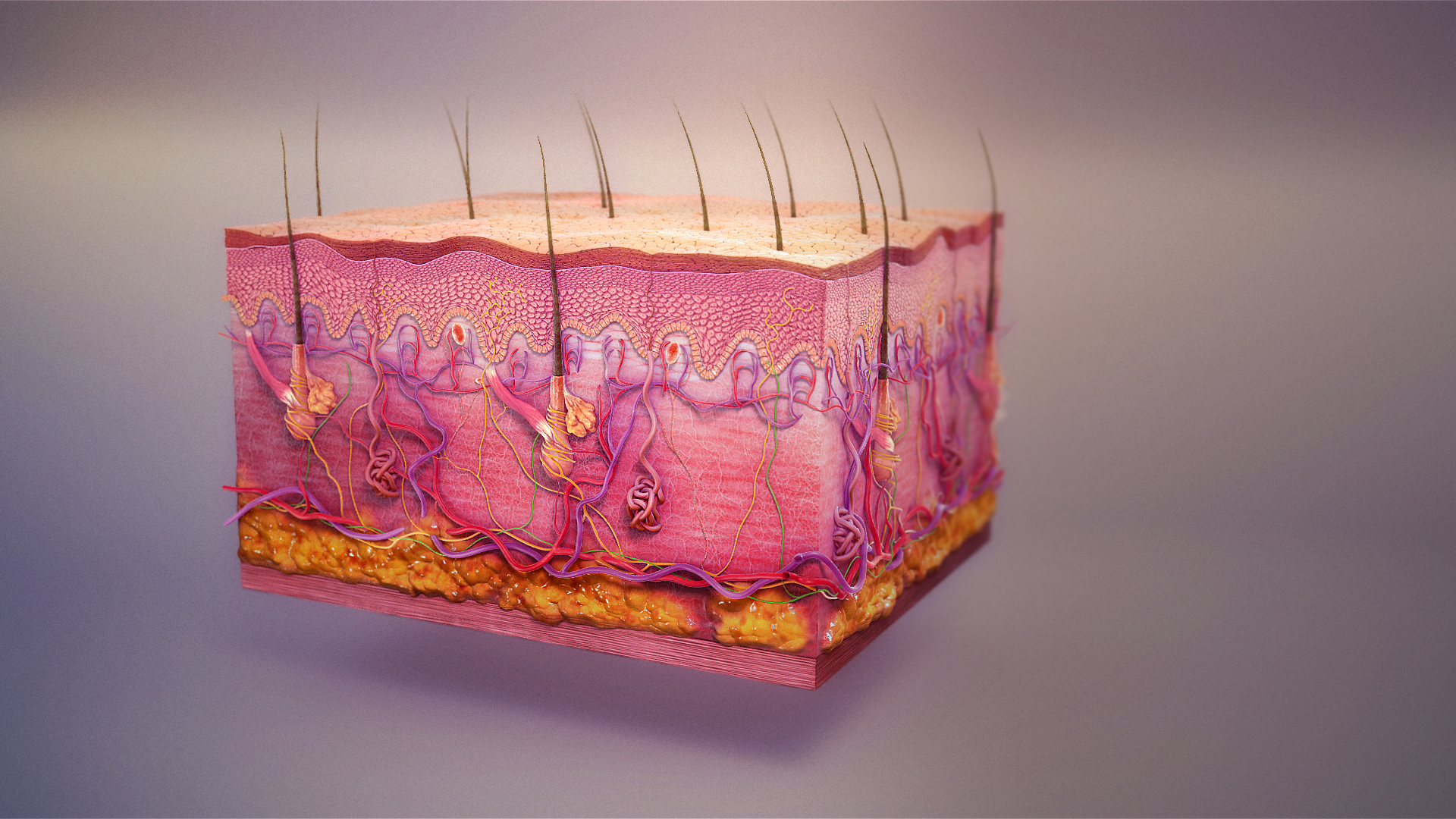
The integumentary system is the set of organs forming the outermost layer of an animal's body. It comprises the skin and its appendages, which act as a physical barrier between the external environment and the internal environment that it serves to protect and maintain the body of the animal. Mainly it is the body's outer skin. The integumentary system includes hair, scales, feathers, hooves, and nails. It has a variety of additional functions: it may serve to maintain water balance, protect the deeper tissues, excrete wastes, and regulate body temperature, and is the attachment site for sensory receptors which detect pain, sensation, pressure, and temperature. Structure Skin The skin is one of the largest organs of the body. In humans, it accounts for about 12 to 15 percent of total body weight and covers 1.5 to 2 m2 of surface area. The skin (integument) is a composite organ, made up of at least two major layers of tissue: the epidermis and the dermis. The epidermis is ... [...More Info...] [...Related Items...] OR: [Wikipedia] [Google] [Baidu] |
Keratin
Keratin () is one of a family of structural fibrous proteins also known as ''scleroproteins''. Alpha-keratin (α-keratin) is a type of keratin found in vertebrates. It is the key structural material making up scales, hair, nails, feathers, horns, claws, hooves, and the outer layer of skin among vertebrates. Keratin also protects epithelial cells from damage or stress. Keratin is extremely insoluble in water and organic solvents. Keratin monomers assemble into bundles to form intermediate filaments, which are tough and form strong unmineralized epidermal appendages found in reptiles, birds, amphibians, and mammals. Excessive keratinization participate in fortification of certain tissues such as in horns of cattle and rhinos, and armadillos' osteoderm. The only other biological matter known to approximate the toughness of keratinized tissue is chitin. Keratin comes in two types, the primitive, softer forms found in all vertebrates and harder, derived forms found only amon ... [...More Info...] [...Related Items...] OR: [Wikipedia] [Google] [Baidu] |
Stratum Granulosum
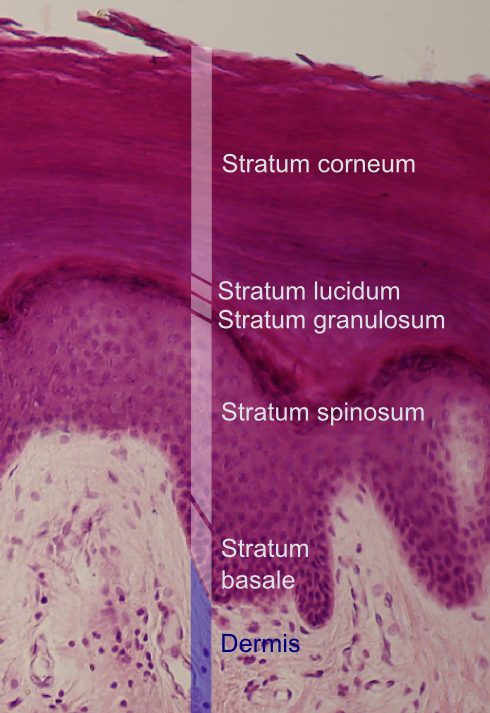
The stratum granulosum (or granular layer) is a thin layer of cells in the epidermis lying above the stratum spinosum and below the stratum corneum (stratum lucidum on the soles and palms).James, William; Berger, Timothy; Elston, Dirk (2005) ''Andrews' Diseases of the Skin: Clinical Dermatology'' (10th ed.). Saunders. Page 2. . Keratinocytes migrating from the underlying stratum spinosum become known as granular cells in this layer. These cells contain keratohyalin granules, which are filled with histidine- and cysteine-rich proteins that appear to bind the keratin filaments together. Therefore, the main function of keratohyalin granules is to bind intermediate keratin filaments together.Marks, James G; Miller, Jeffery (2006). ''Lookingbill and Marks' Principles of Dermatology'' (4th ed.). Elsevier Inc. Page 7. . At the transition between this layer and the stratum corneum, cells secrete lamellar bodies (containing lipids and proteins) into the extracellular space. This results ... [...More Info...] [...Related Items...] OR: [Wikipedia] [Google] [Baidu] |
Cornea
The cornea is the transparent front part of the eye that covers the iris, pupil, and anterior chamber. Along with the anterior chamber and lens, the cornea refracts light, accounting for approximately two-thirds of the eye's total optical power. In humans, the refractive power of the cornea is approximately 43 dioptres. The cornea can be reshaped by surgical procedures such as LASIK. While the cornea contributes most of the eye's focusing power, its focus is fixed. Accommodation (the refocusing of light to better view near objects) is accomplished by changing the geometry of the lens. Medical terms related to the cornea often start with the prefix "'' kerat-''" from the Greek word κέρας, ''horn''. Structure The cornea has unmyelinated nerve endings sensitive to touch, temperature and chemicals; a touch of the cornea causes an involuntary reflex to close the eyelid. Because transparency is of prime importance, the healthy cornea does not have or need blood vessels with ... [...More Info...] [...Related Items...] OR: [Wikipedia] [Google] [Baidu] |
Stratum Basale
The ''stratum basale'' (basal layer, sometimes referred to as ''stratum germinativum'') is the deepest layer of the five layers of the epidermis, the external covering of skin in mammals. The ''stratum basale'' is a single layer of columnar or cuboidal basal cells. The cells are attached to each other and to the overlying stratum spinosum cells by desmosomes and hemidesmosomes. The nucleus is large, ovoid and occupies most of the cell. Some basal cells can act like stem cells with the ability to divide and produce new cells, and these are sometimes called basal keratinocyte stem cells. Others serve to anchor the epidermis glabrous skin (hairless), and hyper-proliferative epidermis (from a skin disease).McGrath, J.A.; Eady, R.A.; Pope, F.M. (2004). ''Rook's Textbook of Dermatology'' (Seventh Edition). Blackwell Publishing. Pages 3.7. . They divide to form the keratinocytes of the stratum spinosum, which migrate superficially. Other types of cells found within the ''stratum bas ... [...More Info...] [...Related Items...] OR: [Wikipedia] [Google] [Baidu] |
Merkel Cell
Merkel cells, also known as Merkel-Ranvier cells or tactile epithelial cells, are oval-shaped mechanoreceptors essential for light touch sensation and found in the skin of vertebrates. They are abundant in highly sensitive skin like that of the fingertips in humans, and make synaptic contacts with somatosensory afferent nerve fibers. Though it has been reported that Merkel cells are derived from neural crest cells, more recent experiments in mammals have indicated that they are in fact epithelial in origin. Structure Merkel cells are found in the skin and some parts of the mucosa of all vertebrates. In mammalian skin, they are clear cells found in the ''stratum basale'' (at the bottom of sweat duct ridges) of the epidermis approximately 10 μm in diameter. They also occur in epidermal invaginations of the plantar foot surface called rete ridges. Most often, they are associated with sensory nerve endings, when they are known as Merkel nerve endings (also called a Merkel ... [...More Info...] [...Related Items...] OR: [Wikipedia] [Google] [Baidu] |
List Of Cutaneous Conditions Caused By Mutations In Keratins
There are many different keratin proteins normally expressed in the human integumentary system. Mutations in keratin proteins in the skin can cause disease. Of note, other structural proteins in the epidermis of the skin that are closely related to keratins may also cause disease if mutated. Examples include: Footnotes See also * List of keratins expressed in the human integumentary system * List of cutaneous conditions caused by problems with junctional proteins * List of target antigens in pemphigoid * List of target antigens in pemphigus * Cutaneous conditions with immunofluorescence findings * List of cutaneous conditions * List of genes mutated in cutaneous conditions * List of histologic stains that aid in diagnosis of cutaneous conditions * Keratoderma Keratoderma is a hornlike skin condition. Classification The keratodermas are classified into the following subgroups:Freedberg, et al. (2003). ''Fitzpatrick's Dermatology in General Medicine''. (6th ed.). ... [...More Info...] [...Related Items...] OR: [Wikipedia] [Google] [Baidu] |
List Of Target Antigens In Pemphigoid
Circulating auto-antibodies in the human body can target normal parts of the skin leading to disease. This is a list of antigens in the skin that may become targets of circulating auto-antibodies leading to the various types of pemphigoid. Of note, there are also several other diseases that are caused by auto-antibodies that target the same anatomic area of the skin which is termed the basement membrane zone. These diseases include: Footnotes See also * List of target antigens in pemphigus * List of immunofluorescence findings for autoimmune bullous conditions * List of cutaneous conditions * List of genes mutated in cutaneous conditions * List of histologic stains that aid in diagnosis of cutaneous conditions A number of histologic stains are used in the field of dermatology that aid in the diagnosis of conditions of or affecting the human integumentary system. Footnotes See also * List of conditions associated with café au lait macules * ... Refer ... [...More Info...] [...Related Items...] OR: [Wikipedia] [Google] [Baidu] |
List Of Target Antigens In Pemphigus
Circulating auto-antibodies in the human body can target normal parts of the skin leading to disease. This is a list of antigens in the skin that may become targets of circulating auto-antibodies leading to the various types of pemphigus. Footnotes See also * List of target antigens in pemphigoid * List of conditions caused by problems with junctional proteins * List of immunofluorescence findings for autoimmune bullous conditions * List of cutaneous conditions * List of genes mutated in cutaneous conditions * List of histologic stains that aid in diagnosis of cutaneous conditions A number of histologic stains are used in the field of dermatology that aid in the diagnosis of conditions of or affecting the human integumentary system. Footnotes See also * List of conditions associated with café au lait macules * ... References * * {{DEFAULTSORT:Target antigens in pemphigus Cutaneous conditions Dermatology-related lists ... [...More Info...] [...Related Items...] OR: [Wikipedia] [Google] [Baidu] |
Cutaneous Conditions With Immunofluorescence Findings
Several cutaneous conditions can be diagnosed with the aid of immunofluorescence studies. Cutaneous conditions with positive direct or indirect immunofluorescence when using salt-split skin include: For several subtypes of pemphigus a variety of substrates are used for indirect immunofluorescence: See also * List of cutaneous conditions * List of genes mutated in cutaneous conditions * List of cutaneous conditions caused by mutations in keratins There are many different keratin proteins normally expressed in the human integumentary system. Mutations in keratin proteins in the skin can cause disease. Of note, other structural proteins in the epidermis of the skin that are closely rel ... References * * {{DEFAULTSORT:Immunofluorescence findings for autoimmune bullous conditions Cutaneous conditions Dermatology-related lists ... [...More Info...] [...Related Items...] OR: [Wikipedia] [Google] [Baidu] |
List Of Cutaneous Conditions
Many skin conditions affect the human integumentary system—the organ system covering the entire surface of the body and composed of skin, hair, nails, and related muscle and glands. The major function of this system is as a barrier against the external environment. The skin weighs an average of four kilograms, covers an area of two square metres, and is made of three distinct layers: the epidermis, dermis, and subcutaneous tissue. The two main types of human skin are: glabrous skin, the hairless skin on the palms and soles (also referred to as the "palmoplantar" surfaces), and hair-bearing skin.Burns, Tony; ''et al''. (2006) ''Rook's Textbook of Dermatology CD-ROM''. Wiley-Blackwell. . Within the latter type, the hairs occur in structures called pilosebaceous units, each with hair follicle, sebaceous gland, and associated arrector pili muscle. In the embryo, the epidermis, hair, and glands form from the ectoderm, which is chemically influenced by the underlying mesoderm th ... [...More Info...] [...Related Items...] OR: [Wikipedia] [Google] [Baidu] |


