|
List Of Histologic Stains That Aid In Diagnosis Of Cutaneous Conditions
A number of histologic stains are used in the field of dermatology that aid in the diagnosis of conditions of or affecting the human integumentary system. Footnotes See also * List of conditions associated with café au lait macules * List of contact allergens *List of cutaneous conditions associated with increased risk of nonmelanoma skin cancer *List of cutaneous conditions associated with internal malignancy *List of cutaneous conditions caused by mutations in keratins *List of cutaneous neoplasms associated with systemic syndromes *List of cutaneous conditions caused by problems with junctional proteins *List of dental abnormalities associated with cutaneous conditions *List of genes mutated in cutaneous conditions *List of genes mutated in pigmented cutaneous lesions *List of human leukocyte antigen alleles associated with cutaneous conditions *List of immunofluorescence findings for autoimmune bullous conditions * List of inclusion bodies that aid in diagnosis of c ... [...More Info...] [...Related Items...] OR: [Wikipedia] [Google] [Baidu] |
Staining
Staining is a technique used to enhance contrast in samples, generally at the microscopic level. Stains and dyes are frequently used in histology (microscopic study of biological tissues), in cytology (microscopic study of cells), and in the medical fields of histopathology, hematology, and cytopathology that focus on the study and diagnoses of diseases at the microscopic level. Stains may be used to define biological tissues (highlighting, for example, muscle fibers or connective tissue), cell populations (classifying different blood cells), or organelles within individual cells. In biochemistry, it involves adding a class-specific ( DNA, proteins, lipids, carbohydrates) dye to a substrate to qualify or quantify the presence of a specific compound. Staining and fluorescent tagging can serve similar purposes. Biological staining is also used to mark cells in flow cytometry, and to flag proteins or nucleic acids in gel electrophoresis. Light microscopes are us ... [...More Info...] [...Related Items...] OR: [Wikipedia] [Google] [Baidu] |
Diffuse Large B-cell Lymphoma Leg Type
Diffuse large B-cell lymphoma (DLBCL) is a cancer of B cells, a type of lymphocyte that is responsible for producing antibodies. It is the most common form of non-Hodgkin lymphoma among adults, with an annual incidence of 7–8 cases per 100,000 people per year in the US and UK. This cancer occurs primarily in older individuals, with a median age of diagnosis at ~70 years, although it can occur in young adults and, in rare cases, children. DLBCL can arise in virtually any part of the body and, depending on various factors, is often a very aggressive malignancy. The first sign of this illness is typically the observation of a rapidly growing mass or tissue infiltration that is sometimes associated with systemic B symptoms, e.g. fever, weight loss, and night sweats. The causes of diffuse large B-cell lymphoma are not well understood. Usually DLBCL arises from normal B cells, but it can also represent a malignant transformation of other types of lymphoma (particularly marginal zone ... [...More Info...] [...Related Items...] OR: [Wikipedia] [Google] [Baidu] |
Langerhans Cell Histiocytosis
Langerhans cell histiocytosis (LCH) is an abnormal clonal proliferation of Langerhans cells, abnormal cells deriving from bone marrow and capable of migrating from skin to lymph nodes. Symptoms range from isolated bone lesions to multisystem disease. LCH is part of a group of syndromes called histiocytoses, which are characterized by an abnormal proliferation of histiocytes (an archaic term for activated dendritic cells and macrophages). These diseases are related to other forms of abnormal proliferation of white blood cells, such as leukemias and lymphomas. The disease has gone by several names, including Hand–Schüller–Christian disease, Abt-Letterer-Siwe disease, Hashimoto-Pritzker disease (a very rare self-limiting variant seen at birth) and histiocytosis X, until it was renamed in 1985 by the Histiocyte Society. Classification The disease spectrum results from clonal accumulation and proliferation of cells resembling the epidermal dendritic cells called La ... [...More Info...] [...Related Items...] OR: [Wikipedia] [Google] [Baidu] |
Langerhans Cell
A Langerhans cell (LC) is a tissue-resident macrophage of the skin. These cells contain organelles called Birbeck granules. They are present in all layers of the epidermis and are most prominent in the stratum spinosum. They also occur in the papillary dermis, particularly around blood vessels, as well as in the mucosa of the mouth, foreskin, and vaginal epithelium. They can be found in other tissues, such as lymph nodes, particularly in association with the condition Langerhans cell histiocytosis (LCH). Function In skin infections, the local Langerhans cells take up and process microbial antigens to become fully functional antigen-presenting cells. Generally, tissue-resident macrophages are involved in immune homeostasis and the uptake of apoptotic bodies. However, Langerhans cells can also take on a dendritic cell-like phenotype and migrate to lymph nodes to interact with naive T-cells. Langerhans cells derive from primitive erythro-myeloid progenitors that arise in ... [...More Info...] [...Related Items...] OR: [Wikipedia] [Google] [Baidu] |
Eccrine Poroma
Poromas are rare, benign, cutaneous adnexal tumors. Cutaneous adnexal tumors are a group of skin tumors consisting of tissues that have differentiated (i.e. matured from stem cells) towards one or more of the four primary adnexal structures found in normal skin: hair follicles, sebaceous sweat glands, apocrine sweat glands, and eccrine sweat glands. Poromas are eccrine or apocrine sweat gland tumors derived from the cells in the terminal portion of these glands' ducts. This part of the sweat gland duct is termed the acrosyringium and had led to grouping poromas in the acrospiroma class of skin tumors (syringofibroadenomas and syringoacanthomas are classified as acrospiromas). Here, poromas are regarded as distinct sweat gland tumors that differ from other sweat gland tumors by their characteristic clinical presentations, microscopic histopathology, and the genetic mutations that their neoplastic cells have recently been found to carry. As currently viewed, there are 4 poroma va ... [...More Info...] [...Related Items...] OR: [Wikipedia] [Google] [Baidu] |
Microcystic Adnexal Carcinoma
Microcystic adnexal carcinoma (MAC) is a rare sweat gland cancer, which often appears as a yellow spot or bump in the skin. It usually occurs in the neck or head, although cases have been documented in other areas of the body. Most diagnosis occur past the age of 50. Although considered an invasive cancer, metastasis rarely occurs. If the tumor spreads, it can grow and invade fat, muscles, and other types of tissue. Main treatments are wide local excision or Mohs micrographic surgery, which ensures that most, if not all, cancer cells are removed surgically. Presentation MACs usually present as a smooth, flesh or yellow colored, slow-growing nodule or bump somewhere on the face or neck with typical development being 3–5 years. The most common location is the mouth (occurring in 74% of cases), however cases have been documented on the scalp, tongue, trunk, upper extremities, and genitals. Patients are more likely to be white, female, and middle aged or elderly, although cases have ... [...More Info...] [...Related Items...] OR: [Wikipedia] [Google] [Baidu] |
Extramammary Paget's Disease
Extramammary Paget's Disease (EMPD) is a rare and slow-growing malignancy which occurs within the epithelium and accounts for 6.5% of all Paget's disease. The clinical presentation of this disease is similar to the characteristics of mammary Paget's disease (MPD). However, unlike MPD, which occurs in large lactiferous ducts and then extends into the epidermis, EMPD originates in glandular regions rich in apocrine secretions outside the mammary glands. EMPD incidence is increasing by 3.2% every year, affecting hormonally-targeted tissues such as the vulva and scrotum. In women, 81.3% of EMPD cases are related to the vulva, while for men, 43.2% of the manifestations present at the scrotum. The disease can be classified as being either primary or secondary depending on the presence or absence of associated malignancies. EMPD presents with typical symptoms such as scaly, erythematous, eczematous lesions accompanied by itchiness. In addition to this, 10% of patients are often asympto ... [...More Info...] [...Related Items...] OR: [Wikipedia] [Google] [Baidu] |
Eccrine Gland
Eccrine sweat glands (; from Greek ''ekkrinein'' ' secrete'; sometimes called merocrine glands) are the major sweat glands of the human body, found in virtually all skin, with the highest density in palm and soles, then on the head, but much less on the torso and the extremities. In other mammals, they are relatively sparse, being found mainly on hairless areas such as foot pads. They reach their peak of development in humans, where they may number 200–400/cm2 of skin surface.Bolognia, J., Jorizzo, J., & Schaffer, J. (2012). Dermatology (3rd ed., pp. 539-544). hiladelphia Elsevier Saunders. They produce a clear, odorless substance, sweat, consisting primarily of water. These are present from birth. Their secretory part is present deep inside the dermis. Eccrine glands are composed of an intraepidermal spiral duct, the "acrosyringium"; a dermal duct, consisting of a straight and coiled portion; and a secretory tubule, coiled deep in the dermis or hypodermis. The eccrine gland o ... [...More Info...] [...Related Items...] OR: [Wikipedia] [Google] [Baidu] |
Carcinoembryonic Antigen
Carcinoembryonic antigen (CEA) describes a set of highly related glycoproteins involved in cell adhesion. CEA is normally produced in gastrointestinal tissue during fetal development, but the production stops before birth. Consequently, CEA is usually present at very low levels in the blood of healthy adults (about 2–4 ng/mL). However, the serum levels are raised in some types of cancer, which means that it can be used as a tumor marker in clinical tests. Serum levels can also be elevated in heavy smokers. CEA are glycosyl phosphatidyl inositol (GPI) cell-surface-anchored glycoproteins whose specialized sialo fucosylated glycoforms serve as functional colon carcinoma L-selectin and E-selectin ligands, which may be critical to the metastatic dissemination of colon carcinoma cells. Immunologically they are characterized as members of the CD66 cluster of differentiation. The proteins include CD66a, CD66b, CD66c, CD66d, CD66e, CD66f. History CEA was first identi ... [...More Info...] [...Related Items...] OR: [Wikipedia] [Google] [Baidu] |
Melanoma
Melanoma, also redundantly known as malignant melanoma, is a type of skin cancer that develops from the pigment-producing cells known as melanocytes. Melanomas typically occur in the skin, but may rarely occur in the mouth, intestines, or eye (uveal melanoma). In women, they most commonly occur on the legs, while in men, they most commonly occur on the back. About 25% of melanomas develop from moles. Changes in a mole that can indicate melanoma include an increase in size, irregular edges, change in color, itchiness, or skin breakdown. The primary cause of melanoma is ultraviolet light (UV) exposure in those with low levels of the skin pigment melanin. The UV light may be from the sun or other sources, such as tanning devices. Those with many moles, a history of affected family members, and poor immune function are at greater risk. A number of rare genetic conditions, such as xeroderma pigmentosum, also increase the risk. Diagnosis is by biopsy and analysis of any skin lesio ... [...More Info...] [...Related Items...] OR: [Wikipedia] [Google] [Baidu] |
Merkel Cell Carcinoma
Merkel cell carcinoma (MCC) is a rare and aggressive skin cancer occurring in about 3 people per 1,000,000 members of the population. It is also known as cutaneous APUDoma, primary neuroendocrine carcinoma of the skin, primary small cell carcinoma of the skin, and trabecular carcinoma of the skin. Factors involved in the development of MCC include the Merkel cell polyomavirus (MCPyV or MCV), a weakened immune system, and exposure to ultraviolet radiation. Merkel-cell carcinoma usually arises on the head, neck, and extremities, as well as in the perianal region and on the eyelid. It is more common in people over 60 years old, Caucasian people, and males. MCC is less common in children. Signs and symptoms Merkel cell carcinoma (MCC) usually presents as a firm nodule (up to 2 cm diameter) or mass (>2 cm diameter). These flesh-colored, red, or blue tumors typically vary in size from 0.5 cm (less than one-quarter of an inch) to more than 5 cm (2 inches) in diam ... [...More Info...] [...Related Items...] OR: [Wikipedia] [Google] [Baidu] |
Eumycotic Mycetoma
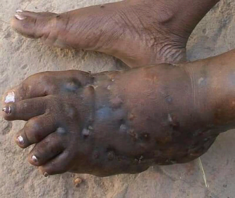
Eumycetoma, also known as Madura foot, is a persistent fungal infection of the skin and the tissues just under the skin, affecting most commonly the feet, although it can occur in hands and other body parts. It starts as a painless wet nodule, which may be present for years before ulceration, swelling, grainy discharge and weeping from sinuses and fistulae, followed by bone deformity. Several fungi can cause eumycetoma, including: ''Madurella mycetomatis'', ''Madurella grisea'', '' Leptosphaeria senegalensis'', ''Curvularia lunata'', ''Scedosporium apiospermum'', '' Neotestudina rosatii'', and '' Acremonium'' and ''Fusarium'' species. Diagnosis is by biopsy, visualising the fungi under the microscope and culture. Medical imaging may reveal extent of bone involvement. Other tests include ELISA, immunodiffusion, and DNA Barcoding. Treatment includes surgical removal of affected tissue and antifungal medicines. After treatment, recurrence is common. Sometimes, amputation is ... [...More Info...] [...Related Items...] OR: [Wikipedia] [Google] [Baidu] |


