|
Leukocyte Adhesion Deficiency-2
Congenital disorder of glycosylation type IIc or Leukocyte adhesion deficiency-2 (LAD2) is a type of leukocyte adhesion deficiency attributable to the absence of neutrophil sialyl-LewisX, a ligand of P- and E-selectin on vascular endothelium. It is associated with ''SLC35C1''. This disorder was discovered in two unrelated Israeli boys 3 and 5 years of age, each the offspring of consanguineous parents. Both had severe mental retardation, short stature, a distinctive facial appearance, and the Bombay (hh) blood phenotype, and both were secretor- and Lewis-negative. They both had had recurrent severe bacterial infections similar to those seen in patients with LAD1, including pneumonia, periodontitis, otitis media, and localized cellulitis. Similar to that in patients with LAD1, their infections were accompanied by pronounced leukocytosis (30,000 to 150,000/mm3) but an absence of pus formation at sites of recurrent cellulitis. In vitro studies revealed a pronounced defect in n ... [...More Info...] [...Related Items...] OR: [Wikipedia] [Google] [Baidu] |
Leukocyte Adhesion Deficiency
Leukocyte adhesion deficiency (LAD) is a rare autosomal recessive disorder characterized by immunodeficiency resulting in recurrent infections. LAD is currently divided into three subtypes: LAD1, LAD2, and the recently described LAD3, also known as LAD-1/variant. In LAD3, the immune defects are supplemented by a Glanzmann thrombasthenia-like bleeding tendency. Signs and symptoms LAD was first recognized as a distinct clinical entity in the 1970s. The classic descriptions of LAD included recurrent bacterial infections, defects in neutrophil adhesion, and a delay in umbilical cord sloughing. The adhesion defects result in poor leukocyte chemotaxis, particularly neutrophil, inability to form pus and neutrophilia. Individuals with LAD suffer from bacterial infections beginning in the neonatal period. Infections such as omphalitis, pneumonia, gingivitis, and peritonitis are common and often life-threatening due to the infant's inability to properly destroy the invading pathogens. ... [...More Info...] [...Related Items...] OR: [Wikipedia] [Google] [Baidu] |
Leukocytosis
Leukocytosis is a condition in which the white cell (leukocyte count) is above the normal range in the blood. It is frequently a sign of an inflammatory response, most commonly the result of infection, but may also occur following certain parasitic infections or bone tumors as well as leukemia. It may also occur after strenuous exercise, convulsions such as epilepsy, emotional stress, pregnancy and labor, anesthesia, as a side effect of medication (e.g., lithium), and epinephrine administration. There are five principal types of leukocytosis: # Neutrophilia (the most common form) # Lymphocytosis # Monocytosis # Eosinophilia # Basophilia This increase in leukocyte (primarily neutrophils) is usually accompanied by a "left upper shift" in the ratio of immature to mature neutrophils and macrophages. The proportion of immature leukocytes increases due to proliferation and inhibition of granulocyte and monocyte precursors in the bone marrow which is stimulated by several products of infl ... [...More Info...] [...Related Items...] OR: [Wikipedia] [Google] [Baidu] |
Autosomal Recessive Disorders
An autosome is any chromosome that is not a sex chromosome. The members of an autosome pair in a diploid cell have the same morphology, unlike those in allosomal (sex chromosome) pairs, which may have different structures. The DNA in autosomes is collectively known as atDNA or auDNA. For example, humans have a diploid genome that usually contains 22 pairs of autosomes and one allosome pair (46 chromosomes total). The autosome pairs are labeled with numbers (1–22 in humans) roughly in order of their sizes in base pairs, while allosomes are labelled with their letters. By contrast, the allosome pair consists of two X chromosomes in females or one X and one Y chromosome in males. Unusual combinations of XYY, XXY, XXX, XXXX, XXXXX or XXYY, among other Salome combinations, are known to occur and usually cause developmental abnormalities. Autosomes still contain sexual determination genes even though they are not sex chromosomes. For example, the SRY gene on the Y chromosome e ... [...More Info...] [...Related Items...] OR: [Wikipedia] [Google] [Baidu] |
Leukocyte Adhesion Deficiency
Leukocyte adhesion deficiency (LAD) is a rare autosomal recessive disorder characterized by immunodeficiency resulting in recurrent infections. LAD is currently divided into three subtypes: LAD1, LAD2, and the recently described LAD3, also known as LAD-1/variant. In LAD3, the immune defects are supplemented by a Glanzmann thrombasthenia-like bleeding tendency. Signs and symptoms LAD was first recognized as a distinct clinical entity in the 1970s. The classic descriptions of LAD included recurrent bacterial infections, defects in neutrophil adhesion, and a delay in umbilical cord sloughing. The adhesion defects result in poor leukocyte chemotaxis, particularly neutrophil, inability to form pus and neutrophilia. Individuals with LAD suffer from bacterial infections beginning in the neonatal period. Infections such as omphalitis, pneumonia, gingivitis, and peritonitis are common and often life-threatening due to the infant's inability to properly destroy the invading pathogens. ... [...More Info...] [...Related Items...] OR: [Wikipedia] [Google] [Baidu] |
Congenital Disorder Of Glycosylation
A congenital disorder of glycosylation (previously called carbohydrate-deficient glycoprotein syndrome) is one of several rare inborn errors of metabolism in which glycosylation of a variety of tissue proteins and/or lipids is deficient or defective. Congenital disorders of glycosylation are sometimes known as CDG syndromes. They often cause serious, sometimes fatal, malfunction of several different organ systems (especially the nervous system, muscles, and intestines) in affected infants. The most common sub-type is PMM2-CDG (formally known as CDG-Ia) where the genetic defect leads to the loss of phosphomannomutase 2 (PMM2), the enzyme responsible for the conversion of mannose-6-phosphate into mannose-1-phosphate. Presentation The specific problems produced differ according to the particular abnormal synthesis involved. Common manifestations include ataxia; seizures; retinopathy; liver disease; coagulopathies; failure to thrive (FTT); dysmorphic features (''e.g.,'' inverted nip ... [...More Info...] [...Related Items...] OR: [Wikipedia] [Google] [Baidu] |
CDNA
In genetics, complementary DNA (cDNA) is DNA synthesized from a single-stranded RNA (e.g., messenger RNA (mRNA) or microRNA (miRNA)) template in a reaction catalyzed by the enzyme reverse transcriptase. cDNA is often used to express a specific protein in a cell that does not normally express that protein (i.e., heterologous expression), or to sequence or quantify mRNA molecules using DNA based methods (qPCR, RNA-seq). cDNA that codes for a specific protein can be transferred to a recipient cell for expression, often bacterial or yeast expression systems. cDNA is also generated to analyze transcriptomic profiles in bulk tissue, single cells, or single nuclei in assays such as microarrays, qPCR, and RNA-seq. cDNA is also produced naturally by retroviruses (such as HIV-1, HIV-2, simian immunodeficiency virus, etc.) and then integrated into the host's genome, where it creates a provirus. The term ''cDNA'' is also used, typically in a bioinformatics context, to refer to an mR ... [...More Info...] [...Related Items...] OR: [Wikipedia] [Google] [Baidu] |
Golgi Apparatus
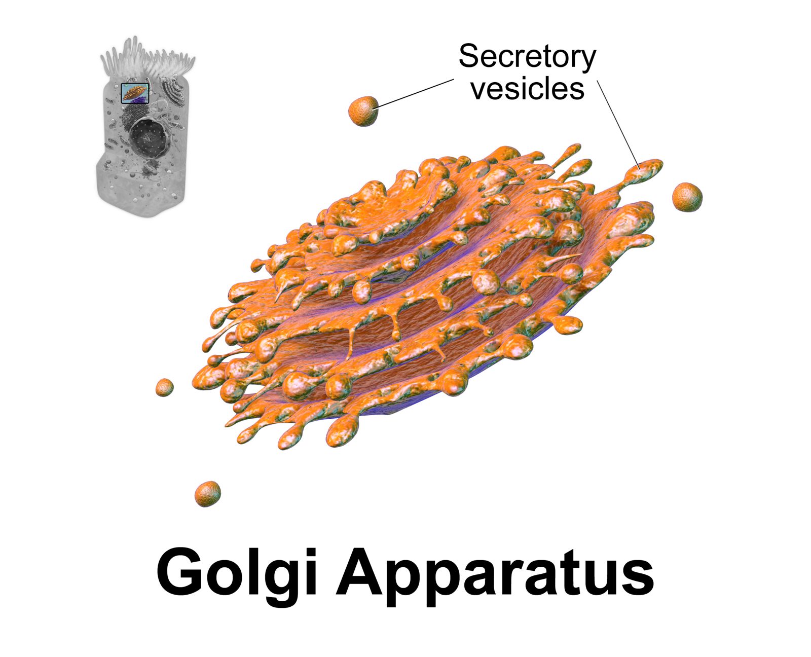
The Golgi apparatus (), also known as the Golgi complex, Golgi body, or simply the Golgi, is an organelle found in most eukaryotic cells. Part of the endomembrane system in the cytoplasm, it packages proteins into membrane-bound vesicles inside the cell before the vesicles are sent to their destination. It resides at the intersection of the secretory, lysosomal, and endocytic pathways. It is of particular importance in processing proteins for secretion, containing a set of glycosylation enzymes that attach various sugar monomers to proteins as the proteins move through the apparatus. It was identified in 1897 by the Italian scientist Camillo Golgi and was named after him in 1898. Discovery Owing to its large size and distinctive structure, the Golgi apparatus was one of the first organelles to be discovered and observed in detail. It was discovered in 1898 by Italian physician Camillo Golgi during an investigation of the nervous system. After first observing it under his ... [...More Info...] [...Related Items...] OR: [Wikipedia] [Google] [Baidu] |
Fucose
Fucose is a hexose deoxy sugar with the chemical formula C6H12O5. It is found on ''N''-linked glycans on the mammalian, insect and plant cell surface. Fucose is the fundamental sub-unit of the seaweed polysaccharide fucoidan. The α(1→3) linked core of fucose is a suspected carbohydrate antigen for IgE-mediated allergy. Two structural features distinguish fucose from other six-carbon sugars present in mammals: the lack of a hydroxyl group on the carbon at the 6-position (C-6) (thereby making it a deoxy sugar) and the L -configuration. It is equivalent to 6-deoxy--galactose. In the fucose-containing glycan structures, fucosylated glycans, fucose can exist as a terminal modification or serve as an attachment point for adding other sugars. In human ''N''-linked glycans, fucose is most commonly linked α-1,6 to the reducing terminal β-''N''-acetylglucosamine. However, fucose at the non-reducing termini linked α-1,2 to galactose forms the H antigen, the substructure of the A an ... [...More Info...] [...Related Items...] OR: [Wikipedia] [Google] [Baidu] |
Fucosyltransferase
A fucosyltransferase is an enzyme that transfers an L-fucose sugar from a GDP-fucose (guanosine diphosphate-fucose) donor substrate to an acceptor substrate. The acceptor substrate can be another sugar such as the transfer of a fucose to a core GlcNAc (N-acetylglucosamine) sugar as in the case of N-linked glycosylation, or to a protein, as in the case of ''O''-linked glycosylation produced by ''O''-fucosyltransferase. There are various fucosyltransferases in mammals, the vast majority of which, are located in the Golgi apparatus. The ''O''-fucosyltransferases have recently been shown to localize to the endoplasmic reticulum (ER). Some of the proteins in this group are responsible for the molecular basis of the blood group antigens, surface markers on the outside of the red blood cell membrane. Most of these markers are proteins, but some are carbohydrates attached to lipids or proteins eid M.E., Lomas-Francis C. The Blood Group Antigen FactsBook Academic Press, London / San Di ... [...More Info...] [...Related Items...] OR: [Wikipedia] [Google] [Baidu] |
H Antigen
H antigen can refer to one of various types of antigens having diverse biological functions. H antigen is located on the 19th chromosome in humans, and has a variety of functions and definitions as follows: * Also known as substance H, H antigen is a precursor to each of the ABO blood group antigens, apparently present in all people except those with the Bombay Blood phenotype (see hh blood group) * Histocompatibility antigen, a major factor in graft rejection. Even when Major Histocompatibility Complex genotype is perfectly matched, can cause slow rejection of a graft. ** major H antigens "encode molecules that present foreign peptides to T cells" ** minor H antigens "present polymorphic self peptides to T cells". Includes, e.g. the H-Y antigen H-Y antigen is a male tissue specific antigen. Originally thought to trigger the formation of testes (via loci, an autosomal gene that generates the antigen and one that generates the receptor) it is now known that it does not tri ... [...More Info...] [...Related Items...] OR: [Wikipedia] [Google] [Baidu] |
Cellulitis
Cellulitis is usually a bacterial infection involving the inner layers of the skin. It specifically affects the dermis and subcutaneous fat. Signs and symptoms include an area of redness which increases in size over a few days. The borders of the area of redness are generally not sharp and the skin may be swollen. While the redness often turns white when pressure is applied, this is not always the case. The area of infection is usually painful. Lymphatic vessels may occasionally be involved, and the person may have a fever and feel tired. The legs and face are the most common sites involved, although cellulitis can occur on any part of the body. The leg is typically affected following a break in the skin. Other risk factors include obesity, leg swelling, and old age. For facial infections, a break in the skin beforehand is not usually the case. The bacteria most commonly involved are streptococci and '' Staphylococcus aureus''. In contrast to cellulitis, erysipelas is a bacte ... [...More Info...] [...Related Items...] OR: [Wikipedia] [Google] [Baidu] |
Neutrophil
Neutrophils (also known as neutrocytes or heterophils) are the most abundant type of granulocytes and make up 40% to 70% of all white blood cells in humans. They form an essential part of the innate immune system, with their functions varying in different animals. They are formed from stem cells in the bone marrow and Cellular differentiation, differentiated into #Subpopulations, subpopulations of neutrophil-killers and neutrophil-cagers. They are short-lived and highly mobile, as they can enter parts of tissue where other cells/molecules cannot. Neutrophils may be subdivided into segmented neutrophils and banded neutrophils (or Band cell, bands). They form part of the polymorphonuclear cells family (PMNs) together with basophils and eosinophils. The name ''neutrophil'' derives from staining characteristics on hematoxylin and eosin (H&E stain, H&E) histology, histological or cell biology, cytological preparations. Whereas basophilic white blood cells stain dark blue and eosinoph ... [...More Info...] [...Related Items...] OR: [Wikipedia] [Google] [Baidu] |


