|
Letterer–Siwe Disease
Letterer–Siwe disease, (LSD) or Abt-Letterer-Siwe disease, is one of the four recognized clinical syndromes of Langerhans cell histiocytosis (LCH) and is the most severe form, involving multiple organ systems such as the skin, bone marrow, spleen, liver, and lung. Oral cavity and gastrointestinal involvement may also be seen. Shahlaee, A. H. and Arceci, R. J. (2006). Histiocytic disorders, in LCH and all its subtypes are characterized by monoclonal migration and proliferation of specific dendritic cells. The subcategorization of Letterer-Siwe disease is a historical eponym. Designating the four subtypes of LCH as separate entities are mostly of historical significance, because they are varied manifestations of the same underlying disease process, and patients also often exhibit symptoms from more than one of the four syndromes. Letterer-Siwe causes approximately 10% of LCH disease. Prevalence is estimated at 1:500,000 and the disease almost exclusively occurs in children les ... [...More Info...] [...Related Items...] OR: [Wikipedia] [Google] [Baidu] |
Langerhans Cell Histiocytosis
Langerhans cell histiocytosis (LCH) is an abnormal clonal proliferation of Langerhans cells, abnormal cells deriving from bone marrow and capable of migrating from skin to lymph nodes. Symptoms range from isolated bone lesions to multisystem disease. LCH is part of a group of syndromes called histiocytoses, which are characterized by an abnormal proliferation of histiocytes (an archaic term for activated dendritic cells and macrophages). These diseases are related to other forms of abnormal proliferation of white blood cells, such as leukemias and lymphomas. The disease has gone by several names, including Hand–Schüller–Christian disease, Abt-Letterer-Siwe disease, Hashimoto-Pritzker disease (a very rare self-limiting variant seen at birth) and histiocytosis X, until it was renamed in 1985 by the Histiocyte Society. Classification The disease spectrum results from clonal accumulation and proliferation of cells resembling the epidermal dendritic cells called Langerhan ... [...More Info...] [...Related Items...] OR: [Wikipedia] [Google] [Baidu] |
Dendritic Cells
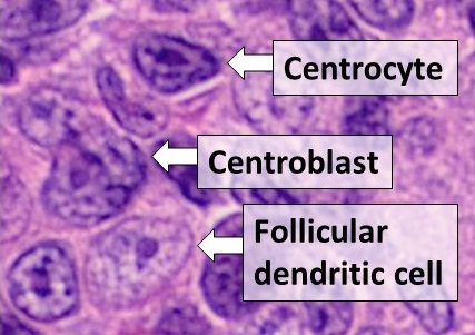
Dendritic cells (DCs) are antigen-presenting cells (also known as ''accessory cells'') of the mammalian immune system. Their main function is to process antigen material and present it on the cell surface to the T cells of the immune system. They act as messengers between the innate and the adaptive immune systems. Dendritic cells are present in those tissues that are in contact with the external environment, such as the skin (where there is a specialized dendritic cell type called the Langerhans cell) and the inner lining of the nose, lungs, stomach and intestines. They can also be found in an immature state in the blood. Once activated, they migrate to the lymph nodes where they interact with T cells and B cells to initiate and shape the adaptive immune response. At certain development stages they grow branched projections, the ''dendrites'' that give the cell its name (δένδρον or déndron being Greek for 'tree'). While similar in appearance, these are structures disti ... [...More Info...] [...Related Items...] OR: [Wikipedia] [Google] [Baidu] |
Granulomatous Lesions
A granuloma is an aggregation of macrophages that forms in response to chronic inflammation. This occurs when the immune system The immune system is a network of biological processes that protects an organism from diseases. It detects and responds to a wide variety of pathogens, from viruses to parasitic worms, as well as cancer cells and objects such as wood splint ... attempts to isolate foreign substances that it is otherwise unable to eliminate. Such substances include infectious organisms including bacteria and fungi, as well as other materials such as Foreign body, foreign objects, keratin, and Surgical suture, suture fragments. Definition In pathology, a granuloma is an organized collection of macrophages. In medical practice, doctors occasionally use the term ''granuloma'' in its more literal meaning: "a small nodule". Since a small nodule (medicine), nodule can represent any tissue from a harmless nevus to a malignant tumor, this use of the term is not very sp ... [...More Info...] [...Related Items...] OR: [Wikipedia] [Google] [Baidu] |
Histiocytes
A histiocyte is a vertebrate cell that is part of the mononuclear phagocyte system (also known as the reticuloendothelial system or lymphoreticular system). The mononuclear phagocytic system is part of the organism's immune system. The histiocyte is a tissue macrophage or a dendritic cell (histio, diminutive of histo, meaning ''tissue'', and cyte, meaning ''cell''). Part of their job is to clear out neutrophils once they've reached the end of their lifespan. Development Histiocytes are derived from the bone marrow by multiplication from a stem cell. The derived cells migrate from the bone marrow to the blood as monocytes. They circulate through the body and enter various organs, where they undergo differentiation into histiocytes, which are part of the mononuclear phagocytic system (MPS). However, the term ''histiocyte'' has been used for multiple purposes in the past, and some cells called "histocytes" do not appear to derive from monocytic-macrophage lines. The term Histioc ... [...More Info...] [...Related Items...] OR: [Wikipedia] [Google] [Baidu] |
Oncogene
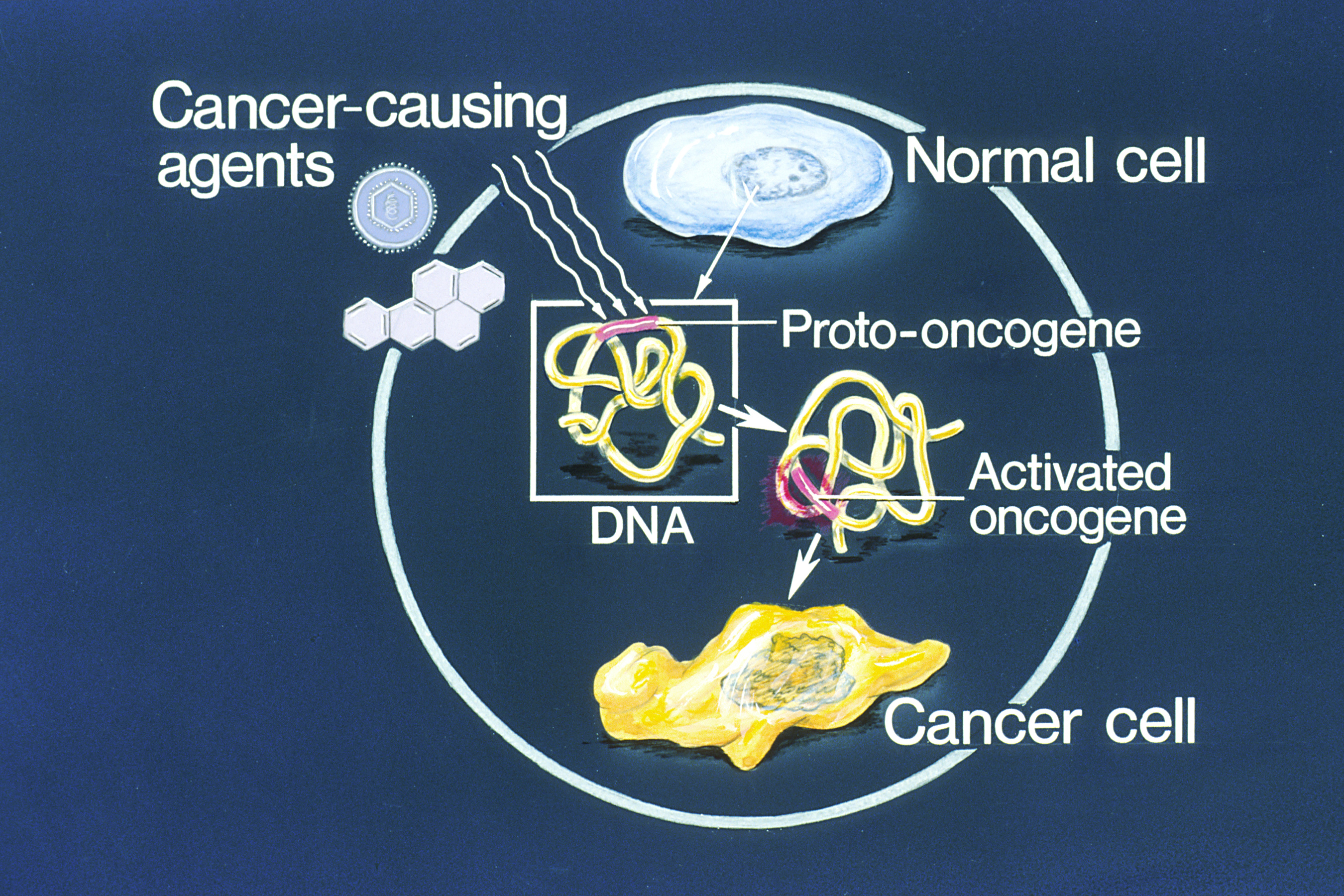
An oncogene is a gene that has the potential to cause cancer. In tumor cells, these genes are often mutated, or expressed at high levels.Kimball's Biology Pages. "Oncogenes" Free full text Most normal cells will undergo a programmed form of rapid cell death () when critical functions are altered and malfunctioning. Activated oncogenes can cause those cells designated for apoptosis to survive and proliferate instead. Most oncogenes began as proto-oncogenes: normal genes involved in cell growth and proliferation or inhibition of apoptosis. If, through mutation, normal genes promoting cellular growth are up-regulated (gain-of-function mutation), they will predisp ... [...More Info...] [...Related Items...] OR: [Wikipedia] [Google] [Baidu] |
X-rays
An X-ray, or, much less commonly, X-radiation, is a penetrating form of high-energy electromagnetic radiation. Most X-rays have a wavelength ranging from 10 Picometre, picometers to 10 Nanometre, nanometers, corresponding to frequency, frequencies in the range 30 Hertz, petahertz to 30 Hertz, exahertz ( to ) and energies in the range 145 electronvolt, eV to 124 keV. X-ray wavelengths are shorter than those of ultraviolet, UV rays and typically longer than those of gamma rays. In many languages, X-radiation is referred to as Röntgen radiation, after the German scientist Wilhelm Röntgen, Wilhelm Conrad Röntgen, who discovered it on November 8, 1895. He named it ''X-radiation'' to signify an unknown type of radiation.Novelline, Robert (1997). ''Squire's Fundamentals of Radiology''. Harvard University Press. 5th edition. . Spellings of ''X-ray(s)'' in English include the variants ''x-ray(s)'', ''xray(s)'', and ''X ray(s)''. The most familiar use of X-ra ... [...More Info...] [...Related Items...] OR: [Wikipedia] [Google] [Baidu] |
CT Scan
A computed tomography scan (CT scan; formerly called computed axial tomography scan or CAT scan) is a medical imaging technique used to obtain detailed internal images of the body. The personnel that perform CT scans are called radiographers or radiology technologists. CT scanners use a rotating X-ray tube and a row of detectors placed in a gantry (medical), gantry to measure X-ray Attenuation#Radiography, attenuations by different tissues inside the body. The multiple X-ray measurements taken from different angles are then processed on a computer using tomographic reconstruction algorithms to produce Tomography, tomographic (cross-sectional) images (virtual "slices") of a body. CT scans can be used in patients with metallic implants or pacemakers, for whom magnetic resonance imaging (MRI) is Contraindication, contraindicated. Since its development in the 1970s, CT scanning has proven to be a versatile imaging technique. While CT is most prominently used in medical diagnosis, ... [...More Info...] [...Related Items...] OR: [Wikipedia] [Google] [Baidu] |
Needle Biopsy
Fine-needle aspiration (FNA) is a diagnostic procedure used to investigate lumps or masses. In this technique, a thin (23–25 gauge (0.52 to 0.64 mm outer diameter)), hollow needle is inserted into the mass for sampling of cells that, after being stained, are examined under a microscope (biopsy). The sampling and biopsy considered together are called fine-needle aspiration biopsy (FNAB) or fine-needle aspiration cytology (FNAC) (the latter to emphasize that any aspiration biopsy involves cytopathology, not histopathology). Fine-needle aspiration biopsies are very safe minor surgical procedures. Often, a major surgical (excisional or open) biopsy can be avoided by performing a needle aspiration biopsy instead, eliminating the need for hospitalization. In 1981, the first fine-needle aspiration biopsy in the United States was done at Maimonides Medical Center. Today, this procedure is widely used in the diagnosis of cancer and inflammatory conditions. Aspiration is safer and ... [...More Info...] [...Related Items...] OR: [Wikipedia] [Google] [Baidu] |
Rare Diseases
A rare disease is any disease that affects a small percentage of the population. In some parts of the world, an orphan disease is a rare disease whose rarity means there is a lack of a market large enough to gain support and resources for discovering treatments for it, except by the government granting economically advantageous conditions to creating and selling such treatments. Orphan drugs are ones so created or sold. Most rare diseases are genetic and thus are present throughout the person's entire life, even if symptoms do not immediately appear. Many rare diseases appear early in life, and about 30% of children with rare diseases will die before reaching their fifth birthdays. With only four diagnosed patients in 27 years, ribose-5-phosphate isomerase deficiency is considered the rarest known genetic disease. No single cut-off number has been agreed upon for which a disease is considered rare. A disease may be considered rare in one part of the world, or in a particular gro ... [...More Info...] [...Related Items...] OR: [Wikipedia] [Google] [Baidu] |
Autosomal Recessive Disorders
An autosome is any chromosome that is not a sex chromosome. The members of an autosome pair in a diploid cell have the same morphology, unlike those in allosomal (sex chromosome) pairs, which may have different structures. The DNA in autosomes is collectively known as atDNA or auDNA. For example, humans have a diploid genome that usually contains 22 pairs of autosomes and one allosome pair (46 chromosomes total). The autosome pairs are labeled with numbers (1–22 in humans) roughly in order of their sizes in base pairs, while allosomes are labelled with their letters. By contrast, the allosome pair consists of two X chromosomes in females or one X and one Y chromosome in males. Unusual combinations of XYY, XXY, XXX, XXXX, XXXXX or XXYY, among other Salome combinations, are known to occur and usually cause developmental abnormalities. Autosomes still contain sexual determination genes even though they are not sex chromosomes. For example, the SRY gene on the Y chromosome e ... [...More Info...] [...Related Items...] OR: [Wikipedia] [Google] [Baidu] |


