|
Language Center
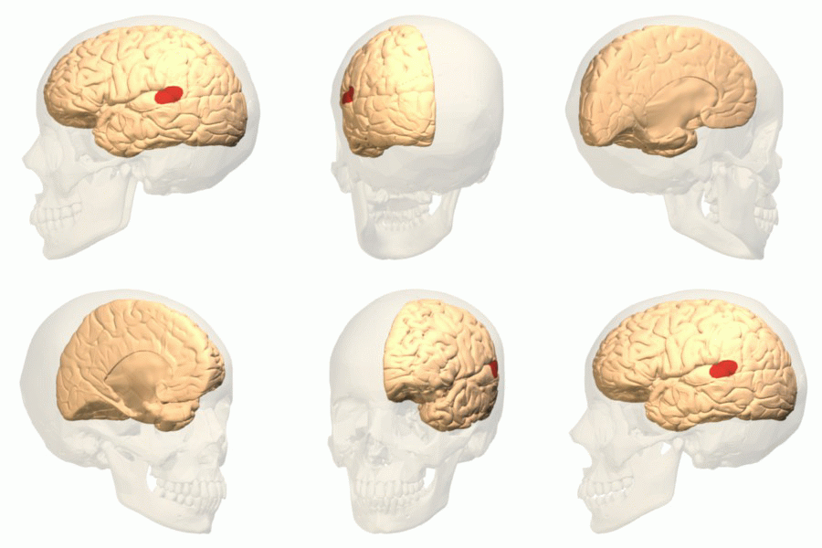
In neuroscience and psychology, the term language center refers collectively to the areas of the brain which serve a particular function for speech processing and production. Language is a core system, which gives humans the capacity to solve difficult problems and provides them with a unique type of social interaction. Language allows individuals to attribute symbols (e.g. words or signs) to specific concepts and display them through sentences and phrases that follow proper grammatical rules. Moreover, speech is the mechanism in which language is orally expressed. Information is exchanged in a larger system including language-related regions. These regions are connected by white matter fiber tracts that make possible the transmission of information between regions. The white matter fiber bunches were recognized to be important for language production after suggesting that it is possible to make a connection between multiple language centers. The three classical language areas ... [...More Info...] [...Related Items...] OR: [Wikipedia] [Google] [Baidu] |
Neuroscience
Neuroscience is the scientific study of the nervous system (the brain, spinal cord, and peripheral nervous system), its functions and disorders. It is a multidisciplinary science that combines physiology, anatomy, molecular biology, developmental biology, cytology, psychology, physics, computer science, chemistry, medicine, statistics, and Mathematical Modeling, mathematical modeling to understand the fundamental and emergent properties of neurons, glia and neural circuits. The understanding of the biological basis of learning, memory, behavior, perception, and consciousness has been described by Eric Kandel as the "epic challenge" of the Biology, biological sciences. The scope of neuroscience has broadened over time to include different approaches used to study the nervous system at different scales. The techniques used by neuroscientists have expanded enormously, from molecular biology, molecular and cell biology, cellular studies of individual neurons to neuroimaging, imaging ... [...More Info...] [...Related Items...] OR: [Wikipedia] [Google] [Baidu] |
Paul Broca
Pierre Paul Broca (, also , , ; 28 June 1824 – 9 July 1880) was a French physician, anatomist and anthropologist. He is best known for his research on Broca's area, a region of the frontal lobe that is named after him. Broca's area is involved with language. His work revealed that the brains of patients with aphasia contained lesions in a particular part of the cortex, in the left frontal region. This was the first anatomical proof of localization of brain function. Broca's work also contributed to the development of physical anthropology, advancing the science of anthropometry. Biography Paul Broca was born on 28 June 1824 in Sainte-Foy-la-Grande, Bordeaux, France, the son of Jean Pierre "Benjamin" Broca, a medical practitioner and former surgeon in Napoleon's service, and Annette Thomas, a well-educated daughter of a Calvinist, Reformed Protestant, preacher. Huguenot Broca received basic education in the school in his hometown, earning a bachelor's degree at the age of 16. H ... [...More Info...] [...Related Items...] OR: [Wikipedia] [Google] [Baidu] |
Brodmann Area 6
Brodmann area 6 (BA6) is part of the frontal cortex in the human brain. Situated just anterior to the primary motor cortex ( BA4), it is composed of the premotor cortex and, medially, the supplementary motor area (SMA). This large area of the frontal cortex is believed to play a role in planning complex, coordinated movements. Brodmann area 6 is also called agranular frontal area 6 in humans because it lacks an internal granular cortical layer (layer IV). It is a subdivision of the cytoarchitecturally defined precentral region of cerebral cortex. In the human brain, it is located on the portions of the precentral gyrus that are not occupied by Brodmann area 4; furthermore, BA6 extends onto the caudal portions of the superior frontal and middle frontal gyri. It extends from the cingulate sulcus on the medial aspect of the hemisphere to the lateral sulcus on the lateral aspect. It is bounded rostrally by the granular frontal region and caudally by the gigantopyramidal area 4 (Brod ... [...More Info...] [...Related Items...] OR: [Wikipedia] [Google] [Baidu] |
Caudate Nucleus
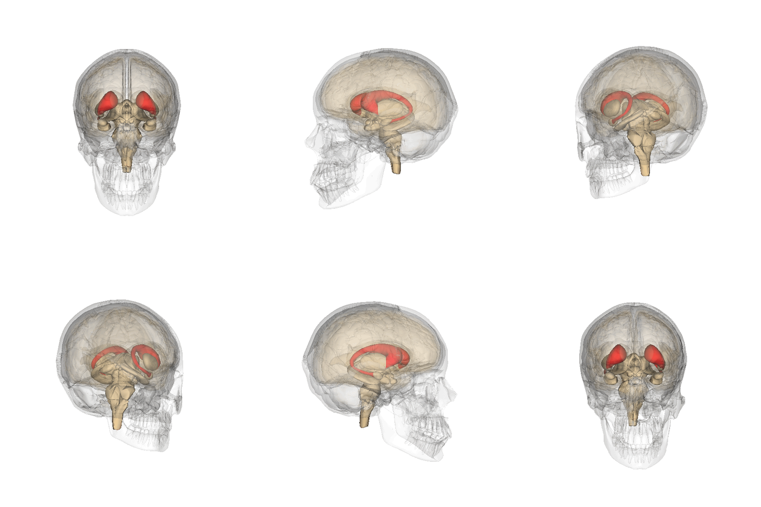
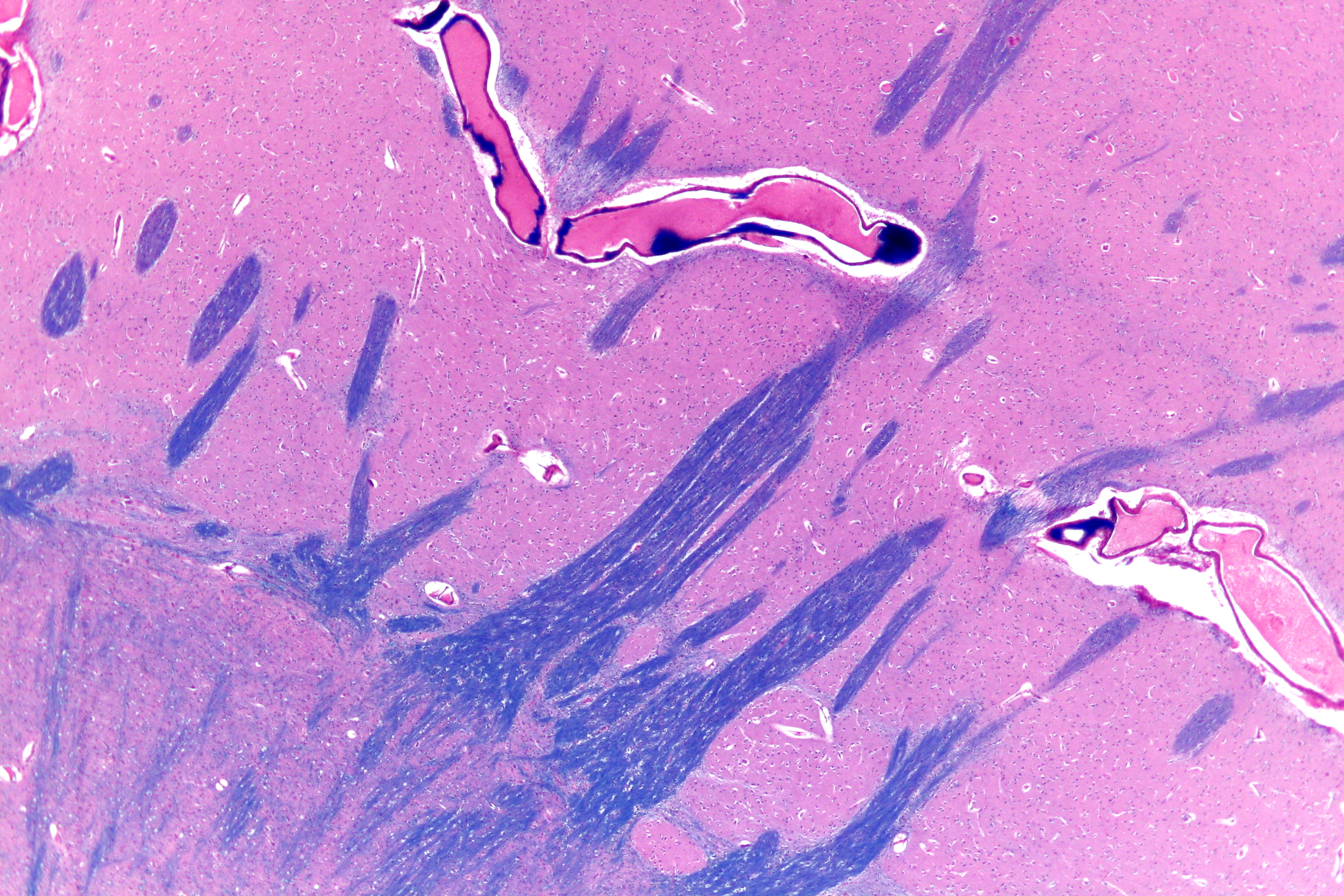
The caudate nucleus is one of the structures that make up the corpus striatum, which is a component of the basal ganglia in the human brain. While the caudate nucleus has long been associated with motor processes due to its role in Parkinson's disease, it plays important roles in various other nonmotor functions as well, including procedural learning, associative learning and inhibitory control of action, among other functions. The caudate is also one of the brain structures which compose the reward system and functions as part of the cortico–basal ganglia– thalamic loop. Structure Together with the putamen, the caudate forms the dorsal striatum, which is considered a single functional structure; anatomically, it is separated by a large white matter tract, the internal capsule, so it is sometimes also referred to as two structures: the medial dorsal striatum (the caudate) and the lateral dorsal striatum (the putamen). In this vein, the two are functionally distinct not a ... [...More Info...] [...Related Items...] OR: [Wikipedia] [Google] [Baidu] |
Putamen
The putamen (; from Latin, meaning "nutshell") is a round structure located at the base of the forebrain (telencephalon). The putamen and caudate nucleus together form the dorsal striatum. It is also one of the structures that compose the basal nuclei. Through various pathways, the putamen is connected to the substantia nigra, the globus pallidus, the claustrum, and the thalamus, in addition to many regions of the cerebral cortex. A primary function of the putamen is to regulate movements at various stages (e.g. preparation and execution) and influence various types of learning. It employs GABA, acetylcholine, and enkephalin to perform its functions. The putamen also plays a role in degenerative neurological disorders, such as Parkinson's disease. History The word "putamen" is from Latin, referring to that which "falls off in pruning", from "putare", meaning "to prune, to think, or to consider". Until recently, most MRI research focused broadly on the basal ganglia as a who ... [...More Info...] [...Related Items...] OR: [Wikipedia] [Google] [Baidu] |
Cerebral Cortex
The cerebral cortex, also known as the cerebral mantle, is the outer layer of neural tissue of the cerebrum of the brain in humans and other mammals. The cerebral cortex mostly consists of the six-layered neocortex, with just 10% consisting of allocortex. It is separated into two cortices, by the longitudinal fissure that divides the cerebrum into the left and right cerebral hemispheres. The two hemispheres are joined beneath the cortex by the corpus callosum. The cerebral cortex is the largest site of neural integration in the central nervous system. It plays a key role in attention, perception, awareness, thought, memory, language, and consciousness. The cerebral cortex is part of the brain responsible for cognition. In most mammals, apart from small mammals that have small brains, the cerebral cortex is folded, providing a greater surface area in the confined volume of the cranium. Apart from minimising brain and cranial volume, cortical folding is crucial for the brain ... [...More Info...] [...Related Items...] OR: [Wikipedia] [Google] [Baidu] |
Functional Magnetic Resonance Imaging
Functional magnetic resonance imaging or functional MRI (fMRI) measures brain activity by detecting changes associated with blood flow. This technique relies on the fact that cerebral blood flow and neuronal activation are coupled. When an area of the brain is in use, blood flow to that region also increases. The primary form of fMRI uses the blood-oxygen-level dependent (BOLD) contrast, discovered by Seiji Ogawa in 1990. This is a type of specialized brain and body scan used to map neuron, neural activity in the brain or spinal cord of humans or other animals by imaging the change in blood flow (hemodynamic response) related to energy use by brain cells. Since the early 1990s, fMRI has come to dominate brain mapping research because it does not involve the use of injections, surgery, the ingestion of substances, or exposure to ionizing radiation. This measure is frequently corrupted by noise from various sources; hence, statistical procedures are used to extract the underlying si ... [...More Info...] [...Related Items...] OR: [Wikipedia] [Google] [Baidu] |
Positron Emission Tomography
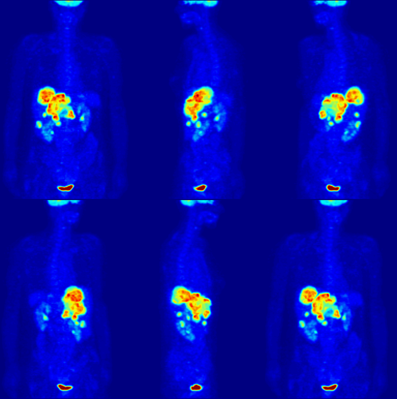
Positron emission tomography (PET) is a functional imaging technique that uses radioactive substances known as radiotracers to visualize and measure changes in Metabolism, metabolic processes, and in other physiological activities including blood flow, regional chemical composition, and absorption. Different tracers are used for various imaging purposes, depending on the target process within the body. For example, 18F-FDG, -FDG is commonly used to detect cancer, Sodium fluoride#Medical imaging, NaF is widely used for detecting bone formation, and Isotopes of oxygen#Oxygen-15, oxygen-15 is sometimes used to measure blood flow. PET is a common medical imaging, imaging technique, a Scintigraphy#Process, medical scintillography technique used in nuclear medicine. A radiopharmaceutical, radiopharmaceutical — a radioisotope attached to a drug — is injected into the body as a radioactive tracer, tracer. When the radiopharmaceutical undergoes beta plus decay, a positron is ... [...More Info...] [...Related Items...] OR: [Wikipedia] [Google] [Baidu] |
Temporal Lobe
The temporal lobe is one of the four Lobes of the brain, major lobes of the cerebral cortex in the brain of mammals. The temporal lobe is located beneath the lateral fissure on both cerebral hemispheres of the mammalian brain. The temporal lobe is involved in processing sensory input into derived meanings for the appropriate retention of visual memory, language comprehension, and emotion association. ''Temporal'' refers to the head's Temple (anatomy), temples. Structure The Temple (anatomy)#Etymology, temporal Lobe (anatomy), lobe consists of structures that are vital for declarative or long-term memory. Declarative memory, Declarative (denotative) or Explicit memory, explicit memory is conscious memory divided into semantic memory (facts) and episodic memory (events). Medial temporal lobe structures that are critical for long-term memory include the hippocampus, along with the surrounding Hippocampal formation, hippocampal region consisting of the Perirhinal cortex, perirhinal, ... [...More Info...] [...Related Items...] OR: [Wikipedia] [Google] [Baidu] |
Inferior Temporal Gyrus
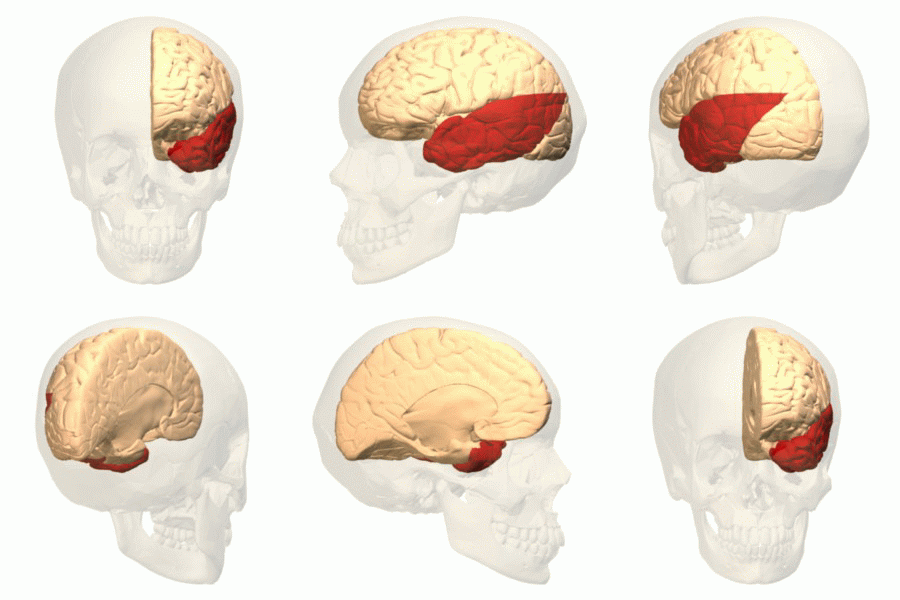
The inferior temporal gyrus is one of three gyri of the temporal lobe and is located below the middle temporal gyrus, connected behind with the inferior occipital gyrus; it also extends around the infero-lateral border on to the inferior surface of the temporal lobe, where it is limited by the inferior Sulcus (neuroanatomy), sulcus. This region is one of the higher levels of the ventral stream of visual processing, associated with the representation of objects, places, faces, and colors. It may also be involved in face perception, and in the recognition of numbers and words. The inferior temporal gyrus is the anterior region of the temporal lobe located underneath the central temporal sulcus. The primary function of the occipital temporal gyrus – otherwise referenced as IT cortex – is associated with visual stimuli processing, namely visual object recognition, and has been suggested by recent experimental results as the final location of the ventral cortical visual system. The ... [...More Info...] [...Related Items...] OR: [Wikipedia] [Google] [Baidu] |
Aphasia
Aphasia is an inability to comprehend or formulate language because of damage to specific brain regions. The major causes are stroke and head trauma; prevalence is hard to determine but aphasia due to stroke is estimated to be 0.1–0.4% in the Global North. Aphasia can also be the result of brain tumors, brain infections, or neurodegenerative diseases (such as dementias). To be diagnosed with aphasia, a person's speech or language must be significantly impaired in one (or more) of the four aspects of communication following acquired brain injury. Alternatively, in the case of progressive aphasia, it must have significantly declined over a short period of time. The four aspects of communication are auditory comprehension, verbal expression, reading and writing, and functional communication. The difficulties of people with aphasia can range from occasional trouble finding words, to losing the ability to speak, read, or write; intelligence, however, is unaffected. Expressive lan ... [...More Info...] [...Related Items...] OR: [Wikipedia] [Google] [Baidu] |
Carl Wernicke
Carl (or Karl) Wernicke (; ; 15 May 1848 – 15 June 1905) was a German physician, anatomist, psychiatrist and neuropathologist. He is known for his influential research into the pathological effects of specific forms of encephalopathy and also the study of receptive aphasia, both of which are commonly associated with Wernicke's name and referred to as Wernicke encephalopathy and Wernicke's aphasia, respectively. His research, along with that of Paul Broca, led to groundbreaking realizations of the localization of brain function, specifically in speech. As such, Wernicke's area (a.k.a. Wernicke's Speech Area) has been named after the scientist. Biography Wernicke was born on May 15, 1848, in Tarnowitz, a small town in Upper Silesia, Prussia, now Tarnowskie Góry, Poland. He obtained his secondary education at the gymnasium in Oppeln, which is a school near the university of Breslau. Wernicke then studied medicine at the University of Breslau and did graduate work studying langu ... [...More Info...] [...Related Items...] OR: [Wikipedia] [Google] [Baidu] |

_(14782023652).jpg)
