|
Lacuna (histology)
In histology, a lacuna is a small space, containing an osteocyte in bone, or chondrocyte in cartilage. Bone The lacunae are situated between the lamellae, and consist of a number of oblong spaces. In an ordinary microscopic section, viewed by transmitted light, they appear as fusiform opaque spots. Each lacuna is occupied during life by a branched cell, termed an osteocyte, bone-cell or bone-corpuscle. Lacunae are connected to one another by small canals called canaliculi. A lacuna never contains more than one osteocyte. Sinuses are an example of lacuna. Cartilage The cartilage cells or chondrocytes are contained in cavities in the matrix, called cartilage lacunae; around these, the matrix is arranged in concentric lines as if it had been formed in successive portions around the cartilage cells. This constitutes the so-called capsule of the space. Each lacuna is generally occupied by a single cell, but during the division of the cells, it may contain two, four, or eight cells. L ... [...More Info...] [...Related Items...] OR: [Wikipedia] [Google] [Baidu] |
Femur
The femur (; ), or thigh bone, is the proximal bone of the hindlimb in tetrapod vertebrates. The head of the femur articulates with the acetabulum in the pelvic bone forming the hip joint, while the distal part of the femur articulates with the tibia (shinbone) and patella (kneecap), forming the knee joint. By most measures the two (left and right) femurs are the strongest bones of the body, and in humans, the largest and thickest. Structure The femur is the only bone in the upper leg. The two femurs converge medially toward the knees, where they articulate with the proximal ends of the tibiae. The angle of convergence of the femora is a major factor in determining the femoral-tibial angle. Human females have thicker pelvic bones, causing their femora to converge more than in males. In the condition ''genu valgum'' (knock knee) the femurs converge so much that the knees touch one another. The opposite extreme is ''genu varum'' (bow-leggedness). In the general populatio ... [...More Info...] [...Related Items...] OR: [Wikipedia] [Google] [Baidu] |
Haversian Canals
Haversian canals (sometimes canals of Havers) are a series of microscopic tubes in the outermost region of bone called cortical bone. They allow blood vessels and nerves to travel through them to supply the osteocytes. Structure Each Haversian canal generally contains one or two capillaries and many nerve fibres. The channels are formed by concentric layers called lamellae, which are approximately 50 µm in diameter. The Haversian canals surround blood vessels and nerve cells throughout bones and communicate with osteocytes (contained in spaces within the dense bone matrix called lacunae) through connections called canaliculi. This unique arrangement is conducive to mineral salt deposits and storage which gives bone tissue its strength. Active transport is used to move most substances between the blood vessels and the osteocytes. Haversian canals are contained within osteons, which are typically arranged along the long axis of the bone in parallel to the surface. The canals a ... [...More Info...] [...Related Items...] OR: [Wikipedia] [Google] [Baidu] |
Lamella (zoology)
Lamellae on a gecko's foot. In surface anatomy, a lamella is a thin plate-like structure, often one amongst many lamellae very close to one another, with open space between. Aside from respiratory organs, they appear in other biological roles including filter feeding and the traction surfaces of geckos. In fish, gill lamellae are used to increase the surface area in contact with the environment to maximize gas exchange (both to attain oxygen and to expel carbon dioxide) between the water and the blood. In fish gills there are two types of lamellae, primary and secondary. The primary gill lamellae (also called gill filament) extends from the gill arch, and the secondary gill lamellae extends from the primary gill lamellae. Gas exchange primarily occurs at the secondary gill lamellae, where the tissue is notably only one cell layer thick. Furthermore, countercurrent gas exchange at the secondary gill lamellae further maximizes oxygen uptake and carbon dioxide release. See also ... [...More Info...] [...Related Items...] OR: [Wikipedia] [Google] [Baidu] |
Canaliculus (bone)
Bone canaliculi are microscopic canals between the lacunae of ossified bone. The radiating processes of the osteocytes (called filopodia) project into these canals. These cytoplasmic processes are joined together by gap junctions. Osteocytes do not entirely fill up the canaliculi. The remaining space is known as the periosteocytic space, which is filled with periosteocytic fluid. This fluid contains substances too large to be transported through the gap junctions that connect the osteocytes. In cartilage, the lacunae and hence, the chondrocytes, are isolated from each other. Materials picked up by osteocytes adjacent to blood vessels are distributed throughout the bone matrix via the canaliculi. Dental canaliculi The dental canaliculi (sometimes called dentinal tubules) are the blood supply of a tooth. Odontoblast process run in the canaliculi that transverse the dentin layer and are referred as dentinal tubules. The number and size of the canaliculi decrease as the tubul ... [...More Info...] [...Related Items...] OR: [Wikipedia] [Google] [Baidu] |
Vertebra
The spinal column, a defining synapomorphy shared by nearly all vertebrates,Hagfish are believed to have secondarily lost their spinal column is a moderately flexible series of vertebrae (singular vertebra), each constituting a characteristic irregular bone whose complex structure is composed primarily of bone, and secondarily of hyaline cartilage. They show variation in the proportion contributed by these two tissue types; such variations correlate on one hand with the cerebral/caudal rank (i.e., location within the backbone), and on the other with phylogenetic differences among the vertebrate taxa. The basic configuration of a vertebra varies, but the bone is its ''body'', with the central part of the body constituting the ''centrum''. The upper (closer to) and lower (further from), respectively, the cranium and its central nervous system surfaces of the vertebra body support attachment to the intervertebral discs. The posterior part of a vertebra forms a vertebral arch ... [...More Info...] [...Related Items...] OR: [Wikipedia] [Google] [Baidu] |
Mouse
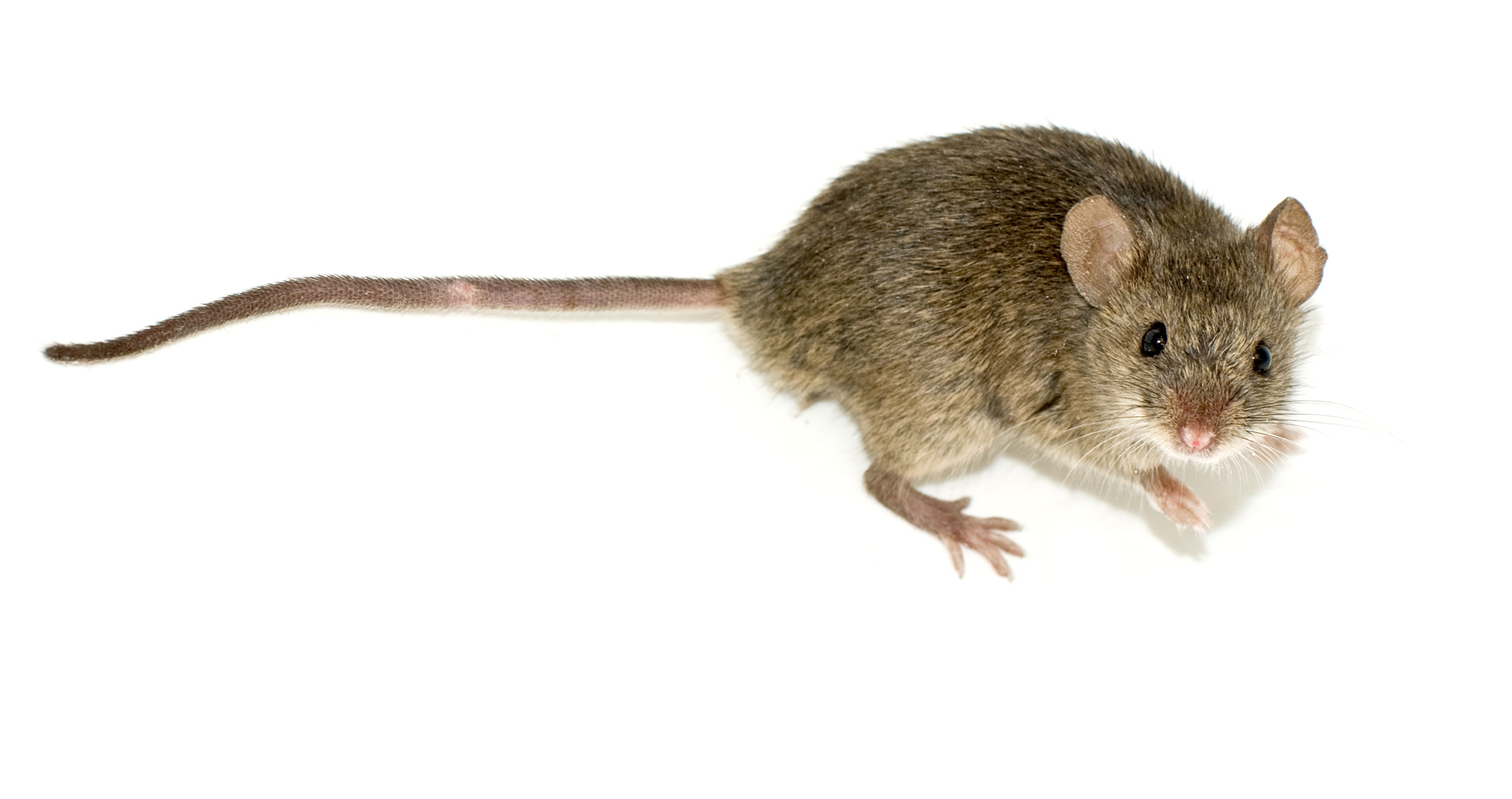
A mouse ( : mice) is a small rodent. Characteristically, mice are known to have a pointed snout, small rounded ears, a body-length scaly tail, and a high breeding rate. The best known mouse species is the common house mouse (''Mus musculus''). Mice are also popular as pets. In some places, certain kinds of field mice are locally common. They are known to invade homes for food and shelter. Mice are typically distinguished from rats by their size. Generally, when a muroid rodent is discovered, its common name includes the term ''mouse'' if it is smaller, or ''rat'' if it is larger. The common terms ''rat'' and ''mouse'' are not taxonomically specific. Typical mice are classified in the genus '' Mus'', but the term ''mouse'' is not confined to members of ''Mus'' and can also apply to species from other genera such as the deer mouse, ''Peromyscus''. Domestic mice sold as pets often differ substantially in size from the common house mouse. This is attributable to breeding a ... [...More Info...] [...Related Items...] OR: [Wikipedia] [Google] [Baidu] |
Histology
Histology, also known as microscopic anatomy or microanatomy, is the branch of biology which studies the microscopic anatomy of biological tissues. Histology is the microscopic counterpart to gross anatomy, which looks at larger structures visible without a microscope. Although one may divide microscopic anatomy into ''organology'', the study of organs, ''histology'', the study of tissues, and ''cytology'', the study of cells, modern usage places all of these topics under the field of histology. In medicine, histopathology is the branch of histology that includes the microscopic identification and study of diseased tissue. In the field of paleontology, the term paleohistology refers to the histology of fossil organisms. Biological tissues Animal tissue classification There are four basic types of animal tissues: muscle tissue, nervous tissue, connective tissue, and epithelial tissue. All animal tissues are considered to be subtypes of these four principal tissue types ... [...More Info...] [...Related Items...] OR: [Wikipedia] [Google] [Baidu] |
Osteocyte
An osteocyte, an oblate shaped type of bone cell with dendritic processes, is the most commonly found cell in mature bone. It can live as long as the organism itself. The adult human body has about 42 billion of them. Osteocytes do not divide and have an average half life of 25 years. They are derived from osteoprogenitor cells, some of which differentiate into active osteoblasts (which may further differentiate to osteocytes). Osteoblasts/osteocytes develop in mesenchyme. In mature bones, osteocytes and their processes reside inside spaces called lacunae (Latin for a ''pit'') and canaliculi, respectively. Osteocytes are simply osteoblasts trapped in the matrix that they secrete. They are networked to each other via long cytoplasmic extensions that occupy tiny canals called canaliculi, which are used for exchange of nutrients and waste through gap junctions. Although osteocytes have reduced synthetic activity and (like osteoblasts) are not capable of mitotic division, they are a ... [...More Info...] [...Related Items...] OR: [Wikipedia] [Google] [Baidu] |
Chondrocyte
Chondrocytes (, from Greek χόνδρος, ''chondros'' = cartilage + κύτος, ''kytos'' = cell) are the only cells found in healthy cartilage. They produce and maintain the cartilaginous matrix, which consists mainly of collagen and proteoglycans. Although the word ''chondroblast'' is commonly used to describe an immature chondrocyte, the term is imprecise, since the progenitor of chondrocytes (which are mesenchymal stem cells) can differentiate into various cell types, including osteoblasts. Development From least- to terminally-differentiated, the chondrocytic lineage is: # Colony-forming unit-fibroblast # Mesenchymal stem cell / marrow stromal cell # Chondrocyte # Hypertrophic chondrocyte Mesenchymal (mesoderm origin) stem cells are undifferentiated, meaning they can differentiate into a variety of generative cells commonly known as osteochondrogenic (or osteogenic, chondrogenic, osteoprogenitor, etc.) cells. When referring to bone, or in this case cartilage, the origin ... [...More Info...] [...Related Items...] OR: [Wikipedia] [Google] [Baidu] |
Lamella Of Osteon
In osteology, the osteon or haversian system (; named for Clopton Havers) is the fundamental functional unit of much compact bone. Osteons are roughly cylindrical structures that are typically between 0.25 mm and 0.35 mm in diameter. Their length is often hard to define, but estimates vary from several millimeters"Osteon," ''Encyclopædia Britannica Online'' (2009); retrieved 23 June 2009. to around 1 centimeter. They are present in many bones of most mammals and some bird, reptile, and amphibian species. Structure Each osteon consists of concentric layers, or ''lamellae'', of compact bone tissue that surround a central canal, the haversian canal. The haversian canal contains the bone's blood supplies. The boundary of an osteon is the cement line. Each haversian canal is surrounded by varying number (5-20) of concentrically arranged lamellae of bone matrix. Near the surface of the compact bone, the lamellae are arranged parallel to the surface; these are called circumf ... [...More Info...] [...Related Items...] OR: [Wikipedia] [Google] [Baidu] |
Chondrocytes
Chondrocytes (, from Greek χόνδρος, ''chondros'' = cartilage + κύτος, ''kytos'' = cell) are the only cells found in healthy cartilage. They produce and maintain the cartilaginous matrix, which consists mainly of collagen and proteoglycans. Although the word ''chondroblast'' is commonly used to describe an immature chondrocyte, the term is imprecise, since the progenitor of chondrocytes (which are mesenchymal stem cells) can differentiate into various cell types, including osteoblasts. Development From least- to terminally-differentiated, the chondrocytic lineage is: # Colony-forming unit-fibroblast # Mesenchymal stem cell / marrow stromal cell # Chondrocyte # Hypertrophic chondrocyte Mesenchymal (mesoderm origin) stem cells are undifferentiated, meaning they can differentiate into a variety of generative cells commonly known as osteochondrogenic (or osteogenic, chondrogenic, osteoprogenitor, etc.) cells. When referring to bone, or in this case cartilage, the origin ... [...More Info...] [...Related Items...] OR: [Wikipedia] [Google] [Baidu] |
Lacunar Stroke
Lacunar stroke or lacunar cerebral infarct (LACI) is the most common type of ischemic stroke, resulting from the occlusion of small penetrating arteries that provide blood to the brain's deep structures. Patients who present with symptoms of a lacunar stroke, but who have not yet had diagnostic imaging performed, may be described as having lacunar stroke syndrome (LACS). Much of the current knowledge of lacunar strokes comes from C. Miller Fisher's cadaver dissections of post-mortem stroke patients. He observed "lacunae" (empty spaces) in the deep brain structures after occlusion of 200–800 μm penetrating arteries and connected them with five classic syndromes. These syndromes are still noted today, though lacunar infarcts are diagnosed based on clinical judgment and radiologic imaging. Signs and symptoms Each of the five classical lacunar syndromes has a relatively distinct symptom complex. Symptoms may occur suddenly, progressively, or in a fluctuating (e.g., the cap ... [...More Info...] [...Related Items...] OR: [Wikipedia] [Google] [Baidu] |


