|
Lysosomal Storage Disorder
Lysosomal storage diseases (LSDs; ) are a group of over 70 rare inherited metabolic disorders that result from defects in lysosomal function. Lysosomes are sacs of enzymes within cells that digest large molecules and pass the fragments on to other parts of the cell for recycling. This process requires several critical enzymes. If one of these enzymes is defective due to a mutation, the large molecules accumulate within the cell, eventually killing it. Lysosomal storage disorders are caused by lysosomal dysfunction usually as a consequence of deficiency of a single enzyme required for the metabolism of lipids, glycoproteins (sugar-containing proteins), or so-called mucopolysaccharides. Individually, lysosomal storage diseases occur with incidences of less than 1:100,000; however, as a group, the incidence is about 1:5,000 – 1:10,000. Most of these disorders are autosomal recessively inherited such as Niemann–Pick disease, type C, but a few are X-linked recessively inherited, su ... [...More Info...] [...Related Items...] OR: [Wikipedia] [Google] [Baidu] |
Micrograph
A micrograph or photomicrograph is a photograph or digital image taken through a microscope or similar device to show a magnify, magnified image of an object. This is opposed to a macrograph or photomacrograph, an image which is also taken on a microscope but is only slightly magnified, usually less than 10 times. Micrography is the practice or art of using microscopes to make photographs. A micrograph contains extensive details of microstructure. A wealth of information can be obtained from a simple micrograph like behavior of the material under different conditions, the phases found in the system, failure analysis, grain size estimation, elemental analysis and so on. Micrographs are widely used in all fields of microscopy. Types Photomicrograph A light micrograph or photomicrograph is a micrograph prepared using an optical microscope, a process referred to as ''photomicroscopy''. At a basic level, photomicroscopy may be performed simply by connecting a camera to a micros ... [...More Info...] [...Related Items...] OR: [Wikipedia] [Google] [Baidu] |
Protein
Proteins are large biomolecules and macromolecules that comprise one or more long chains of amino acid residues. Proteins perform a vast array of functions within organisms, including catalysing metabolic reactions, DNA replication, responding to stimuli, providing structure to cells and organisms, and transporting molecules from one location to another. Proteins differ from one another primarily in their sequence of amino acids, which is dictated by the nucleotide sequence of their genes, and which usually results in protein folding into a specific 3D structure that determines its activity. A linear chain of amino acid residues is called a polypeptide. A protein contains at least one long polypeptide. Short polypeptides, containing less than 20–30 residues, are rarely considered to be proteins and are commonly called peptides. The individual amino acid residues are bonded together by peptide bonds and adjacent amino acid residues. The sequence of amino acid resid ... [...More Info...] [...Related Items...] OR: [Wikipedia] [Google] [Baidu] |
Cystinosis
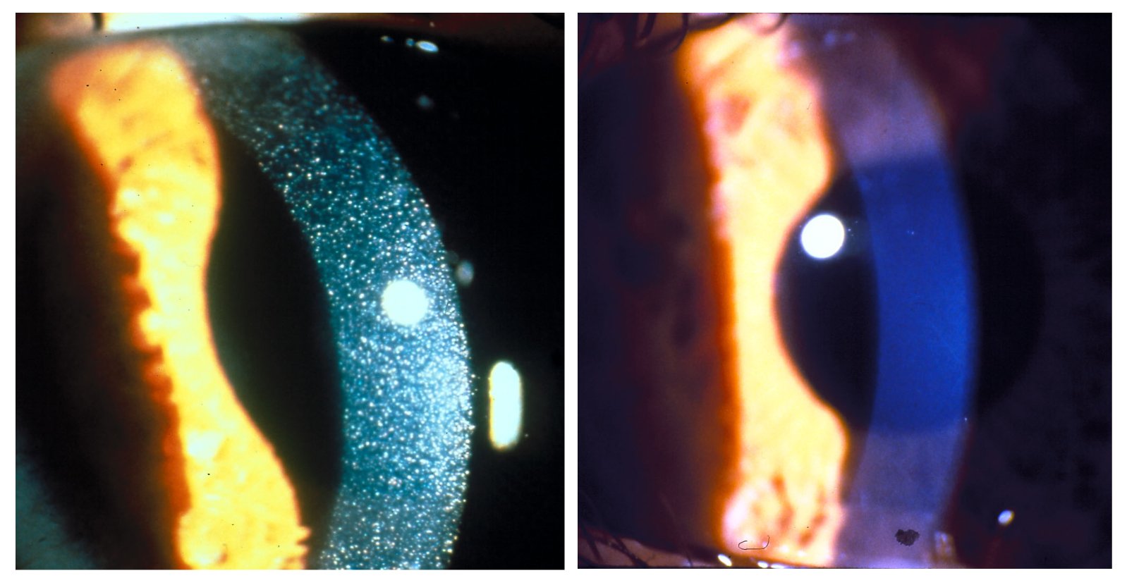
Cystinosis is a lysosomal storage disease characterized by the abnormal accumulation of cystine, the oxidized dimer of the amino acid cysteine. It is a genetic disorder that follows an autosomal recessive inheritance pattern. It is a rare autosomal recessive disorder resulting from accumulation of free cystine in lysosomes, eventually leading to intracellular crystal formation throughout the body. Cystinosis is the most common cause of Fanconi syndrome in the pediatric age group. Fanconi syndrome occurs when the function of cells in renal tubules is impaired, leading to abnormal amounts of carbohydrates and amino acids in the urine, excessive urination, and low blood levels of potassium and phosphates. Cystinosis was the first documented genetic disease belonging to the group of lysosomal storage disease disorders.Nesterova G, Gahl WA. Cystinosis: the evolution of a treatable disease. Pediatr Nephrol 2012;28:51–9. Cystinosis is caused by mutations in the '' CTNS'' gene that cod ... [...More Info...] [...Related Items...] OR: [Wikipedia] [Google] [Baidu] |
Glycogen Storage Disease Type II
Glycogen storage disease type II, also called Pompe disease, is an autosomal recessive metabolic disorder which damages muscle and nerve cells throughout the body. It is caused by an accumulation of glycogen in the lysosome due to deficiency of the lysosomal acid alpha-glucosidase enzyme. It is the only glycogen storage disease with a defect in lysosomal metabolism, and the first glycogen storage disease to be identified, in 1932 by the Dutch pathologist J. C. Pompe. The build-up of glycogen causes progressive muscle weakness (myopathy) throughout the body and affects various body tissues, particularly in the heart, skeletal muscles, liver and the nervous system. Signs and symptoms Newborn The infantile form usually comes to medical attention within the first few months of life. The usual presenting features are cardiomegaly (92%), hypotonia (88%), cardiomyopathy (88%), respiratory distress (78%), muscle weakness (63%), feeding difficulties (57%) and failure to thrive (50%). ... [...More Info...] [...Related Items...] OR: [Wikipedia] [Google] [Baidu] |
Mucolipidosis
Mucolipidosis is a group of inherited metabolic disorders that affect the body's ability to carry out the normal turnover of various materials within cells. When originally named, the mucolipidoses derived their name from the similarity in presentation to both mucopolysaccharidoses and sphingolipidoses. A biochemical understanding of these conditions has changed how they are classified. Four conditions (types I, II, III, and IV) were historically labeled as mucolipidoses. However, type I ( sialidosis) is now classified as a glycoproteinosis, and type IV ( Mucolipidosis type IV) is now classified as a gangliosidosis. ML II and III The other two types are closely related. Mucolipidosis types II and III (ML II and ML III) result from a deficiency of the enzyme N-acetylglucosamine-1-phosphotransferase, which phosphorylates target carbohydrate residues on N-linked glycoproteins. Without this phosphorylation, the glycoproteins are not destined for lysosomes, and they escape ou ... [...More Info...] [...Related Items...] OR: [Wikipedia] [Google] [Baidu] |
Glycoproteinosis
Glycoproteinosis are lysosomal storage diseases affecting glycoproteins, resulting from defects in lysosomal function. The term is sometimes reserved for conditions involving degradation of glycoproteins. Types * (E77.0) Defects in post-translational modification of lysosomal enzymes ** Mucolipidosis II (I-cell disease) ** Mucolipidosis III (pseudo-Hurler polydystrophy) * (E77.1) Defects in glycoprotein degradation ** Aspartylglucosaminuria ** Fucosidosis ** Mannosidosis ** Sialidosis (mucolipidosis Mucolipidosis is a group of inherited metabolic disorders that affect the body's ability to carry out the normal turnover of various materials within cells. When originally named, the mucolipidoses derived their name from the similarity in pr ... I) Another type, recently characterized, is galactosialidosis. References External links NIH Glycoprotein metabolism disorders {{endocrine-disease-stub ... [...More Info...] [...Related Items...] OR: [Wikipedia] [Google] [Baidu] |
Hurler Disease
Hurler syndrome, also known as mucopolysaccharidosis Type IH (MPS-IH), Hurler's disease, and formerly gargoylism, is a genetic disorder that results in the buildup of large sugar molecules called glycosaminoglycans (GAGs) in lysosomes. The inability to break down these molecules results in a wide variety of symptoms caused by damage to several different organ systems, including but not limited to the nervous system, skeletal system, eyes, and heart. The underlying mechanism is a deficiency of alpha-L iduronidase, an enzyme responsible for breaking down GAGs. Without this enzyme, a buildup of dermatan sulfate and heparan sulfate occurs in the body. Symptoms appear during childhood, and early death usually occurs. Other, less severe forms of MPS Type I include Hurler-Scheie Syndrome (MPS-IHS) and Scheie Syndrome (MPS-IS). Hurler syndrome is classified as a lysosomal storage disease. It is clinically related to Hunter syndrome (MPS II); however, Hunter syndrome is X-linked, whi ... [...More Info...] [...Related Items...] OR: [Wikipedia] [Google] [Baidu] |
Mucopolysaccharidosis
Mucopolysaccharidoses are a group of metabolic disorders caused by the absence or malfunctioning of lysosomal enzymes needed to break down molecules called glycosaminoglycans (GAGs). These long chains of sugar carbohydrates occur within the cells that help build bone, cartilage, tendons, corneas, skin and connective tissue. GAGs (formerly called mucopolysaccharides) are also found in the fluids that lubricate joints. Individuals with mucopolysaccharidosis either do not produce enough of one of the eleven enzymes required to break down these sugar chains into simpler molecules, or they produce enzymes that do not work properly. Over time, these GAGs collect in the cells, blood and connective tissues. The result is permanent, progressive cellular damage which affects appearance, physical abilities, organ and system functioning. The mucopolysaccharidoses are part of the lysosomal storage disease family, a group of more than 40 genetic disorders that result when the lysosome o ... [...More Info...] [...Related Items...] OR: [Wikipedia] [Google] [Baidu] |
Leukodystrophy
Leukodystrophies are a group of usually inherited disorders characterized by degeneration of the white matter in the brain. The word ''leukodystrophy'' comes from the Greek roots ''leuko'', "white", ''dys'', "abnormal" and ''troph'', "growth". The leukodystrophies are caused by imperfect growth or development of the myelin sheath, the fatty insulating covering around nerve fibers. Leukodystrophies may be classified as hypomyelinating or demyelinating diseases, depending on whether the damage is present before birth or occurs after. Other demyelinating diseases are usually not congenital and have a toxic or autoimmune cause. When damage occurs to white matter, immune responses can lead to inflammation in the central nervous system (CNS), along with loss of myelin. The degeneration of white matter can be seen in an MRI scan and used to diagnose leukodystrophy. Leukodystrophy is characterized by specific symptoms including decreased motor function, muscle rigidity, and eventua ... [...More Info...] [...Related Items...] OR: [Wikipedia] [Google] [Baidu] |
Tay–Sachs Disease
Tay–Sachs disease is a genetic disorder that results in the destruction of nerve cells in the brain and spinal cord. The most common form is infantile Tay–Sachs disease, which becomes apparent around three to six months of age, with the baby losing the ability to turn over, sit, or crawl. This is then followed by seizures, hearing loss, and inability to move, with death usually occurring by the age of three to five. Less commonly, the disease may occur in later childhood or adulthood (juvenile or late-onset). These forms tend to be less severe, but the juvenile form typically results in death by age 15. Tay–Sachs disease is caused by a genetic mutation in the '' HEXA'' gene on chromosome 15, which codes form a subunit of the hexosaminidase enzyme known as hexosaminidase A. It is inherited from a person's parents in an autosomal recessive manner. The mutation disrupts the activity of the enzyme, which results in the build-up of the molecule GM2 ganglioside within cells, l ... [...More Info...] [...Related Items...] OR: [Wikipedia] [Google] [Baidu] |
Gangliosidosis
Gangliosidosis contains different types of lipid storage disorders caused by the accumulation of lipids known as ganglioside A ganglioside is a molecule composed of a glycosphingolipid ( ceramide and oligosaccharide) with one or more sialic acids (e.g. ''N''-acetylneuraminic acid, NANA) linked on the sugar chain. NeuNAc, an acetylated derivative of the carbohydrate ...s. There are two distinct genetic causes of the disease. Both are autosomal recessive and affect males and females equally. Types * GM1 gangliosidoses - GM1 * GM2 gangliosidoses - GM2 See also * Sphingolipidoses#Overview References External links Autosomal recessive disorders Lipid storage disorders Rare diseases {{endocrine-disease-stub ... [...More Info...] [...Related Items...] OR: [Wikipedia] [Google] [Baidu] |
Niemann–Pick Disease
Niemann–Pick disease is a group of severe inherited metabolic disorders, in which sphingomyelin accumulates in lysosomes in cells (the lysosomes normally degrade material that comes from out of cells). These disorders involve the dysfunctional metabolism of sphingolipids, which are fats found in cell membranes. They can be considered as a kind of sphingolipidosis, which is included in the larger family of lysosomal storage diseases. Signs and symptoms Symptoms are related to the organs in which sphingomyelin accumulates. Enlargement of the liver and spleen ( hepatosplenomegaly) may cause reduced appetite, abdominal distension, and pain. Enlargement of the spleen (splenomegaly) may also cause low levels of platelets in the blood (thrombocytopenia). Accumulation of sphingomyelin in the central nervous system (including the cerebellum) results in unsteady gait (ataxia), slurring of speech (dysarthria), and difficulty swallowing (dysphagia). Basal ganglia dysfunction causes abno ... [...More Info...] [...Related Items...] OR: [Wikipedia] [Google] [Baidu] |


.jpg)


