|
Lymphocyte-variant Eosinophilia
Lymphocyte-variant hypereosinophila is a rare disorder in which eosinophilia or hypereosinophilia (i.e. a large or extremely large increase in the number of eosinophils in the blood circulation) is caused by an aberrant population of lymphocytes. These aberrant lymphocytes function abnormally by stimulating the proliferation and maturation of bone marrow eosinophil-precursor cells termed colony forming unit-Eosinophils or CFU-Eos. The overly stimulated CFU-Eos cells mature to apparently normal appearing but possibly overactive eosinophils which enter the circulation and may accumulate in and damage various tissues. The disorder is usually indolent or slowly progressive but may proceed to a leukemic phase sometimes classified as acute eosinophilic leukemia. Lymphocyte-variant hypereosinophilia can therefore be regarded as a precancerous disorder. The disorder merits therapeutic intervention to avoid or reduce eosinophil-induced tissue injury and treat its leukemic phase. The latt ... [...More Info...] [...Related Items...] OR: [Wikipedia] [Google] [Baidu] |
Eosinophilia
Eosinophilia is a condition in which the eosinophil count in the peripheral blood exceeds . Hypereosinophilia is an elevation in an individual's circulating blood eosinophil count above 1.5 x 10⁹/ L (i.e. 1,500/μL). The hypereosinophilic syndrome is a sustained elevation in this count above 1.5 x 10⁹/L (i.e. 1,500/μL) that is also associated with evidence of eosinophil-based tissue injury. Eosinophils usually account for less than 7% of the circulating leukocytes. A marked increase in non-blood tissue eosinophil count noticed upon histopathologic examination is diagnostic for tissue eosinophilia. Several causes are known, with the most common being some form of allergic reaction or parasitic infection. Diagnosis of eosinophilia is via a complete blood count (CBC), but diagnostic procedures directed at the underlying cause vary depending on the suspected condition(s). An absolute eosinophil count is not generally needed if the CBC shows marked eosinophilia. The location of the c ... [...More Info...] [...Related Items...] OR: [Wikipedia] [Google] [Baidu] |
Gleich's Syndrome
Gleich's syndrome is a rare disease in which the body swells up episodically (angioedema), associated with raised antibodies of the IgM type and increased numbers of eosinophil granulocytes, a type of white blood cells, in the blood ( eosinophilia). It was first described in 1984. Its cause is unknown, but it is unrelated to capillary leak syndrome (which may cause similar swelling episodes) and eosinophilia-myalgia syndrome (which features eosinophilia but alternative symptoms). Some studies have shown that edema attacks are associated with degranulation (release of enzymes and mediators from eosinophils), and others have demonstrated antibodies against endothelium (cells lining blood vessels) in the condition. Gleich's syndrome is not a form of the idiopathic hypereosinophilic syndrome in that there is little or no evidence that it leads to organ damage. Rather, recent studies report that a subset of T cells (a special form of lymphocyte blood cell) found in several Gleich syndr ... [...More Info...] [...Related Items...] OR: [Wikipedia] [Google] [Baidu] |
Hodgkin's Lymphoma
Hodgkin lymphoma (HL) is a type of lymphoma, in which cancer originates from a specific type of white blood cell called lymphocytes, where multinucleated Reed–Sternberg cells (RS cells) are present in the patient's lymph nodes. The condition was named after the English physician Thomas Hodgkin, who first described it in 1832. Symptoms may include fever, night sweats, and weight loss. Often, nonpainful enlarged lymph nodes occur in the neck, under the arm, or in the groin. Those affected may feel tired or be itchy. The two major types of Hodgkin lymphoma are classic Hodgkin lymphoma and nodular lymphocyte-predominant Hodgkin lymphoma. About half of cases of Hodgkin lymphoma are due to Epstein–Barr virus (EBV) and these are generally the classic form. Other risk factors include a family history of the condition and having HIV/AIDS. Diagnosis is conducted by confirming the presence of cancer and identifying RS cells in lymph node biopsies. The virus-positive cases are classified ... [...More Info...] [...Related Items...] OR: [Wikipedia] [Google] [Baidu] |
B Cell
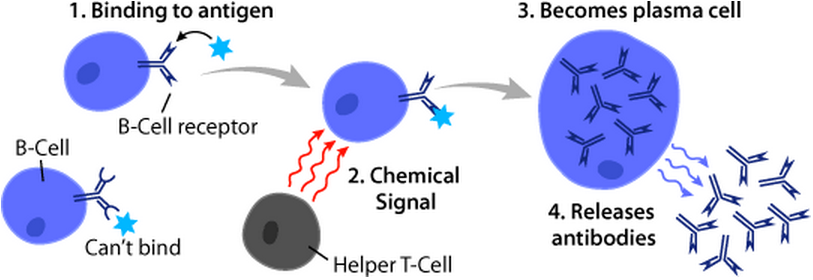
B cells, also known as B lymphocytes, are a type of white blood cell of the lymphocyte subtype. They function in the humoral immunity component of the adaptive immune system. B cells produce antibody molecules which may be either secreted or inserted into the plasma membrane where they serve as a part of B-cell receptors. When a naïve or memory B cell is activated by an antigen, it proliferates and differentiates into an antibody-secreting effector cell, known as a plasmablast or plasma cell. Additionally, B cells present antigens (they are also classified as professional antigen-presenting cells (APCs)) and secrete cytokines. In mammals, B cells mature in the bone marrow, which is at the core of most bones. In birds, B cells mature in the bursa of Fabricius, a lymphoid organ where they were first discovered by Chang and Glick, which is why the 'B' stands for bursa and not bone marrow as commonly believed. B cells, unlike the other two classes of lymphocytes, T cells and ... [...More Info...] [...Related Items...] OR: [Wikipedia] [Google] [Baidu] |
Angioimmunoblastic T Cell Lymphoma
Angioimmunoblastic T-cell lymphoma (AITL, sometimes misspelled AILT, formerly known as "angioimmunoblastic lymphadenopathy with dysproteinemia") is a mature T-cell lymphoma of blood or lymph vessel immunoblasts characterized by a polymorphous lymph node infiltrate showing a marked increase in follicular dendritic cells (FDCs) and high endothelial venules (HEVs) and systemic involvement. Signs and symptoms Patients with AITL usually present at an advanced stage and show systemic involvement. The clinical findings typically include a pruritic skin rash and possibly edema, ascites, pleural effusions, and arthritis. Sites of involvement Due to the systemic nature of AITL, neoplastic cells can be found in lymph nodes, liver, spleen, skin, and bone marrow. Causes AITL was originally thought to be a premalignant condition, termed angioimmunoblastic lymphadenopathy, and this atypical reactive lymphadenopathy carried a risk for transformation into a lymphoma. It is postulated that the o ... [...More Info...] [...Related Items...] OR: [Wikipedia] [Google] [Baidu] |
Adult T-cell Leukemia/lymphoma
Adult T-cell leukemia/lymphoma (ATL or ATLL) is a rare cancer of the immune system's T-cells caused by human T cell leukemia/lymphotropic virus type 1 (HTLV-1). All ATL cells contain integrated HTLV-1 provirus further supporting that causal role of the virus in the cause of the neoplasm. A small amount of HTLV-1 individuals progress to develop ATL with a long latency period between infection and ATL development. ATL is categorized into 4 subtypes: acute, smoldering, lymphoma-type, chronic. Acute and Lymphoma-type are known to particularity be aggressive with poorer prognosis. Globally, the retrovirus HTLV-1 is estimated to infect 20 million people with the incidence of ATL approximately 0.05 per 100,000 with endemic regions such as regions of Japan, as high as 27 per 100,000. However, cases have increased in non-endemic regions with highest incidence of HTLV-1 in southern/northern islands of Japan, Caribbean, Central and South America, intertropical Africa, Romania, northern Iran ... [...More Info...] [...Related Items...] OR: [Wikipedia] [Google] [Baidu] |
Cutaneous T Cell Lymphoma
Cutaneous T-cell lymphoma (CTCL) is a class of non-Hodgkin lymphoma, which is a type of cancer of the immune system. Unlike most non-Hodgkin lymphomas (which are generally B-cell-related), CTCL is caused by a mutation of T cells. The cancerous T cells in the body initially migrate to the skin, causing various lesions to appear. These lesions change shape as the disease progresses, typically beginning as what appears to be a rash which can be very itchy and eventually forming plaques and tumors before spreading to other parts of the body. Signs and symptoms The presentation depends if it is mycosis fungoides or Sézary syndrome, the most common, though not the only types. Among the symptoms for the aforementioned types are: enlarged lymph nodes, an enlarged liver and spleen, and non-specific dermatitis. Cause The cause of CTCL is unknown. Diagnosis A point-based algorithm for the diagnosis for early forms of cutaneous T-cell lymphoma was proposed by the International Societ ... [...More Info...] [...Related Items...] OR: [Wikipedia] [Google] [Baidu] |
Neoplasm
A neoplasm () is a type of abnormal and excessive growth of tissue. The process that occurs to form or produce a neoplasm is called neoplasia. The growth of a neoplasm is uncoordinated with that of the normal surrounding tissue, and persists in growing abnormally, even if the original trigger is removed. This abnormal growth usually forms a mass, when it may be called a tumor. ICD-10 classifies neoplasms into four main groups: benign neoplasms, in situ neoplasms, malignant neoplasms, and neoplasms of uncertain or unknown behavior. Malignant neoplasms are also simply known as cancers and are the focus of oncology. Prior to the abnormal growth of tissue, as neoplasia, cells often undergo an abnormal pattern of growth, such as metaplasia or dysplasia. However, metaplasia or dysplasia does not always progress to neoplasia and can occur in other conditions as well. The word is from Ancient Greek 'new' and 'formation, creation'. Types A neoplasm can be benign, potentially ma ... [...More Info...] [...Related Items...] OR: [Wikipedia] [Google] [Baidu] |
Thrombosis
Thrombosis (from Ancient Greek "clotting") is the formation of a blood clot inside a blood vessel, obstructing the flow of blood through the circulatory system. When a blood vessel (a vein or an artery) is injured, the body uses platelets (thrombocytes) and fibrin to form a blood clot to prevent blood loss. Even when a blood vessel is not injured, blood clots may form in the body under certain conditions. A clot, or a piece of the clot, that breaks free and begins to travel around the body is known as an embolus. Thrombosis may occur in veins (venous thrombosis) or in arteries (arterial thrombosis). Venous thrombosis (sometimes called DVT, deep vein thrombosis) leads to a blood clot in the affected part of the body, while arterial thrombosis (and, rarely, severe venous thrombosis) affects the blood supply and leads to damage of the tissue supplied by that artery (ischemia and necrosis). A piece of either an arterial or a venous thrombus can break off as an embolus, which could ... [...More Info...] [...Related Items...] OR: [Wikipedia] [Google] [Baidu] |
Eosinophilic Myocarditis
Eosinophilic myocarditis is inflammation in the heart muscle that is caused by the infiltration and destructive activity of a type of white blood cell, the eosinophil. Typically, the disorder is associated with hypereosinophilia, i.e. an eosinophil blood cell count greater than 1,500 per microliter (normal 100 to 400 per microliter). It is distinguished from non-eosinophilic myocarditis, which is heart inflammation caused by other types of white blood cells, i.e. lymphocytes and monocytes, as well as the respective descendants of these cells, NK cells and macrophages. This distinction is important because the eosinophil-based disorder is due to a particular set of underlying diseases and its preferred treatments differ from those for non-eosinophilic myocarditis. Eosinophilic myocarditis is often viewed as a disorder that has three progressive stages. The first stage of eosinophilic myocarditis involves acute inflammation and cardiac cell necrosis (i.e. areas of dead cells); it is ... [...More Info...] [...Related Items...] OR: [Wikipedia] [Google] [Baidu] |
Splenomegaly
Splenomegaly is an enlargement of the spleen. The spleen usually lies in the left upper quadrant (LUQ) of the human abdomen. Splenomegaly is one of the four cardinal signs of ''hypersplenism'' which include: some reduction in number of circulating blood cells affecting granulocytes, erythrocytes or platelets in any combination; a compensatory proliferative response in the bone marrow; and the potential for correction of these abnormalities by splenectomy. Splenomegaly is usually associated with increased workload (such as in hemolytic anemias), which suggests that it is a response to hyperfunction. It is therefore not surprising that splenomegaly is associated with any disease process that involves abnormal red blood cells being destroyed in the spleen. Other common causes include congestion due to portal hypertension and infiltration by leukemias and lymphomas. Thus, the finding of an enlarged spleen, along with caput medusae, is an important sign of portal hypertension. Definiti ... [...More Info...] [...Related Items...] OR: [Wikipedia] [Google] [Baidu] |
Arthralgia
Arthralgia (from Greek ''arthro-'', joint + ''-algos'', pain) literally means ''joint pain''. Specifically, arthralgia is a symptom of injury, infection, illness (in particular arthritis), or an allergic reaction to medication. According to MeSH, the term "arthralgia" should only be used when the condition is non-inflammatory, and the term "arthritis" should be used when the condition is inflammatory. Causes The causes of ''arthralgia'' are varied and range, from a joints perspective, from degenerative and destructive processes such as osteoarthritis and sports injuries to inflammation of tissues surrounding the joints, such as bursitis. These might be triggered by other things, such as infections or vaccinations. Diagnosis Diagnosis involves interviewing the patient and performing physical exams. When attempting to establish the cause of the arthralgia, the emphasis is on the interview. The patient is asked questions intended to narrow the number of potential causes. Given th ... [...More Info...] [...Related Items...] OR: [Wikipedia] [Google] [Baidu] |
.jpg)
_mixed_cellulary_type.jpg)


