|
Lower Motor Neurons
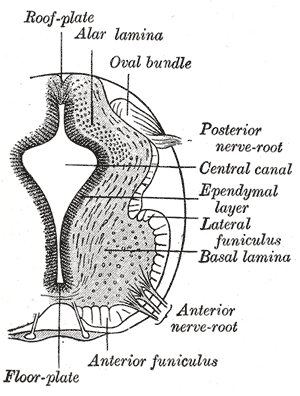
Lower motor neurons (LMNs) are motor neurons located in either the anterior grey column, anterior nerve roots (spinal lower motor neurons) or the cranial nerve nuclei of the brainstem and cranial nerves with motor function (cranial nerve lower motor neurons). Many voluntary movements rely on spinal lower motor neurons, which innervate skeletal muscle fibers and act as a link between upper motor neurons and muscles. Cranial nerve lower motor neurons also control some voluntary movements of the eyes, face and tongue, and contribute to chewing, swallowing and vocalization. Damage to the lower motor neurons can lead to flaccid paralysis, absent deep tendon reflexes and muscle atrophy. Classification Lower motor neurons are classified based on the type of muscle fiber they innervate: * Alpha motor neurons (α-MNs) innervate extrafusal muscle fibers, the most numerous type of muscle fiber and the one involved in muscle contraction. * Beta motor neurons (β-MNs) innervate intrafu ... [...More Info...] [...Related Items...] OR: [Wikipedia] [Google] [Baidu] |
Motor Neuron
A motor neuron (or motoneuron or efferent neuron) is a neuron whose cell body is located in the motor cortex, brainstem or the spinal cord, and whose axon (fiber) projects to the spinal cord or outside of the spinal cord to directly or indirectly control effector organs, mainly muscles and glands. There are two types of motor neuron – upper motor neurons and lower motor neurons. Axons from upper motor neurons synapse onto interneurons in the spinal cord and occasionally directly onto lower motor neurons. The axons from the lower motor neurons are efferent nerve fibers that carry signals from the spinal cord to the effectors. Types of lower motor neurons are alpha motor neurons, beta motor neurons, and gamma motor neurons. A single motor neuron may innervate many muscle fibres and a muscle fibre can undergo many action potentials in the time taken for a single muscle twitch. Innervation takes place at a neuromuscular junction and twitches can become superimposed as a resul ... [...More Info...] [...Related Items...] OR: [Wikipedia] [Google] [Baidu] |
Gamma Motor Neuron
A gamma motor neuron (γ motor neuron), also called gamma motoneuron, or fusimotor neuron, is a type of lower motor neuron that takes part in the process of muscle contraction, and represents about 30% of ( Aγ) fibers going to the muscle. Like alpha motor neurons, their cell bodies are located in the anterior grey column of the spinal cord. They receive input from the reticular formation of the pons in the brainstem. Their axons are smaller than those of the alpha motor neurons, with a diameter of only 5 μm. Unlike the alpha motor neurons, gamma motor neurons do not directly adjust the lengthening or shortening of muscles. However, their role is important in keeping muscle spindles taut, thereby allowing the continued firing of alpha neurons, leading to muscle contraction. These neurons also play a role in adjusting the sensitivity of muscle spindles. The presence of myelination in gamma motor neurons allows a conduction velocity of 4 to 24 meters per second, signif ... [...More Info...] [...Related Items...] OR: [Wikipedia] [Google] [Baidu] |
Reflex
In biology, a reflex, or reflex action, is an involuntary, unplanned sequence or action and nearly instantaneous response to a stimulus. Reflexes are found with varying levels of complexity in organisms with a nervous system. A reflex occurs via neural pathways in the nervous system called reflex arcs. A stimulus initiates a neural signal, which is carried to a synapse. The signal is then transferred across the synapse to a motor neuron which evokes a target response. These neural signals do not always travel to the brain, so many reflexes are an automatic response to a stimulus that does not receive or need conscious thought. Many reflexes are fine-tuned to increase organism survival and self-defense. This is observed in reflexes such as the startle reflex, which provides an automatic response to an unexpected stimuli, and the feline righting reflex, which reorients a cat's body when falling to ensure safe landing. The simplest type of reflex, a short-latency reflex, has ... [...More Info...] [...Related Items...] OR: [Wikipedia] [Google] [Baidu] |
Muscle Wasting
Muscle atrophy is the loss of skeletal muscle mass. It can be caused by immobility, aging, malnutrition, medications, or a wide range of injuries or diseases that impact the musculoskeletal or nervous system. Muscle atrophy leads to muscle weakness and causes disability. Disuse causes rapid muscle atrophy and often occurs during injury or illness that requires immobilization of a limb or bed rest. Depending on the duration of disuse and the health of the individual, this may be fully reversed with activity. Malnutrition first causes fat loss but may progress to muscle atrophy in prolonged starvation and can be reversed with nutritional therapy. In contrast, cachexia is a wasting syndrome caused by an underlying disease such as cancer that causes dramatic muscle atrophy and cannot be completely reversed with nutritional therapy. Sarcopenia is age-related muscle atrophy and can be slowed by exercise. Finally, diseases of the muscles such as muscular dystrophy or myopathies can ca ... [...More Info...] [...Related Items...] OR: [Wikipedia] [Google] [Baidu] |
Lower Motor Neuron Lesion
A lower motor neuron lesion is a lesion which affects nerve fibers traveling from the lower motor neuron(s) in the anterior horn/ anterior grey column of the spinal cord, or in the motor nuclei of the cranial nerves, to the relevant muscle(s). One major characteristic used to identify a lower motor neuron lesion is flaccid paralysis – paralysis accompanied by loss of muscle tone. This is in contrast to an upper motor neuron lesion, which often presents with spastic paralysis – paralysis accompanied by severe hypertonia. Signs and symptoms * Muscle paresis or paralysis * Fibrillations * Fasciculations – caused by increased receptor concentration on muscles to compensate for lack of innervation. * Hypotonia or atonia – Tone is not velocity dependent. * Hyporeflexia - Along with deep reflexes even cutaneous reflexes are also decreased or absent. * Strength – weakness is limited to segmental or focal pattern, Root innervated pattern The extensor plantar reflex is ... [...More Info...] [...Related Items...] OR: [Wikipedia] [Google] [Baidu] |
Muscle Cell
A muscle cell is also known as a myocyte when referring to either a cardiac muscle cell (cardiomyocyte), or a smooth muscle cell as these are both small cells. A skeletal muscle cell is long and threadlike with many nuclei and is called a muscle fiber. Muscle cells (including myocytes and muscle fibers) develop from embryonic precursor cells called myoblasts. Myoblasts fuse to form multinucleated skeletal muscle cells known as syncytia in a process known as myogenesis. Skeletal muscle cells and cardiac muscle cells both contain myofibrils and sarcomeres and form a striated muscle tissue. Cardiac muscle cells form the cardiac muscle in the walls of the heart chambers, and have a single central nucleus. Cardiac muscle cells are joined to neighboring cells by intercalated discs, and when joined in a visible unit they are described as a ''cardiac muscle fiber''. Smooth muscle cells control involuntary movements such as the peristalsis contractions in the esophagus ... [...More Info...] [...Related Items...] OR: [Wikipedia] [Google] [Baidu] |
Synaptic Cleft
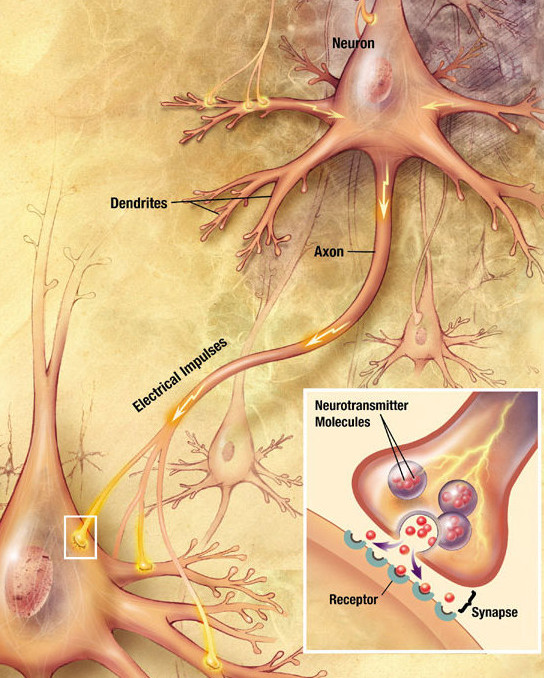
Chemical synapses are biological junctions through which neurons' signals can be sent to each other and to non-neuronal cells such as those in muscles or glands. Chemical synapses allow neurons to form circuits within the central nervous system. They are crucial to the biological computations that underlie perception and thought. They allow the nervous system to connect to and control other systems of the body. At a chemical synapse, one neuron releases neurotransmitter molecules into a small space (the synaptic cleft) that is adjacent to another neuron. The neurotransmitters are contained within small sacs called synaptic vesicles, and are released into the synaptic cleft by exocytosis. These molecules then bind to neurotransmitter receptors on the postsynaptic cell. Finally, the neurotransmitters are cleared from the synapse through one of several potential mechanisms including enzymatic degradation or re-uptake by specific transporters either on the presynaptic cell or ... [...More Info...] [...Related Items...] OR: [Wikipedia] [Google] [Baidu] |
Acetylcholine
Acetylcholine (ACh) is an organic chemical that functions in the brain and body of many types of animals (including humans) as a neurotransmitter. Its name is derived from its chemical structure: it is an ester of acetic acid and choline. Parts in the body that use or are affected by acetylcholine are referred to as cholinergic. Substances that increase or decrease the overall activity of the cholinergic system are called cholinergics and anticholinergics, respectively. Acetylcholine is the neurotransmitter used at the neuromuscular junction—in other words, it is the chemical that motor neurons of the nervous system release in order to activate muscles. This property means that drugs that affect cholinergic systems can have very dangerous effects ranging from paralysis to convulsions. Acetylcholine is also a neurotransmitter in the autonomic nervous system, both as an internal transmitter for the sympathetic nervous system and as the final product released by the para ... [...More Info...] [...Related Items...] OR: [Wikipedia] [Google] [Baidu] |
Neuromuscular Junction
A neuromuscular junction (or myoneural junction) is a chemical synapse between a motor neuron and a muscle fiber. It allows the motor neuron to transmit a signal to the muscle fiber, causing muscle contraction. Muscles require innervation to function—and even just to maintain muscle tone, avoiding atrophy. In the neuromuscular system nerves from the central nervous system and the peripheral nervous system are linked and work together with muscles. Synaptic transmission at the neuromuscular junction begins when an action potential reaches the presynaptic terminal of a motor neuron, which activates voltage-gated calcium channels to allow calcium ions to enter the neuron. Calcium ions bind to sensor proteins ( synaptotagmins) on synaptic vesicles, triggering vesicle fusion with the cell membrane and subsequent neurotransmitter release from the motor neuron into the synaptic cleft. In vertebrates, motor neurons release acetylcholine (ACh), a small molecule neurotransmitter, ... [...More Info...] [...Related Items...] OR: [Wikipedia] [Google] [Baidu] |
Axon
An axon (from Greek ἄξων ''áxōn'', axis), or nerve fiber (or nerve fibre: see spelling differences), is a long, slender projection of a nerve cell, or neuron, in vertebrates, that typically conducts electrical impulses known as action potentials away from the nerve cell body. The function of the axon is to transmit information to different neurons, muscles, and glands. In certain sensory neurons ( pseudounipolar neurons), such as those for touch and warmth, the axons are called afferent nerve fibers and the electrical impulse travels along these from the periphery to the cell body and from the cell body to the spinal cord along another branch of the same axon. Axon dysfunction can be the cause of many inherited and acquired neurological disorders that affect both the peripheral and central neurons. Nerve fibers are classed into three types group A nerve fibers, group B nerve fibers, and group C nerve fibers. Groups A and B are myelinated, and group C are unmyelin ... [...More Info...] [...Related Items...] OR: [Wikipedia] [Google] [Baidu] |
Depolarization
In biology, depolarization or hypopolarization is a change within a cell, during which the cell undergoes a shift in electric charge distribution, resulting in less negative charge inside the cell compared to the outside. Depolarization is essential to the function of many cells, communication between cells, and the overall physiology of an organism. Most cells in higher organisms maintain an internal environment that is negatively charged relative to the cell's exterior. This difference in charge is called the cell's membrane potential. In the process of depolarization, the negative internal charge of the cell temporarily becomes more positive (less negative). This shift from a negative to a more positive membrane potential occurs during several processes, including an action potential. During an action potential, the depolarization is so large that the potential difference across the cell membrane briefly reverses polarity, with the inside of the cell becoming positively char ... [...More Info...] [...Related Items...] OR: [Wikipedia] [Google] [Baidu] |
Glutamate
Glutamic acid (symbol Glu or E; the ionic form is known as glutamate) is an α-amino acid that is used by almost all living beings in the biosynthesis of proteins. It is a non-essential nutrient for humans, meaning that the human body can synthesize enough for its use. It is also the most abundant excitatory neurotransmitter in the vertebrate nervous system. It serves as the precursor for the synthesis of the inhibitory gamma-aminobutyric acid (GABA) in GABA-ergic neurons. Its molecular formula is . Glutamic acid exists in three optically isomeric forms; the dextrorotatory -form is usually obtained by hydrolysis of gluten or from the waste waters of beet-sugar manufacture or by fermentation.Webster's Third New International Dictionary of the English Language Unabridged, Third Edition, 1971. Its molecular structure could be idealized as HOOC−CH()−()2−COOH, with two carboxyl groups −COOH and one amino group −. However, in the solid state and mildly acidic water solu ... [...More Info...] [...Related Items...] OR: [Wikipedia] [Google] [Baidu] |


