|
Low-grade Myofibroblastic Sarcoma
Low-grade myofibroblastic sarcoma (LGMS) is a subtype of the malignant sarcomas. As it is currently recognized, LGMS was first described as a rare, atypical myofibroblastic tumor (i.e. a tumor consisting of cells with the microscopic features of fibroblasts and smooth muscle cells) by Mentzel et al. in 1998. Myofibroblastic sarcomas had been divided into low-grade myofibroblastic sarcomas, intermediate‐grade myofibroblasic sarcomas, i.e. IGMS, and high‐grade myofibroblasic sarcomas, i.e. HGMS (also termed undifferentiated pleomorphic sarcoma and pleomorphic myofibrosarcoma [and formerly termed malignant fibrous histiocytoma]) based on their microscopic morphology (biology), morphological, immunophenotype, immunophenotypic, and malignancy features. LGMS and IGMS are now classified together by the World Health Organization (WHO), 2020, in the category of intermediate (rarely metastasizing) fibroblastic and myofibroblastic tumors. WHO, 2020, classifies HGMS (preferred name: undiff ... [...More Info...] [...Related Items...] OR: [Wikipedia] [Google] [Baidu] |
Metastasizes
Metastasis is a pathogenic agent's spread from an initial or primary site to a different or secondary site within the host's body; the term is typically used when referring to metastasis by a cancerous tumor. The newly pathological sites, then, are metastases (mets). It is generally distinguished from cancer invasion, which is the direct extension and penetration by cancer cells into neighboring tissues. Cancer occurs after cells are genetically altered to proliferate rapidly and indefinitely. This uncontrolled proliferation by mitosis produces a primary tumor, primary tumour heterogeneity, heterogeneic tumour. The cells which constitute the tumor eventually undergo metaplasia, followed by dysplasia then anaplasia, resulting in a Malignancy, malignant phenotype. This malignancy allows for invasion into the circulation, followed by invasion to a second site for tumorigenesis. Some cancer cells known as circulating tumor cells acquire the ability to penetrate the walls of lymphat ... [...More Info...] [...Related Items...] OR: [Wikipedia] [Google] [Baidu] |
Mandible
In anatomy, the mandible, lower jaw or jawbone is the largest, strongest and lowest bone in the human facial skeleton. It forms the lower jaw and holds the lower tooth, teeth in place. The mandible sits beneath the maxilla. It is the only movable bone of the skull (discounting the ossicles of the middle ear). It is connected to the temporal bones by the temporomandibular joints. The bone is formed prenatal development, in the fetus from a fusion of the left and right mandibular prominences, and the point where these sides join, the mandibular symphysis, is still visible as a faint ridge in the midline. Like other symphyses in the body, this is a midline articulation where the bones are joined by fibrocartilage, but this articulation fuses together in early childhood.Illustrated Anatomy of the Head and Neck, Fehrenbach and Herring, Elsevier, 2012, p. 59 The word "mandible" derives from the Latin word ''mandibula'', "jawbone" (literally "one used for chewing"), from ''wikt:mandere ... [...More Info...] [...Related Items...] OR: [Wikipedia] [Google] [Baidu] |
Pleura
The pulmonary pleurae (''sing.'' pleura) are the two opposing layers of serous membrane overlying the lungs and the inside of the surrounding chest walls. The inner pleura, called the visceral pleura, covers the surface of each lung and dips between the lobes of the lung as ''fissures'', and is formed by the invagination of lung buds into each thoracic sac during embryonic development. The outer layer, called the parietal pleura, lines the inner surfaces of the thoracic cavity on each side of the mediastinum, and can be subdivided into ''mediastinal'' (covering the side surfaces of the fibrous pericardium, oesophagus and thoracic aorta), ''diaphragmatic'' (covering the upper surface of the diaphragm), ''costal'' (covering the inside of rib cage) and cervical (covering the underside of the suprapleural membrane) pleurae. The visceral and the mediastinal parietal pleurae are connected at the root of the lung ("hilum") through a smooth fold known as ''pleural reflections'', and ... [...More Info...] [...Related Items...] OR: [Wikipedia] [Google] [Baidu] |
Pancreas
The pancreas is an organ of the digestive system and endocrine system of vertebrates. In humans, it is located in the abdomen behind the stomach and functions as a gland. The pancreas is a mixed or heterocrine gland, i.e. it has both an endocrine and a digestive exocrine function. 99% of the pancreas is exocrine and 1% is endocrine. As an endocrine gland, it functions mostly to regulate blood sugar levels, secreting the hormones insulin, glucagon, somatostatin, and pancreatic polypeptide. As a part of the digestive system, it functions as an exocrine gland secreting pancreatic juice into the duodenum through the pancreatic duct. This juice contains bicarbonate, which neutralizes acid entering the duodenum from the stomach; and digestive enzymes, which break down carbohydrates, proteins, and fats in food entering the duodenum from the stomach. Inflammation of the pancreas is known as pancreatitis, with common causes including chronic alcohol use and gallstones. Becaus ... [...More Info...] [...Related Items...] OR: [Wikipedia] [Google] [Baidu] |
Palpitation
Palpitations are perceived abnormalities of the heartbeat characterized by awareness of cardiac muscle contractions in the chest, which is further characterized by the hard, fast and/or irregular beatings of the heart. Symptoms include a rapid pulsation, an abnormally rapid or irregular beating of the heart. Palpitations are a sensory symptom and are often described as a skipped beat, rapid fluttering in the chest, pounding sensation in the chest or neck, or a flip-flopping in the chest. Palpitation can be associated with anxiety and does not necessarily indicate a structural or functional abnormality of the heart, but it can be a symptom arising from an objectively rapid or irregular heartbeat. Palpitation can be intermittent and of variable frequency and duration, or continuous. Associated symptoms include dizziness, shortness of breath, sweating, headaches and chest pain. Palpitation may be associated with coronary heart disease, hyperthyroidism, diseases affecting card ... [...More Info...] [...Related Items...] OR: [Wikipedia] [Google] [Baidu] |
Bowel Obstruction
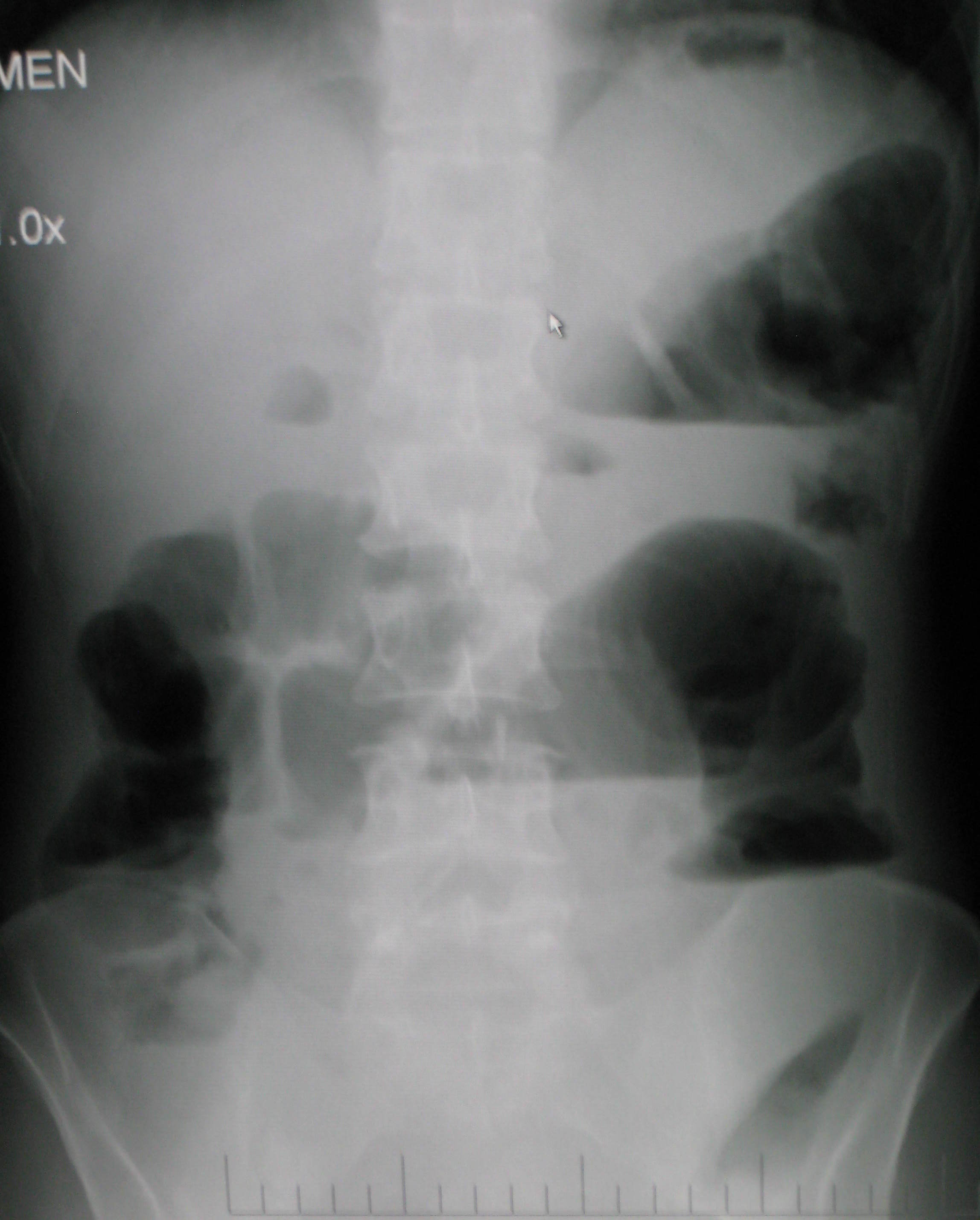
Bowel obstruction, also known as intestinal obstruction, is a mechanical or Ileus, functional obstruction of the Gastrointestinal tract#Lower gastrointestinal tract, intestines which prevents the normal movement of the products of digestion. Either the Small intestine, small bowel or Large intestine, large bowel may be affected. Signs and symptoms include abdominal pain, vomiting, abdominal bloating, bloating and not passing flatulence, gas. Mechanical obstruction is the cause of about 5 to 15% of cases of acute abdomen, severe abdominal pain of sudden onset requiring admission to hospital. Causes of bowel obstruction include Adhesion (medicine), adhesions, hernias, volvulus, endometriosis, inflammatory bowel disease, appendicitis, Neoplasm, tumors, diverticulitis, ischemic colitis, ischemic bowel, tuberculosis and intussusception (medical disorder), intussusception. Small bowel obstructions are most often due to adhesions and hernias while large bowel obstructions are most often ... [...More Info...] [...Related Items...] OR: [Wikipedia] [Google] [Baidu] |
Lesser Omentum
The lesser omentum (small omentum or gastrohepatic omentum) is the double layer of peritoneum that extends from the liver to the lesser curvature of the stomach, and to the first part of the duodenum. The lesser omentum is usually divided into these two connecting parts: the hepatogastric ligament, and the hepatoduodenal ligament. Structure The lesser omentum is extremely thin, and is continuous with the two layers of peritoneum which cover respectively the antero-superior and postero-inferior surfaces of the stomach and first part of the duodenum. When these two layers reach the lesser curvature of the stomach and the upper border of the duodenum, they join and ascend as a double fold to the porta hepatis. To the left of the porta, the fold is attached to the bottom of the fossa for the ductus venosus, along which it is carried to the diaphragm, where the two layers separate to embrace the end of the esophagus. At the right border of the lesser omentum, the two layers are c ... [...More Info...] [...Related Items...] OR: [Wikipedia] [Google] [Baidu] |
Greater Omentum
The greater omentum (also the great omentum, omentum majus, gastrocolic omentum, epiploon, or, especially in animals, caul) is a large apron-like fold of visceral peritoneum that hangs down from the stomach. It extends from the greater curvature of the stomach, passing in front of the small intestines and doubles back to ascend to the transverse colon before reaching to the posterior abdominal wall. The greater omentum is larger than the lesser omentum, which hangs down from the liver to the lesser curvature. The common anatomical term "epiploic" derives from "epiploon", from the Greek ''epipleein'', meaning to float or sail on, since the greater omentum appears to float on the surface of the intestines. It is the first structure observed when the abdominal cavity is opened anteriorly (from the front). Structure The greater omentum is the larger of the two peritoneal folds. It consists of a double sheet of peritoneum, folded on itself so that it has four layers. The two layers o ... [...More Info...] [...Related Items...] OR: [Wikipedia] [Google] [Baidu] |
Small Intestine
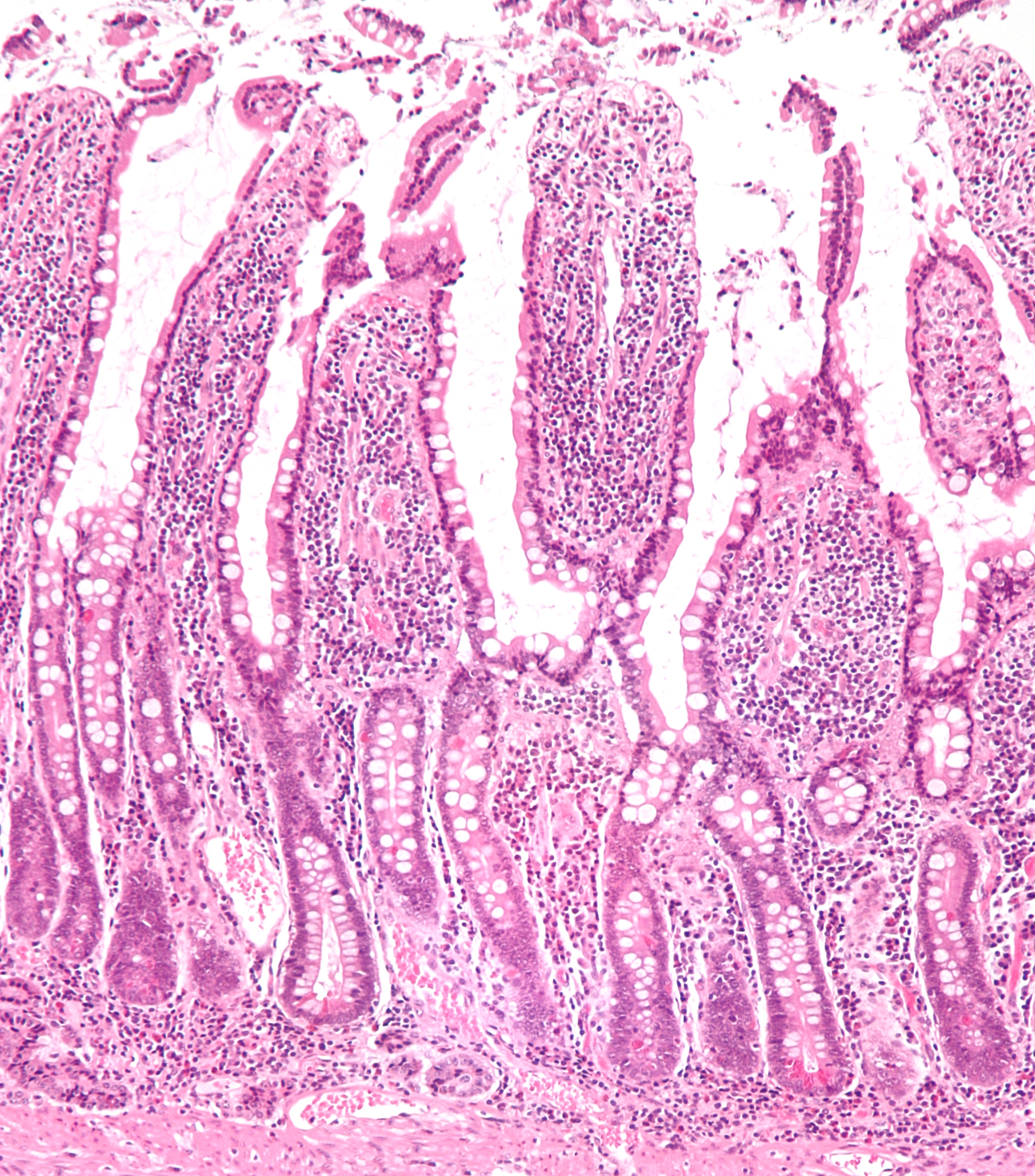
The small intestine or small bowel is an organ in the gastrointestinal tract where most of the absorption of nutrients from food takes place. It lies between the stomach and large intestine, and receives bile and pancreatic juice through the pancreatic duct to aid in digestion. The small intestine is about long and folds many times to fit in the abdomen. Although it is longer than the large intestine, it is called the small intestine because it is narrower in diameter. The small intestine has three distinct regions – the duodenum, jejunum, and ileum. The duodenum, the shortest, is where preparation for absorption through small finger-like protrusions called villi begins. The jejunum is specialized for the absorption through its lining by enterocytes: small nutrient particles which have been previously digested by enzymes in the duodenum. The main function of the ileum is to absorb vitamin B12, bile salts, and whatever products of digestion that were not absorbed by the ... [...More Info...] [...Related Items...] OR: [Wikipedia] [Google] [Baidu] |
Oral Mucosa
The oral mucosa is the mucous membrane lining the inside of the mouth. It comprises stratified squamous epithelium, termed "oral epithelium", and an underlying connective tissue termed ''lamina propria''. The oral cavity has sometimes been described as a mirror that reflects the health of the individual. Changes indicative of disease are seen as alterations in the oral mucosa lining the mouth, which can reveal systemic conditions, such as diabetes or vitamin deficiency, or the local effects of chronic tobacco or alcohol use. The oral mucosa tends to heal faster and with less scar formation compared to the skin. The underlying mechanism remains unknown, but research suggests that extracellular vesicles might be involved. Classification Oral mucosa can be divided into three main categories based on function and histology: *Lining mucosa, nonkeratinized stratified squamous epithelium, found almost everywhere else in the oral cavity, including the: **Alveolar mucosa, the lining betwe ... [...More Info...] [...Related Items...] OR: [Wikipedia] [Google] [Baidu] |
Sacrum
The sacrum (plural: ''sacra'' or ''sacrums''), in human anatomy, is a large, triangular bone at the base of the spine that forms by the fusing of the sacral vertebrae (S1S5) between ages 18 and 30. The sacrum situates at the upper, back part of the pelvic cavity, between the two wings of the pelvis. It forms joints with four other bones. The two projections at the sides of the sacrum are called the alae (wings), and articulate with the ilium at the L-shaped sacroiliac joints. The upper part of the sacrum connects with the last lumbar vertebra (L5), and its lower part with the coccyx (tailbone) via the sacral and coccygeal cornua. The sacrum has three different surfaces which are shaped to accommodate surrounding pelvic structures. Overall it is concave (curved upon itself). The base of the sacrum, the broadest and uppermost part, is tilted forward as the sacral promontory internally. The central part is curved outward toward the posterior, allowing greater room for the pel ... [...More Info...] [...Related Items...] OR: [Wikipedia] [Google] [Baidu] |
Hard Palate
The hard palate is a thin horizontal bony plate made up of two bones of the facial skeleton, located in the roof of the mouth. The bones are the palatine process of the maxilla and the horizontal plate of palatine bone. The hard palate spans the alveolar process, alveolar arch formed by the alveolar process that holds the upper teeth (when these are developed). Structure The hard palate is formed by the palatine process of the maxilla and horizontal plate of palatine bone. It forms a partition between the nasal passages and the mouth. On the anterior portion of the hard palate are the plicae, irregular ridges in the mucous membrane that help facilitate the movement of food backward towards the larynx. This partition is continued deeper into the mouth by a fleshy extension called the soft palate. On the ventral surface of hard palate, some projections or transverse ridges are present which are called as palatine rugae. Function The hard palate is important for feeding and sp ... [...More Info...] [...Related Items...] OR: [Wikipedia] [Google] [Baidu] |


