|
Louis-Charles Malassez
Louis-Charles Malassez (21 September 1842 – 22 December 1909) was a French anatomist and histologist born in Nevers, department of Nièvre. He studied medicine in Paris, where he worked as an ''interne'' from 1867. He served with the 5th Ambulance Corps during the Franco-Prussian War, afterwards returning to Paris, where he worked with distinguished physicians that included Claude Bernard, Jean-Martin Charcot and Pierre Potain. In 1875, he attained the chair of anatomy at Collège de France, and in 1894 he became a member of the ''Académie de Médecine''. He conducted histological research of the blood, and is credited for design of the hemocytometer, a device used to quantitatively measure blood cells. In the field of dentistry, he described residual cells of the epithelial root sheath in the periodontal ligament. These remaining cells are referred to as epithelial cell rests of Malassez (ERM). A genus of fungi called ''Malassezia'' bears his name. The species in the genus ... [...More Info...] [...Related Items...] OR: [Wikipedia] [Google] [Baidu] |
Periodontal Ligament
The periodontal ligament, commonly abbreviated as the PDL, is a group of specialized connective tissue fibers that essentially attach a tooth to the alveolar bone within which it sits. It inserts into root cementum one side and onto alveolar bone on the other. Structure The PDL consists of principal fibres, loose connective tissue, blast and clast cells, oxytalan fibres and Cell Rest of Malassez. Alveolodental ligament The main principal fiber group is the alveolodental ligament, which consists of five fiber subgroups: alveolar crest, horizontal, oblique, apical, and interradicular on multirooted teeth. Principal fibers other than the alveolodental ligament are the transseptal fibers. All these fibers help the tooth withstand the naturally substantial compressive forces that occur during chewing and remain embedded in the bone. The ends of the principal fibers that are within either cementum or alveolar bone proper are considered Sharpey fibers. * Alveolar crest fibers ('' ... [...More Info...] [...Related Items...] OR: [Wikipedia] [Google] [Baidu] |
Otitis Externa
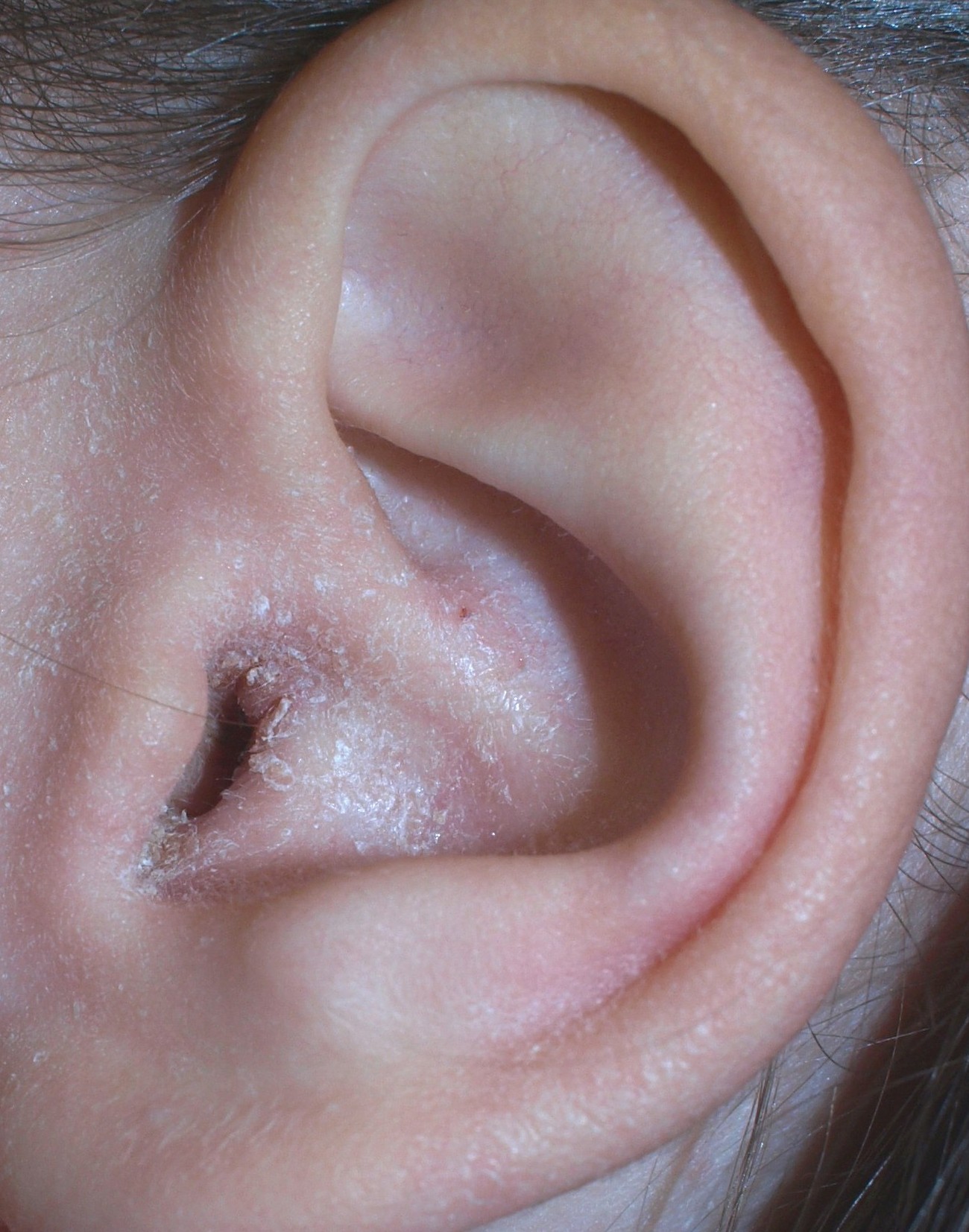
Otitis externa, also called swimmer's ear, is inflammation of the ear canal. It often presents with ear pain, swelling of the ear canal, and occasionally decreased hearing. Typically there is pain with movement of the outer ear. A high fever is typically not present except in severe cases. Otitis externa may be acute (lasting less than six weeks) or chronic (lasting more than three months). Acute cases are typically due to bacterial infection, and chronic cases are often due to allergies and autoimmune disorders. the most common cause of Otitis externa is bacterial. Risk factors for acute cases include swimming, minor trauma from cleaning, using hearing aids and ear plugs, and other skin problems, such as psoriasis and dermatitis. People with diabetes are at risk of a severe form of ''malignant otitis externa''. Diagnosis is based on the signs and symptoms. Culturing the ear canal may be useful in chronic or severe cases. Acetic acid ear drops may be used as a preventive measur ... [...More Info...] [...Related Items...] OR: [Wikipedia] [Google] [Baidu] |
Tinea Versicolor
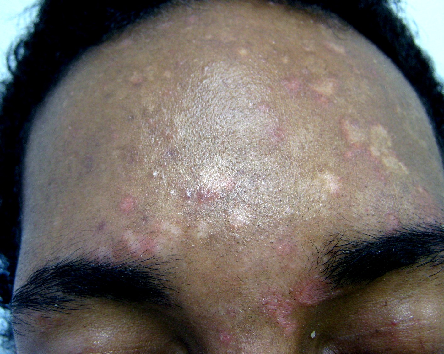
Tinea versicolor (also pityriasis versicolor) is a condition characterized by a skin eruption on the trunk and proximal extremities. The majority of tinea versicolor is caused by the fungus '' Malassezia globosa'', although ''Malassezia furfur'' is responsible for a small number of cases. These yeasts are normally found on the human skin and become troublesome only under certain circumstances, such as a warm and humid environment, although the exact conditions that cause initiation of the disease process are poorly understood. The condition pityriasis versicolor was first identified in 1846. Versicolor comes from the Latin ' 'to turn' + ''color''. It is also commonly referred to as Peter Elam's disease in many parts of South Asia. Signs and symptoms The symptoms of this condition include: * Occasional fine scaling of the skin producing a very superficial ash-like scale * Pale, dark tan, or pink in color, with a reddish undertone that can darken when the patient is overheated, s ... [...More Info...] [...Related Items...] OR: [Wikipedia] [Google] [Baidu] |
Dermatitis
Dermatitis is inflammation of the skin, typically characterized by itchiness, redness and a rash. In cases of short duration, there may be small blisters, while in long-term cases the skin may become thickened. The area of skin involved can vary from small to covering the entire body. Dermatitis is often called eczema, and the difference between those terms is not standardized. The exact cause of the condition is often unclear. Cases may involve a combination of allergy and poor venous return. The type of dermatitis is generally determined by the person's history and the location of the rash. For example, irritant dermatitis often occurs on the hands of those who frequently get them wet. Allergic contact dermatitis occurs upon exposure to an allergen, causing a hypersensitivity reaction in the skin. Prevention of atopic dermatitis is typically with essential fatty acids, and may be treated with moisturizers and steroid creams. The steroid creams should generally be of mid ... [...More Info...] [...Related Items...] OR: [Wikipedia] [Google] [Baidu] |
Seborrhea
A sebaceous gland is a microscopic exocrine gland in the skin that opens into a hair follicle to secrete an oily or waxy matter, called sebum, which lubricates the hair and skin of mammals. In humans, sebaceous glands occur in the greatest number on the face and scalp, but also on all parts of the skin except the palms of the hands and soles of the feet. In the eyelids, meibomian glands, also called tarsal glands, are a type of sebaceous gland that secrete a special type of sebum into tears. Surrounding the female nipple, areolar glands are specialized sebaceous glands for lubricating the nipple. Fordyce spots are benign, visible, sebaceous glands found usually on the lips, gums and inner cheeks, and genitals. Structure Location Sebaceous glands are found throughout all areas of the skin, except the palms of the hands and soles of the feet. There are two types of sebaceous glands, those connected to hair follicles and those that exist independently. Sebaceous glands are fo ... [...More Info...] [...Related Items...] OR: [Wikipedia] [Google] [Baidu] |
Lipophilic
Lipophilicity (from Greek λίπος "fat" and φίλος "friendly"), refers to the ability of a chemical compound to dissolve in fats, oils, lipids, and non-polar solvents such as hexane or toluene. Such non-polar solvents are themselves lipophilic (translated as "fat-loving" or "fat-liking"), and the axiom that "like dissolves like" generally holds true. Thus lipophilic substances tend to dissolve in other lipophilic substances, but hydrophilic ("water-loving") substances tend to dissolve in water and other hydrophilic substances. Lipophilicity, hydrophobicity, and non-polarity may describe the same tendency towards participation in the London dispersion force, as the terms are often used interchangeably. However, the terms "lipophilic" and "hydrophobic" are not synonymous, as can be seen with silicones and fluorocarbons, which are hydrophobic but not lipophilic. __TOC__ Surfactants Hydrocarbon-based surfactants are compounds that are amphiphilic (or amphipathic), having a h ... [...More Info...] [...Related Items...] OR: [Wikipedia] [Google] [Baidu] |
Malassezia Sympodialis
''Malassezia sympodialis'' is a species in the genus ''Malassezia''. It is characterized by a pronounced lipophily, unilateral, percurrent or sympodial budding and an irregular, corrugated cell wall ultrastructure. It is one of the most common species found on the skin of healthy and diseased individuals. It is considered to be part of the skin's normal human microbiota and begins to colonize the skin of humans shortly after birth. ''Malassezia sympodialis'', often has a symbiotic or commensal relationship with its host, but it can act as a pathogen causing a number of different skin diseases, such as atopic dermatitis. History In 1846, Karl Ferdinand Eichstedt was the first to identify the association of fungi with pityriasis versicolor, a common infection associated with the genus ''Malassezia''. The name applied to the fungal agent responsible shifted multiple times over the next 150 years until the genus ''Pityrosporum'' was settled upon for the teleomorph, and ''Malassezia' ... [...More Info...] [...Related Items...] OR: [Wikipedia] [Google] [Baidu] |
Malassezia Pachydermatis
''Malassezia pachydermatis'' is a zoophilic yeast in the division Basidiomycota. It was first isolated in 1925 by Fred Weidman, and it was named ''pachydermatis'' (Greek for 'thick-skin') after the original sample taken from an Indian rhinoceros (''Rhinocerosus unicornis'') with severe exfoliative dermatitis. Within the genus ''Malassezia'', ''M. pachydermatis'' is most closely related to the species '' M. furfur''. A commensal fungus, it can be found within the microflora of healthy mammals such as humans, cats and dogs, However, it is capable of acting as an opportunistic pathogen under special circumstances and has been seen to cause skin and ear infections, most often occurring in canines. Description ''Malassezia pachydermatis'' is a bottle-shaped, non-lipid dependent lipophilic yeast in the genus ''Malassezia''. Colonies are cream or yellowish in colour, smooth to wrinkled and convex with a margin possessing a slightly lobed appearance. Cells are ovoidal in sh ... [...More Info...] [...Related Items...] OR: [Wikipedia] [Google] [Baidu] |
Malassezia Ovale
''Malassezia'' (formerly known as ''Pityrosporum'') is a genus of fungi. It is the sole genus in family Malasseziaceae, which is the only family in order Malasseziales, itself the single member of class Malasseziomycetes. ''Malassezia'' species are naturally found on the skin surfaces of many animals, including humans. In occasional opportunistic infections, some species can cause hypopigmentation or hyperpigmentation on the trunk and other locations in humans. Allergy tests for these fungi are available. Systematics Due to progressive changes in their nomenclature, some confusion exists about the naming and classification of ''Malassezia'' yeast species. Work on these yeasts has been complicated because they require specific growth media and grow very slowly in laboratory culture. ''Malassezia'' were originally identified by the French scientist Louis-Charles Malassez in the late nineteenth century. Raymond Sabouraud identified a dandruff-causing organism in 1904 and called it ... [...More Info...] [...Related Items...] OR: [Wikipedia] [Google] [Baidu] |
Malassezia Furfur
''Malassezia furfur'' (formerly known as ''Pityrosporum ovale'' in its hyphal form) is a species of yeast (a type of fungus) that is naturally found on the skin surfaces of humans and some other mammals. It is associated with a variety of dermatological conditions caused by fungal infections, notably seborrhoeic dermatitis and tinea versicolor. As an opportunistic pathogen, it has further been associated with dandruff, malassezia folliculitis, pityriasis versicolor (alba), and malassezia intertrigo, as well as catheter-related fungemia and pneumonia in patients receiving hematopoietic transplants. The fungus can also affect other animals, including dogs. Background ''Malassezia furfur'' is a fungus that lives on the superficial layers of the dermis. It generally exists as a commensal organism forming a natural part of the human skin microbiota, but it can gain pathogenic capabilities when morphing from a yeast to a hyphal form during its life cycle, through unknown molecular ... [...More Info...] [...Related Items...] OR: [Wikipedia] [Google] [Baidu] |
Malassezia
''Malassezia'' (formerly known as ''Pityrosporum'') is a genus of fungi. It is the sole genus in family Malasseziaceae, which is the only family in order Malasseziales, itself the single member of class Malasseziomycetes. ''Malassezia'' species are naturally found on the skin surfaces of many animals, including humans. In occasional opportunistic infections, some species can cause hypopigmentation or hyperpigmentation on the trunk and other locations in humans. Allergy tests for these fungi are available. Systematics Due to progressive changes in their nomenclature, some confusion exists about the naming and classification of ''Malassezia'' yeast species. Work on these yeasts has been complicated because they require specific growth media and grow very slowly in laboratory culture. ''Malassezia'' were originally identified by the French scientist Louis-Charles Malassez in the late nineteenth century. Raymond Sabouraud identified a dandruff-causing organism in 1904 and called i ... [...More Info...] [...Related Items...] OR: [Wikipedia] [Google] [Baidu] |


